Copyright
©The Author(s) 2017.
World J Gastroenterol. Oct 14, 2017; 23(38): 6931-6941
Published online Oct 14, 2017. doi: 10.3748/wjg.v23.i38.6931
Published online Oct 14, 2017. doi: 10.3748/wjg.v23.i38.6931
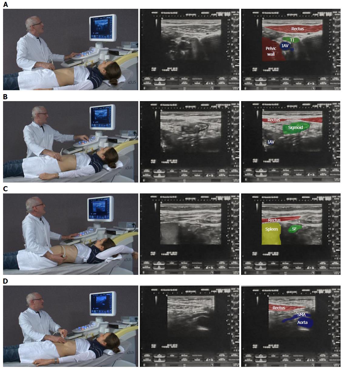
Figure 1 A systematic approach in examining the whole intestine.
A: Examination begins in a relaxed ventral position; B: Beginning medial to the right anterior superior iliac spine, the iliacal vessels (IAV) are identified and the first bowel loop crossing medial-to-lateral is the terminal ileum (TI). The same technique on the right identifies the sigmoid colon; C: Elevating the arm spreads the rib spaces to improve visualisation of the splenic flexure (SF); D: Gentle pressure as the patient breaths out improves visualization of the mesentery and superior mesenteric artery (SMA) to exclude lymphadenopathy. The videos can be accessed via the efsumb website [http://www.efsumb.org/education/cfd-videos001.asp].
- Citation: Atkinson NSS, Bryant RV, Dong Y, Maaser C, Kucharzik T, Maconi G, Asthana AK, Blaivas M, Goudie A, Gilja OH, Nuernberg D, Schreiber-Dietrich D, Dietrich CF. How to perform gastrointestinal ultrasound: Anatomy and normal findings. World J Gastroenterol 2017; 23(38): 6931-6941
- URL: https://www.wjgnet.com/1007-9327/full/v23/i38/6931.htm
- DOI: https://dx.doi.org/10.3748/wjg.v23.i38.6931


