Copyright
©The Author(s) 2016.
World J Gastroenterol. Feb 21, 2016; 22(7): 2349-2356
Published online Feb 21, 2016. doi: 10.3748/wjg.v22.i7.2349
Published online Feb 21, 2016. doi: 10.3748/wjg.v22.i7.2349
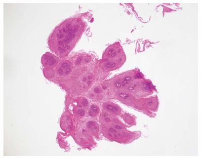
Figure 5 Squamous papilloma of the esophagus is diagnosed pathologically by a hematoxylin and eosin stain.
The lesion is composed of papillary fronds lined by several layers of non-keratinizing squamous epithelium and forming finger-like projections (magnification × 20).
- Citation: Wong MW, Bair MJ, Shih SC, Chu CH, Wang HY, Wang TE, Chang CW, Chen MJ. Using typical endoscopic features to diagnose esophageal squamous papilloma. World J Gastroenterol 2016; 22(7): 2349-2356
- URL: https://www.wjgnet.com/1007-9327/full/v22/i7/2349.htm
- DOI: https://dx.doi.org/10.3748/wjg.v22.i7.2349


