Published online Oct 28, 2016. doi: 10.3748/wjg.v22.i40.9022
Peer-review started: May 17, 2016
First decision: July 12, 2016
Revised: August 25, 2016
Accepted: September 6, 2016
Article in press: September 6, 2016
Published online: October 28, 2016
Processing time: 163 Days and 1.9 Hours
In this report, we present 3 cases of malignant small bowel obstruction, treated with palliative care using endoscopic self-expandable metallic stent (SEMS) placement, with the aim to identify the safety and efficacy of this procedure. Baseline patient characteristics, procedure methods, procedure time, technical and clinical success rates, complications, and patient outcomes were obtained. All 3 patients had pancreatic cancer with small bowel strictures. One patient received the SEMS using colonoscopy, while the other 2 patients received SEMS placement via double balloon endoscopy using the through-the-overtube technique. The median procedure time was 104 min. The technical and clinical success rates were 100%. Post-treatment, obstructive symptoms in all patients improved, and a low-residue diet could be tolerated. All stents remained within the patients until their deaths. The median overall survival time (stent patency time) was 76 d. SEMS placement is safe and effective as a palliative treatment for malignant small bowel obstruction.
Core tip: We present 3 cases of malignant small bowel obstruction, treated with palliative care using endoscopic self-expandable metallic stent (SEMS) placement, and have identified that the procedure is safe and effective. Two patients were treated using the through-the-overtube technique, while the remaining case was the first case of SEMS placement in a malignant distal small bowel obstruction.
- Citation: Tsuboi A, Kuwai T, Nishimura T, Iio S, Mori T, Imagawa H, Yamaguchi T, Yamaguchi A, Kouno H, Kohno H. Safety and efficacy of self-expandable metallic stents in malignant small bowel obstructions. World J Gastroenterol 2016; 22(40): 9022-9027
- URL: https://www.wjgnet.com/1007-9327/full/v22/i40/9022.htm
- DOI: https://dx.doi.org/10.3748/wjg.v22.i40.9022
Malignant small bowel obstructions are primarily treated with surgical intervention. However, palliative surgery is highly invasive in such patients, with poor prognosis; therefore, minimally invasive therapies, such as the endoscopic placement of self-expandable metallic stents (SEMS), have been considered. SEMS has been used successfully to palliate malignant gastrointestinal obstructions, and is widely reported to result in good clinical outcomes for colonic, esophageal, and gastric obstructions[1-4]. However, SEMS placement for malignant small bowel obstructions is little-known and also more challenging. A deep small bowel enteroscopy is limited, and three endoscopy systems are now available: [double balloon endoscopy (DBE), single balloon endoscopy, and spiral endoscopy[5-7]]; however, such endoscope systems do not have working channels large enough for the stent delivery systems to pass through. Therefore, the standard through-the-scope (TTS) technique for stent deployment could not be applied. To mitigate this limitation, we modified the standard over-the-guidewire (OTW) technique for stent deployment. In this report, we present 3 cases of malignant small bowel obstruction treated with palliative SEMS placement.
Of the 3 patients studied, 1 received stent deployment using the standard TTS technique via colonoscopy (CF-H260AI, Olympus, Tokyo, Japan); SEMS placement in the other 2 patients was achieved using the through-the-overtube (TTO) technique via DBE (EN450T5/W, FUJIFILM, Tokyo, Japan). The TTO technique is a modified version of the OTW technique. First, the endoscope with the overtube (TS13140, FUJIFILM, Tokyo, Japan), without its balloon tip, was advanced towards the stricture, and a 0.035-inch guidewire (Jagwire, Boston Scientific Corp., Natick, MA, United States) was passed through the stricture. Subsequently, the guidewire and the overtube were left in place, and the endoscope was removed. Finally, the overtube was utilized as a large channel to advance the stent through the stricture over the guidewire under fluoroscopic guidance. All 3 cases were performed by a single expert endoscopist with experience in over 20 cases of colon stenting.
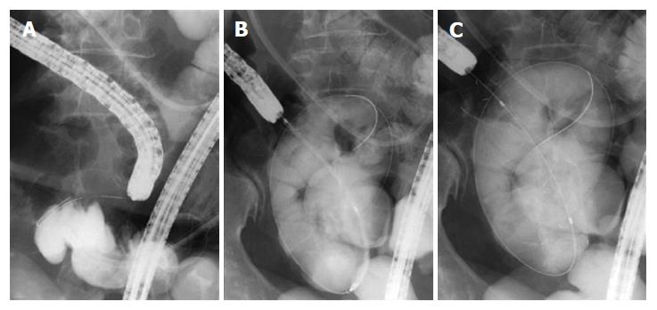
A 60-year-old woman was admitted to our hospital with abdominal pain due to terminal ileum obstruction because of peritoneal dissemination of pancreatic cancer. Although her symptoms improved after insertion of an ileus tube, they recurred following commencement of oral intake. Consequently, the decision was made to attempt SEMS placement as a palliative therapy. A colonoscope was advanced to the stricture, and the standard TTS technique for stenting with a 10 cm × 20 mm uncovered SEMS (Niti-S biliary stent, TaeWoong Medical, Seoul, South Korea) (Figure 1) was used. The patient was discharged 24 d after stenting, and died 109 d after stenting.
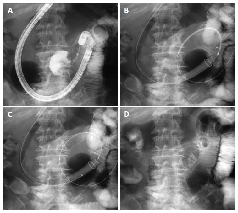
An 87-year-old woman was admitted to our hospital with anorexia, vomiting, and weight loss. An abdominal computed tomography (CT) scan revealed cancer in the head of the pancreas, a metastatic hepatic tumor, and expansion of the stomach and duodenum. We concluded that the obstruction of the distal duodenum/angle of Treitz was secondary to pancreatic cancer invasion. We attempted advancement of a colonoscope (CF-H260AZI, Olympus, Tokyo, Japan) to the stricture, but could not reach the region as it was too deep, and the endoscope position was tortuous. Subsequently, DBE endoscopy was performed, and access was achieved with a stable endoscope position. Unfortunately, we could not use the TTS technique for stent deployment given the position of the stricture. Therefore, we decided to employ the TTO technique for stenting. An endoscope loaded with a balloon overtube was advanced into the stricture for a trans-oral approach. While the guidewire was left in place beyond the stricture, the endoscope was removed, leaving the overtube in place. A 10 cm × 22 mm stent (Niti-S D pyloric/duodenal stent, TaeWoong Medical, Seoul, South Korea) was advanced using the OTW technique through the overtube, and was deployed successfully (Figure 2). The patient was discharged 15 d after the procedure and died 76 d after stenting.
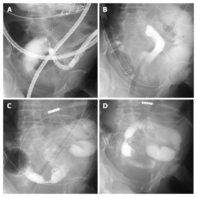
A 69-year-old woman with Stage IV pancreatic cancer who was receiving chemotherapy was admitted due to abdominal distension and vomiting. Abdominal CT revealed an intestinal stricture secondary to peritoneal dissemination. She was initially treated with ileus tube insertion for the obstruction (due to recurrence), but requested palliative SEMS placement. As we were able to reach the stricture with DBE, we decided to place the SEMS using the TTO technique. The endoscope and overtube were advanced to the stricture via the trans-anal approach. The guidewire (Wrangler, PIOLAX medical devices Inc., Kanagawa, Japan) and overtube were left in place while the endoscope was removed. An 8 cm × 18 mm stent (Niti-S D colonic stent, TaeWoong Medical, Seoul, South Korea) was advanced through the overtube, and deployed successfully (Figure 3). The patient was discharged on day 12 after the procedure and died of her primary cancer 29 d after stenting.
All 3 patients in our study tolerated clear fluids the day after stenting, followed by a low residue diet. They were all discharged from the hospital at variable times with no major complications following SEMS placement (Table 1; summary of cases). All stents remained patent until patient death. The technical and clinical success rates were 100%.
| Age/sex | Tumor | Stricture location | Scope | Stent delivery | Stricture length | Type of stent | Procedural time | Stent patency time | Time to oral intake after stent placement |
| 60/F | Pancreatic cancer | Terminal ileum | Olympus | TTS | 40 mm | Niti-S 20 mm × 10 cm | 132 min | 109 d | 5 d |
| Peritoneal | CF-H260A | ||||||||
| dissemination | |||||||||
| 87/F | Pancreatic cancer | Proximal jejunum | FUJIFILM | TTO | 30 mm | Niti-S 22 mm × 10 cm | 46 min | 76 d | 2 d |
| EN-450T5/W | |||||||||
| (trans-oral) | |||||||||
| Pancreatic cancer | Distal ileum | FUJIFILM | TTO | 20 mm | Niti-S 18 mm × 8 cm | 104 min | 29 d | 2 d | |
| Peritoneal dissemination | EN-450T5/W | ||||||||
| 60/F | (trans-anal) |
Malignant small bowel obstructions are typically caused by primary small bowel malignant tumors, local invasion of extrinsic malignant tumors, or metastasis[8]. Although surgical interventions such as gastroenteric bypass or ileostomy are considered the primary treatment for patients with malignant small bowel obstructions, they are not routinely performed in view of poor prognoses. Recently, SEMS placement has been used to treat malignant, non-small bowel gastrointestinal obstructions. Compared with surgery, SEMS placement is much less invasive. Good clinical outcomes have been reported in esophageal, gastroduodenal, and colorectal malignant obstructions[1,9]. While the efficacy and safety of palliative SEMS placement in these types of malignant obstructions are well-established, it remains largely unknown how such parameters measure in malignant small bowel obstructions distal to the ligament of Treitz. In this study, the technical and clinical success rates were 100%, with no major complications observed. Therefore, we propose that SEMS placement for malignant small bowel obstruction is equally effective and safe.
SEMS placement for malignant small bowel obstruction is challenging given the difficulty in accessing the site. Jeurnink et al[10] reported that enteral SEMS placement could be effectively and safely performed for malignant obstructions of the distal duodenum or proximal jejunum with colonoscopy. SEMS placement via colonoscopy is preferable for treatment of these lesions, considering the scope length and working channel, which is large enough for the standard TTS technique. In case 1, we used a colonoscope to deploy the SEMS with the TTS technique, as the scope could be advanced to the malignant stricture of the terminal ileum; the stenting was performed successfully.
Few reports on SEMS placement for malignant small bowel obstructions have been published. Lee et al[11] reported on 19 patients with malignant small bowel obstructions who underwent SEMS insertion. In these patients, SEMS placement was performed with the withdrawal-reinsertion technique using DBE. In their report, the technical and clinical success rates were 95% and 84%, respectively, and no major complications were observed during the procedures. According to the report, SEMS placement appeared to be effective for palliation of malignant small bowel obstructions. However, patients with malignant distal small bowel obstructions were excluded from the report; therefore, the efficacy and safety of SEMS placement in these regions was largely unknown.
As current enteroscopy systems do not possess working channels large enough for stent delivery systems to pass through, SEMS placement utilizing the TTS technique with an enteroscope is not possible. Therefore, the TTO technique was developed by modifying the OTW technique for SEMS placement in malignant distal small bowel obstructions using DBE. Ross et al[5] reported a case of malignant distal duodenal obstruction treated with SEMS placement using DBE with the TTO technique, similar to case 2 of our study. Lennon et al[6] reported a similar technique using SE. The key to this technique is the ability to reach the stricture and lock the overtube in position at the location; this provides a sheath through which the stent could easily pass the stricture and be deployed. Although this technique can potentially treat deeper malignant small bowel obstructions, the few case reports that are available have only used this technique in the distal duodenum, proximal jejunum, or surgically-reconstructed intestines[5,6,11-14]. To the best of our knowledge, case 3 of our study is the first case of SEMS placement in a malignant distal small bowel obstruction.
Shimatani et al[15] recently reported on SEMS placement for malignant afferent-loop obstruction using the TTS technique with a new short-type DBE (EI-580 BT; Fujifilm, Tokyo, Japan). As the new short-type DBE has a 3.2-mm working channel, the 9 Fr SEMS delivery system can be used with the TTS technique. We believe that a long-type DBE (also with a 3.2-mm working channel) will be developed in the near future. This will allow treatment of deeper malignant small bowel obstructions with SEMS placement using the TTS technique.
In conclusion, our study revealed that palliative SEMS placement is safe and effective in malignant small bowel obstructions. However, given our small sample size, further studies are warranted. Nevertheless, we believe that SEMS placement will play a significant role in the primary treatment of malignant small bowel obstructions in the near future, with further development of endoscopy and SEMS delivery systems.
The authors thank Naoko Matsumoto for assistance in collecting data and for office procedures.
Three patients (a 60-year-old woman, an 87-year-old woman, and a 69-year-old woman) presented with small bowel obstruction due to pancreatic cancer.
An abdominal computed tomography (CT) scan revealed the clinical diagnoses in all cases.
Abdominal CT showed small bowel obstruction because of pancreatic cancer.
Endoscopic self-expandable metallic stents (SEMS) were placed in each patient.
Only a few reports regarding SEMS placement for malignant small bowel obstructions have been published. Notably, case 3 in this study may be the first reported case of SEMS placement in a malignant distal small bowel obstruction.
through-the-scope was defined as tube-through-the-scope, over-the-guidewire was defined as over-the-guidewire, and through-the-overtube was defined as through-the-overtube.
The authors present 3 cases of malignant small bowel obstruction that received palliative SEMS placement safely and effectively. The SEMS placement will play a significant role in the primary treatment of malignant small bowel obstruction in the near future, with further development of endoscopy and SEMS delivery systems.
The authors presented 3 cases of malignant small bowel obstruction treated with palliative care by endoscopic SEMS placement and revealed that the procedure is safe and effective. The findings will be of interest to the readership.
Manuscript source: Unsolicited manuscript
Specialty type: Gastroenterology and hepatology
Country of origin: Japan
Peer-review report classification
Grade A (Excellent): 0
Grade B (Very good): B
Grade C (Good): C, C, C
Grade D (Fair): 0
Grade E (Poor): 0
P- Reviewer: Castro FJ, Lakatos PL, Manguso F, Sadik R S- Editor: Qi Y L- Editor: A E- Editor: Zhang FF
| 1. | Sebastian S, Johnston S, Geoghegan T, Torreggiani W, Buckley M. Pooled analysis of the efficacy and safety of self-expanding metal stenting in malignant colorectal obstruction. Am J Gastroenterol. 2004;99:2051-2057. [RCA] [PubMed] [DOI] [Full Text] [Cited by in RCA: 1] [Reference Citation Analysis (0)] |
| 2. | Knyrim K, Wagner HJ, Bethge N, Keymling M, Vakil N. A controlled trial of an expansile metal stent for palliation of esophageal obstruction due to inoperable cancer. N Engl J Med. 1993;329:1302-1307. [RCA] [PubMed] [DOI] [Full Text] [Cited by in Crossref: 543] [Cited by in RCA: 499] [Article Influence: 15.6] [Reference Citation Analysis (0)] |
| 3. | Piesman M, Kozarek RA, Brandabur JJ, Pleskow DK, Chuttani R, Eysselein VE, Silverman WB, Vargo JJ, Waxman I, Catalano MF. Improved oral intake after palliative duodenal stenting for malignant obstruction: a prospective multicenter clinical trial. Am J Gastroenterol. 2009;104:2404-2411. [RCA] [PubMed] [DOI] [Full Text] [Cited by in Crossref: 87] [Cited by in RCA: 83] [Article Influence: 5.2] [Reference Citation Analysis (0)] |
| 4. | van Hooft JE, Uitdehaag MJ, Bruno MJ, Timmer R, Siersema PD, Dijkgraaf MG, Fockens P. Efficacy and safety of the new WallFlex enteral stent in palliative treatment of malignant gastric outlet obstruction (DUOFLEX study): a prospective multicenter study. Gastrointest Endosc. 2009;69:1059-1066. [RCA] [PubMed] [DOI] [Full Text] [Cited by in Crossref: 148] [Cited by in RCA: 156] [Article Influence: 9.8] [Reference Citation Analysis (0)] |
| 5. | Ross AS, Semrad C, Waxman I, Dye C. Enteral stent placement by double balloon enteroscopy for palliation of malignant small bowel obstruction. Gastrointest Endosc. 2006;64:835-837. [RCA] [PubMed] [DOI] [Full Text] [Cited by in Crossref: 59] [Cited by in RCA: 50] [Article Influence: 2.6] [Reference Citation Analysis (0)] |
| 6. | Lennon AM, Chandrasekhara V, Shin EJ, Okolo PI. Spiral-enteroscopy-assisted enteral stent placement for palliation of malignant small-bowel obstruction (with video). Gastrointest Endosc. 2010;71:422-425. [RCA] [PubMed] [DOI] [Full Text] [Cited by in Crossref: 27] [Cited by in RCA: 27] [Article Influence: 1.8] [Reference Citation Analysis (0)] |
| 7. | Espinel J, Pinedo E. A simplified method for stent placement in the distal duodenum: Enteroscopy overtube. World J Gastrointest Endosc. 2011;3:225-227. [RCA] [PubMed] [DOI] [Full Text] [Full Text (PDF)] [Cited by in CrossRef: 17] [Cited by in RCA: 13] [Article Influence: 0.9] [Reference Citation Analysis (0)] |
| 8. | Baron TH. Expandable metal stents for the treatment of cancerous obstruction of the gastrointestinal tract. N Engl J Med. 2001;344:1681-1687. [RCA] [PubMed] [DOI] [Full Text] [Cited by in Crossref: 304] [Cited by in RCA: 280] [Article Influence: 11.7] [Reference Citation Analysis (0)] |
| 9. | Chopita N, Landoni N, Ross A, Villaverde A. Malignant gastroenteric obstruction: therapeutic options. Gastrointest Endosc Clin N Am. 2007;17:533-544, vi-vii. [RCA] [PubMed] [DOI] [Full Text] [Cited by in Crossref: 12] [Cited by in RCA: 13] [Article Influence: 0.7] [Reference Citation Analysis (0)] |
| 10. | Jeurnink SM, Repici A, Luigiano C, Pagano N, Kuipers EJ, Siersema PD. Use of a colonoscope for distal duodenal stent placement in patients with malignant obstruction. Surg Endosc. 2009;23:562-567. [RCA] [PubMed] [DOI] [Full Text] [Cited by in Crossref: 25] [Cited by in RCA: 21] [Article Influence: 1.2] [Reference Citation Analysis (0)] |
| 11. | Lee H, Park JC, Shin SK, Lee SK, Lee YC. Preliminary study of enteroscopy-guided, self-expandable metal stent placement for malignant small bowel obstruction. J Gastroenterol Hepatol. 2012;27:1181-1186. [RCA] [PubMed] [DOI] [Full Text] [Cited by in Crossref: 19] [Cited by in RCA: 22] [Article Influence: 1.7] [Reference Citation Analysis (0)] |
| 12. | Nakahara K, Okuse C, Matsumoto N, Suetani K, Morita R, Michikawa Y, Ozawa S, Hosoya K, Kobayashi S, Otsubo T. Enteral metallic stenting by balloon enteroscopy for obstruction of surgically reconstructed intestine. World J Gastroenterol. 2015;21:7589-7593. [RCA] [PubMed] [DOI] [Full Text] [Full Text (PDF)] [Cited by in CrossRef: 17] [Cited by in RCA: 16] [Article Influence: 1.6] [Reference Citation Analysis (0)] |
| 13. | Park JJ, Cheon JH. Malignant small bowel obstruction: the last frontier for gastrointestinal stenting. J Gastroenterol Hepatol. 2012;27:1136-1137. [RCA] [PubMed] [DOI] [Full Text] [Cited by in Crossref: 9] [Cited by in RCA: 10] [Article Influence: 0.8] [Reference Citation Analysis (0)] |
| 14. | Popa D, Ramesh J, Peter S, Wilcox CM, Mönkemüller K. Small Bowel Stent-in-Stent Placement for Malignant Small Bowel Obstruction Using a Balloon-Assisted Overtube Technique. Clin Endosc. 2014;47:108-111. [RCA] [PubMed] [DOI] [Full Text] [Full Text (PDF)] [Cited by in Crossref: 15] [Cited by in RCA: 15] [Article Influence: 1.4] [Reference Citation Analysis (0)] |
| 15. | Shimatani M, Takaoka M, Tokuhara M, Kato K, Miyoshi H, Ikeura T, Okazaki K. Through-the-scope self-expanding metal stent placement using newly developed short double-balloon endoscope for the effective management of malignant afferent-loop obstruction. Endoscopy. 2016;48 Suppl 1 UCTN:E6-E7. [RCA] [PubMed] [DOI] [Full Text] [Cited by in Crossref: 15] [Cited by in RCA: 16] [Article Influence: 1.8] [Reference Citation Analysis (0)] |


