Copyright
©The Author(s) 2016.
World J Gastroenterol. Oct 21, 2016; 22(39): 8670-8683
Published online Oct 21, 2016. doi: 10.3748/wjg.v22.i39.8670
Published online Oct 21, 2016. doi: 10.3748/wjg.v22.i39.8670
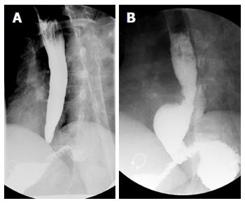
Figure 1 Barium esophagram of a patient before and several months after per oral endoscopic myotomy.
A 45-year-old woman with type II achalasia underwent per oral endoscopic myotomy (POEM). The pre-procedural barium esophagram (A, left panel) demonstrated a dilated esophagus with tapering at the gastroesophageal junction. Following POEM the patient gained 32 lbs over a 9-mo period but had occasional symptoms of regurgitation, which we suspected was from eating too much too quickly. Repeat barium esophagram showed the distal tapering of the gastroesophageal junction has resolved and there was immediate and unimpeded passage contrast passage into the stomach (B, right panel). A distal esophageal diverticulum was incidentally found, which can be seen in patients following POEM with a complete myotomy of the distal esophagus that is carried across the lower esophageal sphincter and into the gastric cardia.
- Citation: Uppal DS, Wang AY. Update on the endoscopic treatments for achalasia. World J Gastroenterol 2016; 22(39): 8670-8683
- URL: https://www.wjgnet.com/1007-9327/full/v22/i39/8670.htm
- DOI: https://dx.doi.org/10.3748/wjg.v22.i39.8670


