Published online Mar 14, 2016. doi: 10.3748/wjg.v22.i10.3052
Peer-review started: May 11, 2015
First decision: September 9, 2015
Revised: September 23, 2015
Accepted: November 24, 2015
Article in press: November 24, 2015
Published online: March 14, 2016
Processing time: 301 Days and 20.3 Hours
Gossypiboma is a surgical sponge that is retained in the body after the operation. A 39-year-old female presented with vague lower abdominal pain, fever, and rectal discharge 15 mo after hysterectomy. The sponge remaining in the abdomen had no radiopaque marker. Therefore a series of radiographic evaluations was fruitless. The surgical sponge was found in the rectosigmoid colon on colonoscopy. The sponge penetrated the sigmoid colon and rectum transmurally, forming an opening on both sides. The patient underwent low anterior resection and was discharged without postoperative complications.
Core tip: This case involved an unusual migration and placement of a retained surgical sponge; the retained sponge penetrated the intestinal submucosa and migrated to the sigmoid colon and rectum, causing formation of a fistula which had two openings. In this case, the importance of the radiopaque marker was reviewed. Surgical materials with radiopaque markers should be used, which make diagnosis significantly in suspected cases of material being left in the abdominal cavity. Without the radiopaque markers, diagnosis of a retained sponge is difficult, as was the situation in this case. We emphasize the importance of using radiopaque-labeled sponges in all abdominal operations and vigilant adherence to surgical material count in all procedures.
- Citation: Shin WY, Im CH, Choi SK, Choe YM, Kim KR. Transmural penetration of sigmoid colon and rectum by retained surgical sponge after hysterectomy. World J Gastroenterol 2016; 22(10): 3052-3055
- URL: https://www.wjgnet.com/1007-9327/full/v22/i10/3052.htm
- DOI: https://dx.doi.org/10.3748/wjg.v22.i10.3052
Reports of retained surgical sponge, or gossypiboma, are infrequent. Due to its relative rarity and medico-legal implications, it is difficult to determine whether there has been an occurrence. Although several cases of complete migration of surgical gauze or sponge into the colon have been reported, to the best of our knowledge, the following case is a rare case of gossypiboma caused by transmural penetration of the colon by a retained surgical sponge. This case was referred from an overseas institution.
A 39-year-old Uzbekistan female presented with complaints of vague lower abdominal pain, febrile sense, and purulent rectal discharge for a month. She had undergone a hysterectomy for myoma uteri in Uzbekistan approximately 15 mo prior to this visit. On physical examination, she appeared to be chronically ill. Vital signs were stable and within normal limits. No particular abdominal findings were observed on physical examination, except mild suprapubic tenderness, and a lower midline laparotomy scar was noted. On digital rectal examination, anal sphincter tone was normal with hemoccult-negative stool. Blood tests included a hemoglobin level of 14.1 g/dL, a white blood cell count of 14600/μL, and a platelet count of 354000/μL. Urinalysis and admission panel including liver function test were negative.
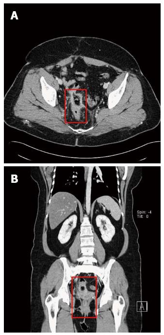
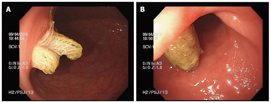
An abdominal plain radiography showed no remarkable findings and an abdominal computed tomography displayed only bowel wall thickening and mucosal enhancement in the rectosigmoid area with pericolic inflammatory change (Figure 1). Colonoscopy showed that the retained sponge had penetrated the sigmoid colon and rectum transmurally, forming an opening on both sides (Figure 2).
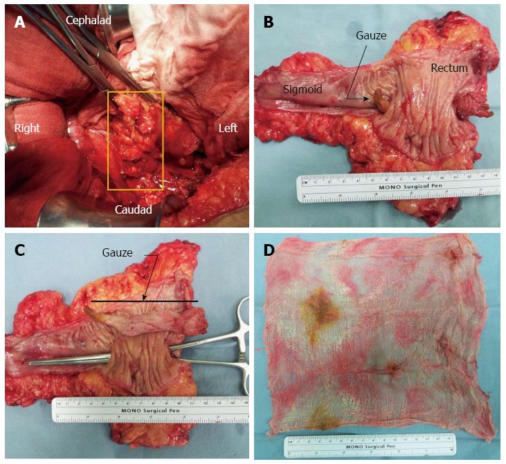
The patient underwent a median laparotomy via a prior surgical incision line. An elongated mass-like lesion was felt within the left lateral wall of the rectosigmoid colon (Figure 3A). There were no other significant findings. Because of the severe local inflammatory process related to the entrance site of the retained sponge, a low anterior resection was performed for removal of this mass. When opened, the specimen revealed an abnormal fistulous communication through the wall of the sigmoid colon and rectum (Figure 3B and C). The foreign body was an undamaged surgical sponge without a radiopaque marker, measuring 20 cm × 20 cm (Figure 3D). The postoperative course was uneventful and the patient was discharged without complications on the 13th postoperative day.
We report on a case in which a surgical sponge completely penetrated the intestinal submucosa and migrated to the lower sigmoid colon and upper rectum, causing formation of a fistula, which had two openings, one to the sigmoid colon and the other to the rectum. To the best of our knowledge, this was an unusual migration and placement of a surgical sponge.
Unintentional retainment of surgical material in the abdomen usually requires a reoperation which increases risk of mortality and morbidity of the patient, cost, and medico-legal implications[1]. The retained sponge occurs commonly at the long operative procedure during which there is a change of the scrubbed nursing staff. The meticulous sponge count do not prevent the retention of surgical sponge, but minimized its occurrence. Diagnosis of a gossypiboma can be difficult if there is no radiopaque marker on the sponge itself. The possibility of retained surgical material should be considered in any postoperative patient who presents with pain, infection, or a palpable mass in the abdomen[2-4].
Appearance of symptoms due to a retained surgical sponge depends on the degree of bacterial contamination at the time of surgery[2]. There are two usual responses to a retained surgical sponge which lead to detection. The sponge can cause an inflammatory reaction which leads to formation of an abscess[2,5]. It can also trigger a fibrotic reaction and development of a mass[6,7]. Formation of an abscess is known to be associated with transmural migration. The bacterial infection and abscess formation leads to intestinal perforation, which initiates the migration of the sponge into the lumen of the bowel. Internal fistulation is a major feature in this phase. Migration of the sponge into the bowel lumen may cause intestinal obstruction or spontaneous passage of the sponge to the rectum to be defecated[2].
Postoperative patients with retained surgical materials might present with obvious signs like a palpable mass in the abdomen, however diagnosis is often difficult. Currently, radiopaque markers are used widely. However, identifying a surgical material on a radiograph can be difficult because the marker may be folded, twisted, or it may disintegrate over a period of time. For example, Kopka et al[8] reported that this marker was seen in only 9 of 13 patients with a radiopaque-labeled retained sponge. Ultrasonography, computed tomography, or magnetic resonance imaging is usually performed when diagnosis cannot be made from a plain X-ray image. Computed tomographic images can reveal air bubbles and whirl-like or spongiform pattern. The sponge may be visualized as a hypodense mass with a thick peripheral rim[8,9].
Retained surgical material should be removed in most cases. However in some cases, attempting to remove the surgical material may cause more harm than the material itself, although it is usually a small piece of surgical material or a needle[6]. Opening the previous operation site can be attempted when the material must be removed. If possible, endoscopic or laparoscopic approaches may be attempted[10].
In this case, the importance of the radiopaque marker was reviewed. Surgical materials with radiopaque markers should be used, which can make diagnosis significantly easier in suspected cases of material left in the abdominal cavity. Without radiopaque markers, diagnosis of retained sponges is difficult, as was the situation in this case. New technologies including an electronic article surveillance system that may decrease the incidence of retained surgical materials are being developed[11]. However, above all, we emphasize the importance of using a radiopaque-labeled sponge in all abdominal operations and vigilant adherence to surgical material count in all procedures.
A 39-year-old Uzbekistan female presented with complaints of vague lower abdominal pain, febrile sense, and purulent rectal discharge for a month.
On physical examination, there was mild suprapubic tenderness, and a lower midline laparotomy scar was noted.
Differential diagnosis included intra-abdominal locally expansile masses such as hematomas, abscesses, neoplastic lesions and fecalomas. Other conditions, such as postoperative adhesion, intussusception and mesenteric panniculitis should be considered among diagnostic possibilities.
Blood tests included a hemoglobin level of 14.1 g/dL, a white blood cell count of 14600/μL and a platelet count of 354000/μL. Urinalysis and admission panel including liver function test were negative.
An abdominal plain radiography showed no remarkable findings and an abdominal computed tomography displayed only bowel wall thickening and mucosal enhancement in the rectosigmoid area with pericolic inflammatory change.
The pathological report revealed a fistulous tract formation between sigmoid colon and rectum with chronic inflammation and foreign body reaction in the sigmoid colon and rectum.
The patient underwent a low anterior resection.
Although several cases of gossypiboma such as complete migration into the bowel have been reported, to the best of our knowledge, this unique type of gossypiboma caused by transmural penetration of sigmoid colon and rectum seems to be the first report.
Gossypiboma or more broadly retained foreign object is the technical term for a surgical complications resulting from foreign materials, such as a surgical sponge, accidentally left inside a patient’s body.
Surgical materials with radiopaque markers should be used, which can make diagnosis significantly easier in suspected cases of material left in the abdominal cavity. Without radiopaque markers, diagnosis of retained sponges is difficult, as was the situation in this case.
This case report in interesting and supported with good figures.
P- Reviewer: Corum CA, Meshikhes AWN S- Editor: Gong ZM L- Editor: A E- Editor: Wang CH
| 1. | Uluçay T, Dizdar MG, SunayYavuz M, Aşirdizer M. The importance of medico-legal evaluation in a case with intraabdominal gossypiboma. Forensic Sci Int. 2010;198:e15-e18. [RCA] [PubMed] [DOI] [Full Text] [Cited by in Crossref: 14] [Cited by in RCA: 17] [Article Influence: 1.1] [Reference Citation Analysis (0)] |
| 2. | Hyslop JW, Maull KI. Natural history of the retained surgical sponge. South Med J. 1982;75:657-660. [RCA] [PubMed] [DOI] [Full Text] [Cited by in Crossref: 82] [Cited by in RCA: 86] [Article Influence: 2.0] [Reference Citation Analysis (0)] |
| 3. | Mefire AC, Tchounzou R, Guifo ML, Fokou M, Pagbe JJ, Essomba A, Malonga EE. Retained sponge after abdominal surgery: experience from a third world country. Pan Afr Med J. 2009;2:10. [PubMed] |
| 4. | Botet del Castillo FX, López S, Reyes G, Salvador R, Llauradó JM, Peñalva F, Trias R. Diagnosis of retained abdominal gauze swabs. Br J Surg. 1995;82:227-228. [PubMed] |
| 5. | Yildirim S, Tarim A, Nursal TZ, Yildirim T, Caliskan K, Torer N, Karagulle E, Noyan T, Moray G, Haberal M. Retained surgical sponge (gossypiboma) after intraabdominal or retroperitoneal surgery: 14 cases treated at a single center. Langenbecks Arch Surg. 2006;391:390-395. [RCA] [PubMed] [DOI] [Full Text] [Cited by in Crossref: 61] [Cited by in RCA: 53] [Article Influence: 2.7] [Reference Citation Analysis (0)] |
| 6. | Gibbs VC, Coakley FD, Reines HD. Preventable errors in the operating room: retained foreign bodies after surgery--Part I. Curr Probl Surg. 2007;44:281-337. [RCA] [PubMed] [DOI] [Full Text] [Cited by in Crossref: 82] [Cited by in RCA: 87] [Article Influence: 4.8] [Reference Citation Analysis (0)] |
| 7. | Sturdy JH, Baird RM, Gerein AN. Surgical sponges: a cause of granuloma and adhesion formation. Ann Surg. 1967;165:128-134. [PubMed] |
| 8. | Kopka L, Fischer U, Gross AJ, Funke M, Oestmann JW, Grabbe E. CT of retained surgical sponges (textilomas): pitfalls in detection and evaluation. J Comput Assist Tomogr. 1996;20:919-923. [RCA] [PubMed] [DOI] [Full Text] [Cited by in Crossref: 113] [Cited by in RCA: 119] [Article Influence: 4.1] [Reference Citation Analysis (0)] |
| 9. | Manzella A, Filho PB, Albuquerque E, Farias F, Kaercher J. Imaging of gossypibomas: pictorial review. AJR Am J Roentgenol. 2009;193:S94-101. [RCA] [PubMed] [DOI] [Full Text] [Cited by in Crossref: 100] [Cited by in RCA: 94] [Article Influence: 6.3] [Reference Citation Analysis (0)] |
| 10. | Karahasanoglu T, Unal E, Memisoglu K, Sahinler I, Atkovar G. Laparoscopic removal of a retained surgical instrument. J Laparoendosc Adv Surg Tech A. 2004;14:241-243. [RCA] [PubMed] [DOI] [Full Text] [Cited by in Crossref: 12] [Cited by in RCA: 14] [Article Influence: 0.7] [Reference Citation Analysis (0)] |
| 11. | Fabian CE. Electronic tagging of surgical sponges to prevent their accidental retention. Surgery. 2005;137:298-301. [RCA] [PubMed] [DOI] [Full Text] [Cited by in Crossref: 53] [Cited by in RCA: 52] [Article Influence: 2.6] [Reference Citation Analysis (0)] |


