Copyright
©The Author(s) 2015.
World J Gastroenterol. Nov 28, 2015; 21(44): 12544-12557
Published online Nov 28, 2015. doi: 10.3748/wjg.v21.i44.12544
Published online Nov 28, 2015. doi: 10.3748/wjg.v21.i44.12544
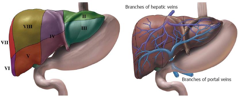
Figure 1 Illustrations of liver and its surrounding stomach and duodenum.
A: The liver can subsequently be divided into 8 segments that is served independently by a secondary or tertiary branch of the portal triad. B: The left hepatic vein divides the left lobe into lateral (II2, III3) and medial (IV4a, IV4b) segments. The right hepatic vein divides the right lobe into anterior (V5, VIII8) and posterior (VI6, VII7) segments. The portal vein divides the liver into upper (II2, IV4a, VIII8, VII7) and lower (III3, IV4b, V5, VI6) segments. The segments are labeled in a clockwise manner. In a normal frontal view segments I1, VI6 and VII7 are not visible.
- Citation: Srinivasan I, Tang SJ, Vilmann AS, Menachery J, Vilmann P. Hepatic applications of endoscopic ultrasound: Current status and future directions. World J Gastroenterol 2015; 21(44): 12544-12557
- URL: https://www.wjgnet.com/1007-9327/full/v21/i44/12544.htm
- DOI: https://dx.doi.org/10.3748/wjg.v21.i44.12544


