Copyright
©The Author(s) 2015.
World J Gastroenterol. Jan 21, 2015; 21(3): 1020-1023
Published online Jan 21, 2015. doi: 10.3748/wjg.v21.i3.1020
Published online Jan 21, 2015. doi: 10.3748/wjg.v21.i3.1020
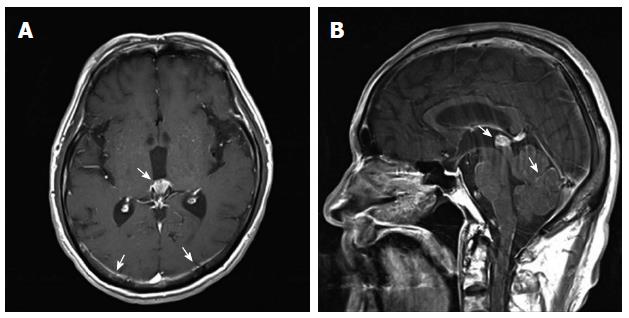
Figure 1 Brain magnetic resonance imaging findings.
A: Pineal gland and parietal leptomeningeal enhancement on a T1 axial image (arrows); B: Pineal gland and cerebellar folia enhancement on a T1 sagittal image (arrows).
- Citation: Yoo IK, Lee HS, Kim CD, Chun HJ, Jeen YT, Keum B, Kim ES, Choi HS, Lee JM, Kim SH, Nam SJ, Hyun JJ. Rare case of pancreatic cancer with leptomeningeal carcinomatosis. World J Gastroenterol 2015; 21(3): 1020-1023
- URL: https://www.wjgnet.com/1007-9327/full/v21/i3/1020.htm
- DOI: https://dx.doi.org/10.3748/wjg.v21.i3.1020


