Published online Jun 28, 2015. doi: 10.3748/wjg.v21.i24.7589
Peer-review started: November 17, 2014
First decision: December 2, 2014
Revised: December 15, 2014
Accepted: March 18, 2015
Article in press: March 19, 2015
Published online: June 28, 2015
Processing time: 226 Days and 21.1 Hours
We present three cases of self-expandable metallic stent (SEMS) placement using a balloon enteroscope (BE) and its overtube (OT) for malignant obstruction of surgically reconstructed intestine. A BE is effective for the insertion of an endoscope into the deep bowel. However, SEMS placement is impossible through the working channel, because the working channel of BE is too small and too long for the stent device. Therefore, we used a technique in which the BE is inserted as far as the stenotic area; thereafter, the BE is removed, leaving only the OT, and then the stent is placed by inserting the stent device through the OT. In the present three cases, a modification of this technique resulted in the successful placement of the SEMS for obstruction of surgically reconstructed intestine, and the procedures were performed without serious complications. We consider that the present procedure is extremely effective as a palliative treatment for distal bowel stenosis, such as in the surgically reconstructed intestine.
Core tip: Self-expandable metallic stent (SEMS) placement in surgically reconstructed intestine is more challenging because of the long length and the bifurcated configuration of the intestine. We present three cases of SEMS placement using a balloon enteroscope and its overtube for malignant obstruction of surgically reconstructed intestine. We consider that the present technique is extremely effective as a palliative treatment for distal bowel stenosis, such as in the surgically reconstructed intestine.
- Citation: Nakahara K, Okuse C, Matsumoto N, Suetani K, Morita R, Michikawa Y, Ozawa SI, Hosoya K, Kobayashi S, Otsubo T, Itoh F. Enteral metallic stenting by balloon enteroscopy for obstruction of surgically reconstructed intestine. World J Gastroenterol 2015; 21(24): 7589-7593
- URL: https://www.wjgnet.com/1007-9327/full/v21/i24/7589.htm
- DOI: https://dx.doi.org/10.3748/wjg.v21.i24.7589
Self-expandable metallic stents (SEMSs) are now widely used for the palliative treatment of malignant gastrointestinal obstructions of the esophagus, stomach, duodenum, and colon. However, there are only a few reports on the use of metallic stents in the small intestine[1-4], particularly in surgically reconstructed small intestine[4,5]. SEMS placement in surgically reconstructed intestine is more challenging because of the long length and the bifurcated of the reconstructed intestine. We present three cases of SEMS placement using a single-balloon enteroscope (SBE) and its overtube (OT) for malignant obstruction of surgically reconstructed intestine.
A 48-year-old man who had undergone pylorus-preserving pancreatoduodenectomy (PPPD) 7 mo before for pancreatic head cancer was referred to our institution because of obstruction of the afferent loop as a result of pancreatic cancer recurrence. Because the tumor had invaded the biliary-jejunal anastomosis causing obstructive jaundice, we first performed percutaneous transhepatic biliary drainage (PTBD) and then placed the enteral SEMS.
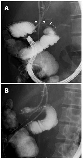
With the patient under general anesthesia, the stenosis was accessed using an SBE (SIF-Q260; Olympus Medical Systems, Tokyo, Japan) with its OT (ST-SB1; Olympus Medical Systems). Contrast medium injection revealed that the stricture was approximately 5-cm long. A 0.035-inch × 550-cm guidewire (RevoWave; Piolax Medical Devices, Kanagawa, Japan) was passed through the stenosis and placed under the SBE (Figure 1A). The SBE was then removed, leaving the OT with an inflated balloon and the guidewire in place. Under fluoroscopic guidance, an enteral stent (22 mm × 12 cm, Niti-S Pyloric/Duodenal stent; TaeWoong Medical, Seoul, South Korea) was advanced over the guidewire and through the OT until it passed through the stenosis. Then, the enteral stent was released in the stenosis (Figure 1B).
We subsequently placed a biliary SEMS via the PTBD route at the site of the biliary-jejunal anastomostic stenosis. Seven days after placement of the enteral stent, the patient was able to consume a diet consisting exclusively of rice porridge and was discharged from the hospital shortly thereafter. He died of primary cancer progression 4 mo after stent placement, but there were no stent problems.
A 76-year-old man who had undergone PPPD 4 years before for middle cholangiocarcinoma was admitted to our institution because of afferent loop obstruction as a result of recurrence of peritoneal dissemination. Blood tests did not indicate elevated total bilirubin levels. Computed tomography (CT) showed a stricture in the afferent loop as well as intestinal dilatation on the distal side, although the intrahepatic bile duct was not dilated.
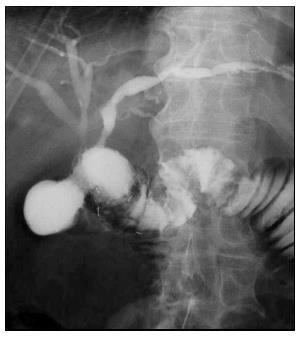
An SBE (SIF-Q260; Olympus Medical Systems) with its OT (ST-SB1; Olympus Medical Systems) was advanced into the stenotic region and an enteral stent (22 mm × 12 cm, Niti-S Pyloric/Duodenal stent; TaeWoong Medical) was placed as described for the abovementioned case (Figure 2).
After stent placement, the patient developed retrograde cholangitis; however, conservative treatment with antibiotics led to an improvement. The patient tolerated a liquid diet a day after stent placement. The diet was advanced and the patient was subsequently discharged. Eventually, there were no stent problems, but the patient died of primary cancer progression 14 mo later.
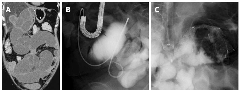
A 51-year-old woman had undergone left hepatic lobectomy and Roux-en-Y (RY) reconstruction 4 years before for hilar cholangiocarcinoma. Eighteen months before, she had undergone distal gastrectomy and RY reconstruction for gastric cancer. She was admitted to our hospital with the chief complaint of abdominal distension and vomiting. Abdominal CT revealed an intestinal stricture as a result of peritoneal dissemination and marked dilation of the small intestine (Figure 3A).
The stenosis was accessed using an SBE (SIF-Q260; Olympus Medical Systems) with its OT (ST-SB1, Olympus Medical Systems). Contrast medium injection revealed the stricture and dilated intestine. Because two surgical procedures indicated complex branching of the intestine and extensive peritoneal dissemination over a wide area, we first placed a 6Fr endoscopic nasal drainage tube into the dilated intestine through the working channel to confirm the drainage effect (Figure 3B).
Because good drainage was subsequently achieved and CT confirmed the amelioration of intestinal dilation, we performed enteral SEMS placement. An SBE (SIF-Q260; Olympus Medical Systems) with its OT (ST-SB1, Olympus Medical Systems) was advanced into the stenotic region and an enteral stent (22 mm × 10 cm, Niti-S Pyloric/Duodenal stent; TaeWoong Medical) was placed as described for the abovementioned cases (Figure 3C).
After this procedure, the patient was unable to consume food because of poor general condition related to the malignancy; however, the abdominal distension and vomiting improved. Although there were no stent problems, the patient eventually died of primary cancer progression 1 mo later.
In recent years, malignant gastrointestinal obstruction of the esophagus, stomach, duodenum, and colon has been widely treated with endoscopic SEMS placement as an effective palliative treatment. However, because there are few studies on obstruction of surgically reconstructed intestine[4,5], its efficacy and safety have not been elucidated.
In cases of surgically reconstructed intestinal obstruction, insertion of the endoscope into the stenotic area is difficult because of the long length and complicated bifurcation of surgically reconstructed intestine. Recent studies have reported that a balloon endoscope (BE) is effective for the insertion of an endoscope into the distal parts of the bowel[6,7]. However, SEMS placement is impossible through the working channel because it is too small and too long for the stent device. Therefore, we used a technique in which the BE is inserted as far as the stenotic area. After this, the BE is removed, leaving only the OT, and then the stent is placed by inserting the stent device through the OT. In the three cases presented above, a modification of this technique resulted in the successful placement of the SEMS for obstruction of surgically reconstructed intestine, and the procedures were performed without serious complications. The technical advantage afforded by the BE and its OT may allow for enteral stent placement in patients with the distal intestinal obstruction that is beyond the reach of conventional endoscopes. Its usefulness is particularly notable in RY cases with long and tortuous intestinal tract reconstruction.
On the other hand, the disadvantage of this technique about which we are concerned is that the kinking of the OT may make the stent delivery system insertion impossible in patients with acutely curved intestine. Moreover, when the obstruction is beyond the reach of BE, or the obstructions are in two or more part of an intestine, this technique may not be suitable.
Our search of PubMed yielded reports of six cases in which SEMSs were placed in intestinal stenoses using a BE and OT in a similar technique as in our procedure[1-5]. Of these, there were only two cases of surgically reconstructed intestinal obstruction[4,5] (Table 1). In all cases, stent placement was successful, good clinical results were obtained, and no serious complications were observed. We believe that the present procedure is extremely effective as a palliative treatment for distal bowel stenosis, such as in the small intestine or surgically reconstructed intestine. In addition, because the stent is difficult to remove once the SEMS is placed, we consider that as in Case 3 in this report, SEMS placement using our method, in which an endoscopic nasal drainage tube is placed in the dilated intestine and then an SEMS is placed after confirming drainage test results, is effective. This method is particularly true in cases in which peritoneal dissemination over a wide area and multiple stenoses cannot be ruled out and in cases of reconstructed intestine with complicated intestinal bifurcation.
| Ref. | Age (yr) | Sex | Disease | Past surgery | Endoscope | Stent | Improve | Complication | Survival |
| Ross et al[1], 2006 | 59 | M | Lymphadenopathyof lung cancer | No | DBE (EN450T5) | Ultraflex | Yes | No | NA |
| Hayashi et al[2], 2006 | 65 | F | Jejunal cancer | No | DBE (EN450P5) | Ultraflex | Yes | No | NA |
| 73 | M | Pancreatic cancer | No | SBE (SIFQ180) | Wallflex | Yes | No | NA | |
| Espinel et al[3], 2011 | 68 | M | Pancreatic cancer | PPPD | DBE (EN450T5) | Niti-S | Yes | No | NA |
| 81 | M | Pancreatic cancer | No | DBE (EC450BI5) | Wallflex | Yes | No | NA | |
| Kida et al[4], 2013 | 80 | F | Pancreatic cancer | Whipple | DBE | NA | Yes | No | 3 mo |
| 48 | M | Pancreatic cancer | PPPD | SBE (SIFQ260) | Niti-S | Yes | No | 4 mo | |
| Popa et al[5], 2014 | 76 | M | Middle cholangiocarcinoma | PPPD | SBE (SIFQ260) | Niti-S | Yes | Cholangitis | 14 mo |
| Our cases | 51 | F | Hilar cholangiocarcinoma, Gastric cancer | LL + RY, DG + RY | SBE (SIFQ260) | Niti-S | Yes | No | 1 mo |
Three cases with a history of intestinal reconstructive surgery presented with symptom due to intestinal obstruction.
The common physical sign of the three cases was abdominal distension, and one case developed jaundice.
Adherent ileus, Anorexia associated with malignancy.
The first patient had elevated serum levels of hepatic and biliary tract enzymes, while the others had no remarkable findings for the laboratory test.
Computed tomography showed intestinal stricture as a result of malignancy and dilated intestine on the distal side.
Past surgical pathological examination revealed malignancy.
Self-expandable metallic stents were placed using an single-balloon enteroscope and its overtube for malignant obstruction of surgically reconstructed intestine.
Only two cases in which self-expandable metallic stent were placed in reconstructed intestinal obstruction using a balloon enteroscope have been reported in the literature.
Although a balloon endoscope is effective tool for insertion into the deep parts of the bowel, the working channel of balloon endoscope is too small and too long for the stent device.
The authors consider that the present technique of enteral metallic stent placement using balloon enteroscope and its overtube is extremely effective as a palliative treatment for deep bowel obstruction, such as in the small intestine or surgically reconstructed intestine.
This work has touched upon an important concept of palliation in patients with advanced malignancy of the gastrointestinal tract. This article is useful as a case report for the development of future treatment.
P- Reviewer: Arihiro S, Nanavati AJ S- Editor: Yu J L- Editor: A E- Editor: Wang CH
| 1. | Ross AS, Semrad C, Waxman I, Dye C. Enteral stent placement by double balloon enteroscopy for palliation of malignant small bowel obstruction. Gastrointest Endosc. 2006;64:835-837. [RCA] [PubMed] [DOI] [Full Text] [Cited by in Crossref: 59] [Cited by in RCA: 60] [Article Influence: 3.2] [Reference Citation Analysis (0)] |
| 2. | Hayashi Y, Yamamoto H, Kita H, Sunada K, Miyata T, Yano T, Sato H, Iwamoto M, Sugano K. Education and imaging. Gastrointestinal: metallic stent for an obstructing jejunal cancer. J Gastroenterol Hepatol. 2006;21:1861. [RCA] [PubMed] [DOI] [Full Text] [Cited by in Crossref: 7] [Cited by in RCA: 5] [Article Influence: 0.3] [Reference Citation Analysis (0)] |
| 3. | Espinel J, Pinedo E. A simplified method for stent placement in the distal duodenum: Enteroscopy overtube. World J Gastrointest Endosc. 2011;3:225-227. [RCA] [PubMed] [DOI] [Full Text] [Full Text (PDF)] [Cited by in CrossRef: 17] [Cited by in RCA: 13] [Article Influence: 0.9] [Reference Citation Analysis (0)] |
| 4. | Kida A, Matsuda K, Noda Y. Endoscopic metallic stenting by double-balloon enteroscopy and its overtube for malignant gastrointestinal obstruction as palliative treatment. Dig Endosc. 2013;25:552-553. [RCA] [PubMed] [DOI] [Full Text] [Cited by in Crossref: 18] [Cited by in RCA: 18] [Article Influence: 1.5] [Reference Citation Analysis (0)] |
| 5. | Popa D, Ramesh J, Peter S, Wilcox CM, Mönkemüller K. Small Bowel Stent-in-Stent Placement for Malignant Small Bowel Obstruction Using a Balloon-Assisted Overtube Technique. Clin Endosc. 2014;47:108-111. [RCA] [PubMed] [DOI] [Full Text] [Full Text (PDF)] [Cited by in Crossref: 15] [Cited by in RCA: 15] [Article Influence: 1.4] [Reference Citation Analysis (0)] |
| 6. | Yamamoto H, Sekine Y, Sato Y, Higashizawa T, Miyata T, Iino S, Ido K, Sugano K. Total enteroscopy with a nonsurgical steerable double-balloon method. Gastrointest Endosc. 2001;53:216-220. [RCA] [PubMed] [DOI] [Full Text] [Cited by in Crossref: 896] [Cited by in RCA: 859] [Article Influence: 35.8] [Reference Citation Analysis (0)] |
| 7. | Tsujikawa T, Saitoh Y, Andoh A, Imaeda H, Hata K, Minematsu H, Senoh K, Hayafuji K, Ogawa A, Nakahara T. Novel single-balloon enteroscopy for diagnosis and treatment of the small intestine: preliminary experiences. Endoscopy. 2008;40:11-15. [RCA] [PubMed] [DOI] [Full Text] [Cited by in Crossref: 215] [Cited by in RCA: 209] [Article Influence: 12.3] [Reference Citation Analysis (0)] |


