Published online Jun 7, 2015. doi: 10.3748/wjg.v21.i21.6754
Peer-review started: November 17, 2014
First decision: December 26, 2014
Revised: January 6, 2014
Accepted: February 12, 2015
Article in press: February 13, 2015
Published online: June 7, 2015
Processing time: 208 Days and 22.3 Hours
A left-sided gallbladder without a right-sided round ligament, which is called a true left-sided gallbladder, is extremely rare. A 71-year-old woman was referred to our hospital due to a gallbladder polyp. Computed tomography (CT) revealed not only a gallbladder polyp but also the gallbladder located to the left of the round ligament connected to the left umbilical portion. CT portography revealed that the main portal vein diverged into the right posterior portal vein and the common trunk of the left portal vein and right anterior portal vein. CT cholangiography revealed that the infraportal bile duct of segment 2 joined the common bile duct. Laparoscopic cholecystectomy was performed for a gallbladder polyp, and the intraoperative finding showed that the cholecystic veins joined the round ligament. A true left-sided gallbladder is closely associated with several anomalies; therefore, surgeons encountering a true left-sided gallbladder should be aware of the potential for these anomalies.
Core tip: A left-sided gallbladder without a right-sided round ligament, which is called a true left-sided gallbladder, is extremely rare. We performed laparoscopic cholecystectomy on a patient with a true left-sided gallbladder which coexisted with an infraportal bile duct of segment 2 and cholecystic venous anomaly. Surgeons encountering a true left-sided gallbladder should be aware of the potential for these anomalies.
- Citation: Ishii H, Noguchi A, Onishi M, Takao K, Maruyama T, Taiyoh H, Araki Y, Shimizu T, Izumi H, Tani N, Yamaguchi M, Yamane T. True left-sided gallbladder with variations of bile duct and cholecystic vein. World J Gastroenterol 2015; 21(21): 6754-6758
- URL: https://www.wjgnet.com/1007-9327/full/v21/i21/6754.htm
- DOI: https://dx.doi.org/10.3748/wjg.v21.i21.6754
A left-sided gallbladder usually denotes that the gallbladder is located to the left side of the round ligament without situs inversus viscerum; however, most reported cases of left-sided gallbladder are associated with a right-sided round ligament, and such a case is called a “false” left-sided gallbladder[1]. A left-sided gallbladder without a right-sided round ligament is called a true left-sided gallbladder[1]. Several variations of infraportal bile duct were reported previously[2-6]; however, the infraportal bile duct of segment 2 has never been reported in detail. Although the cholecystic veins are usually divided into two subgroups, small branches that directly enter the liver through the liver bed and those that run through the Calot’s triangle, cholecystic venous branches entering the round ligament were confirmed intraoperatively. We report an extremely rare case of a true left-sided gallbladder with infraportal bile duct of segment 2 and cholecystic venous anomaly.
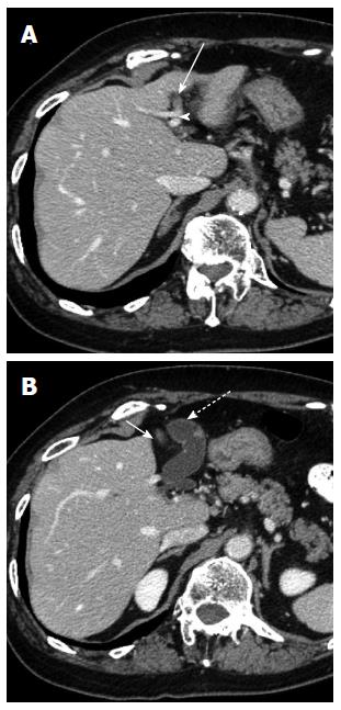
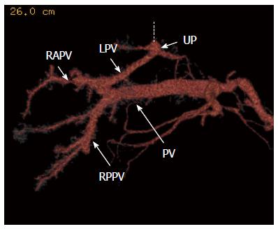
A 71-year-old woman was referred to our hospital due to an asymptomatic gallbladder polyp of 1cm in diameter that enlarged gradually. The laboratory data were within normal limits. An abdominal ultrasonography revealed a gallbladder polyp. An abdominal dynamic computed tomography (CT) examination revealed a gallbladder polyp, without enhancement, of 1 cm in diameter. The gallbladder was located to the left side of the middle hepatic vein and the normal anatomically positioned round ligament connected to the left portal umbilical portion, and was attached to the left lateral section of the liver (Figure 1). CT portography revealed an anomaly of portal venous divergence of the main portal trunk into the right posterior portal vein and the common trunk of the left portal vein and right anterior portal vein (Figure 2). CT arteriography showed that the hepatic artery was of normal type and the cystic artery diverged from the right hepatic artery. Drip infusion cholangiographic-CT (DIC-CT) demonstrated that the cystic duct joined the extrahepatic bile duct on the right side and created a hairpin turn anterior to the extrahepatic bile duct. Furthermore, one of the two bile ducts of segment 2 followed a route that was caudal to the umbilical portion of the left portal vein, that is, an infraportal course, and joined the extrahepatic bile duct (Figure 3). The patient was diagnosed preoperatively as having a polyp of the true left-sided gallbladder with an infraportal bile duct of segment 2.
We performed laparoscopic cholecystectomy with four surgical ports. A good surgical field was obtained by retracting the round ligament using a thread fold via the abdominal wall. The gallbladder was located left of the round ligament and attached to the left lateral section of the liver and the round ligament, and Calot’s triangle was covered by the gallbladder. Therefore, we first separated the gallbladder from the liver bed and the round ligament. Two cholecystic veins which enter the round ligament were confirmed and divided by ultrasonic scalpel. The cystic artery and cystic duct were clipped and divided respectively, and the gallbladder was finally removed. The anatomy was inspected again; we confirmed the liver bed located to the left side of the round ligament and the infraportal bile duct of segment 2 (Figure 4). Pathological examination revealed a cholesterol polyp without malignancy. The postoperative course was uncomplicated.
Since Hochstetter first described a left-sided gallbladder in 1886[7], many cases have been reported in the literature; however, most reported cases of left-sided gallbladder are associated with a right-sided round ligament, and such a case is called a “false” left-sided gallbladder[1]. A left-sided gallbladder without a right-sided round ligament is called a true left-sided gallbladder[1], and is extremely rare. To our knowledge, only two cases of a true left-sided gallbladder have been reported previously[1,8]. A true left-sided gallbladder is an anomaly of the gallbladder; however, a “false” left-sided gallbladder is not an anomaly of the gallbladder, but of the round ligament[9]. Therefore, we think that a true left-sided gallbladder should be clearly distinguished from a “false” left-sided gallbladder. Gross[10] reported that the embryologic development of a left-sided gallbladder may occur in one of two ways. The first way in which this anomaly may develop is as follows: the gallbladder anlage begins as a normal embryologic bud from the hepatic diverticulum and migrates to the left, where it becomes fixed by the developing peritoneum to the undersurface of the left lateral section of the liver. This accounts for the normal entrance of the cystic duct into the extrahepatic bile duct. The second one is as follow: a gallbladder develops on each side, with the left-sided gallbladder persisting on the left and the right-sided one becoming atrophic and disappearing. In such a case, the cystic duct joins the extrahepatic duct or left hepatic duct from the left side. We consider that the true left-sided gallbladder in our case was caused in the first way, because the cystic duct joined the extrahepatic bile duct on the right side.
Although several authors reported that a left-sided gallbladder is not identified preoperatively[11-13], a left-sided gallbladder can be diagnosed preoperatively with improvements in diagnostic imaging methods[1,14,15], and we were able to diagnose this anomaly by CT examination.
It has been reported that a true left-sided gallbladder is closely associated with multiple anomalies of the portal vein and bile duct[1]. In our case, the patient had anomalies of not only the portal vein and bile duct, but also the cholecystic vein. The portal vein anomaly in our case was the same as in the previous report[1]. Several types of infraportal bile duct, such as the infraportal bile duct of the caudate lobe[2], left lateral section[3,4], segment 3[3-6], and right posterior section[4,6], have been reported. To our knowledge, the infraportal bile duct of segment 2 has been described in only two studies[3,4]; therefore, we think that this variation is a rare type. Kitami et al[4] reported that infraportal courses of the bile duct are observed more frequently in patients with portal vein variation, such as our case, than without portal vein variation. The surgeon should initiate the dissection as close to the gallbladder as possible in order to prevent injury of the infraportal bile duct of segment 2 in a patient with true left-sided gallbladder.
The cholecystic veins are usually divided into two subgroups, small branches that directly enter the liver through the liver bed and those that run through Calot’s triangle[16,17]; however, cholecystic venous branches entering the round ligament were confirmed in our case. We think that these cholecystic veins are the specific anomaly of a true left-sided gallbladder. The surgeon should pay special attention to the possibility of this cholecystic venous anomaly when a true left-sided gallbladder is dissected from the round ligament. We reported this extremely rare case to emphasize that laparoscopic cholecystectomy can be performed safely for a true left-sided gallbladder when the surgeon is familiar with the anatomical variations.
The patient had no symptoms.
There were no clinical findings.
Gallbladder carcinoma.
The laboratory data were within normal limits.
Computed tomography revealed that the gallbladder was located to the left side of the middle hepatic vein and the normal anatomically positioned round ligament connected to the left portal umbilical portion, and the infraportal bile duct of segment 2 joined the extrahepatic bile duct.
Cholesterol polyp of the gallbladder.
Laparoscopic cholecystectomy.
To date, only two cases of a true left-sided gallbladder have been reported previously.
A left-sided gallbladder without a right-sided round ligament is called a true left-sided gallbladder.
Laparoscopic cholecystectomy can be performed safely for a true left-sided gallbladder when the surgeon is familiar with the anatomical variations of the bile duct and cholecystic vein.
A rare case of true left-sided gallbladder has been reported. Surgeons need to face different gallbladder anomalies to be ready to manage ambiguities during cholecystectomy.
P- Reviewer: Khorgami Z S- Editor: Yu J L- Editor: A E- Editor: Wang CH
| 1. | Kawai R, Miyata K, Yuasa N, Takeuchi E, Goto Y, Miyake H, Nagai H, Hattori M, Imura J, Hayashi Y. True left-sided gallbladder with a portal anomaly: report of a case. Surg Today. 2012;42:1130-1134. [RCA] [PubMed] [DOI] [Full Text] [Cited by in Crossref: 13] [Cited by in RCA: 14] [Article Influence: 1.0] [Reference Citation Analysis (0)] |
| 2. | Sugiura T, Nagino M, Kamiya J, Nishio H, Ebata T, Yokoyama Y, Igami T, Nimura Y. Infraportal bile duct of the caudate lobe: a troublesome anatomic variation in right-sided hepatectomy for perihilar cholangiocarcinoma. Ann Surg. 2007;246:794-798. [RCA] [PubMed] [DOI] [Full Text] [Cited by in Crossref: 11] [Cited by in RCA: 12] [Article Influence: 0.7] [Reference Citation Analysis (0)] |
| 3. | Kitamura H, Mori T, Arai M, Tsukada K, Numata M, Kawasaki S. Caudal left hepatic duct in relation to the umbilical portion of the portal vein. Hepatogastroenterology. 1999;46:2511-2514. [PubMed] |
| 4. | Kitami M, Takase K, Murakami G, Ko S, Tsuboi M, Saito H, Higano S, Nakajima Y, Takahashi S. Types and frequencies of biliary tract variations associated with a major portal venous anomaly: analysis with multi-detector row CT cholangiography. Radiology. 2006;238:156-166. [RCA] [PubMed] [DOI] [Full Text] [Cited by in Crossref: 67] [Cited by in RCA: 70] [Article Influence: 3.7] [Reference Citation Analysis (0)] |
| 5. | Ozden I, Kamiya J, Nagino M, Uesaka K, Sano T, Nimura Y. Clinicoanatomical study on the infraportal bile ducts of segment 3. World J Surg. 2002;26:1441-1445. [RCA] [PubMed] [DOI] [Full Text] [Cited by in Crossref: 21] [Cited by in RCA: 19] [Article Influence: 0.8] [Reference Citation Analysis (0)] |
| 6. | Ohkubo M, Nagino M, Kamiya J, Yuasa N, Oda K, Arai T, Nishio H, Nimura Y. Surgical anatomy of the bile ducts at the hepatic hilum as applied to living donor liver transplantation. Ann Surg. 2004;239:82-86. [RCA] [PubMed] [DOI] [Full Text] [Cited by in Crossref: 145] [Cited by in RCA: 162] [Article Influence: 7.7] [Reference Citation Analysis (0)] |
| 7. | Hochstetter F. Anomalien der Pfortader und der Nabelvene in Verbindung mit Defect oder Linkslage der Gallenblase. Arch Anat Entwick. 1886;3:369-384. |
| 8. | Jung HS, Huh K, Shin YH, Kim JK, Yun CS, Park CH, Jang JB. Left-sided gallbladder: a complicated percutaneous cholecystostomy and subsequent hepatic embolisation. Br J Radiol. 2009;82:e141-e144. [RCA] [PubMed] [DOI] [Full Text] [Cited by in Crossref: 4] [Cited by in RCA: 5] [Article Influence: 0.3] [Reference Citation Analysis (0)] |
| 9. | Nagai M, Kubota K, Kawasaki S, Takayama T, BandaiY M. Are left-sided gallbladders really located on the left side? Ann Surg. 1997;225:274-280. [PubMed] |
| 10. | Gross RE. Congenital anomalies of the gallbladder. Arch Surg. 1936;32:131-162. [DOI] [Full Text] |
| 11. | Iskandar ME, Radzio A, Krikhely M, Leitman IM. Laparoscopic cholecystectomy for a left-sided gallbladder. World J Gastroenterol. 2013;19:5925-5928. [RCA] [PubMed] [DOI] [Full Text] [Full Text (PDF)] [Cited by in CrossRef: 22] [Cited by in RCA: 23] [Article Influence: 1.9] [Reference Citation Analysis (0)] |
| 12. | Makni A, Magherbi H, Ksantini R, Rebai W, Safta ZB. Left-sided gallbladder: an incidental finding on laparoscopic cholecystectomy. Asian J Surg. 2012;35:93-95. [RCA] [PubMed] [DOI] [Full Text] [Cited by in Crossref: 12] [Cited by in RCA: 14] [Article Influence: 1.1] [Reference Citation Analysis (0)] |
| 13. | Sadhu S, Jahangir TA, Roy MK. Left-sided gallbladder discovered during laparoscopic cholecystectomy in a patient with dextrocardia. Indian J Surg. 2012;74:186-188. [RCA] [PubMed] [DOI] [Full Text] [Cited by in Crossref: 13] [Cited by in RCA: 16] [Article Influence: 1.1] [Reference Citation Analysis (0)] |
| 14. | Matsumura N, Tokumura H, Yasumoto A, Sasaki H, Yamasaki M, Musya H, Fukuyama S, Takahashi K, Toshima T, Funayama Y. Laparoscopic cholecystectomy and common bile duct exploration for cholecystocholedocholithiasis with a left-sided gallbladder: report of a case. Surg Today. 2009;39:252-255. [RCA] [PubMed] [DOI] [Full Text] [Cited by in Crossref: 17] [Cited by in RCA: 17] [Article Influence: 1.1] [Reference Citation Analysis (0)] |
| 15. | Abe T, Kajiyama K, Harimoto N, Gion T, Shirabe K, Nagaie T. Resection of metastatic liver cancer in a patient with a left-sided gallbladder and intrahepatic portal vein and bile duct anomalies: A case report. Int J Surg Case Rep. 2012;3:147-150. [RCA] [PubMed] [DOI] [Full Text] [Cited by in Crossref: 11] [Cited by in RCA: 12] [Article Influence: 0.9] [Reference Citation Analysis (0)] |
| 16. | Yoshimitsu K, Honda H, Kaneko K, Kuroiwa T, Irie H, Chijiiwa K, Takenaka K, Masuda K. Anatomy and clinical importance of cholecystic venous drainage: helical CT observations during injection of contrast medium into the cholecystic artery. AJR Am J Roentgenol. 1997;169:505-510. [PubMed] |
| 17. | Yoshimitsu K, Honda H, Kuroiwa T, Irie H, Aibe H, Shinozaki K, Masuda K. Unusual hemodynamics and pseudolesions of the noncirrhotic liver at CT. Radiographics. 2001;21 Spec No:S81-S96. [RCA] [PubMed] [DOI] [Full Text] [Cited by in RCA: 1] [Reference Citation Analysis (0)] |


