Published online May 7, 2015. doi: 10.3748/wjg.v21.i17.5442
Peer-review started: December 10, 2014
First decision: December 26, 2014
Revised: January 17, 2015
Accepted: February 11, 2015
Article in press: February 11, 2015
Published online: May 7, 2015
Processing time: 153 Days and 11.8 Hours
Hemolymphangioma is a malformation of both lymphatic vessels and blood vessels. Splenic hemolymphangioma is extremely rare. Herein, we present a case of 62-year-old woman with ambiguous upper quadrant abdominal pain for two months who was found to have an occupying lesion in the spleen on computed tomography. She was eventually diagnosed with hemolymphangioma of the spleen. The patient underwent total splenectomy. Neither symptoms nor recurrence was found during the one-year follow-up period.
Core tip: We report an extremely rare case of splenic hemolymphangioma. Computed tomography findings of splenic hemolymphangioma include single or multiple hypodense cysts occupying the spleen, without enhancement, which are correlated with pathologic configuration. However, it is difficult to establish an accurate preoperative diagnosis according to radiological features. Pathologic examination is essential for diagnosis. The most effective treatment for splenic hemolymphangioma is complete resection of the tumor.
- Citation: Mei Y, Peng CJ, Chen L, Li XX, Li WN, Shu DJ, Xie WT. Hemolymphangioma of the spleen: A report of a rare case. World J Gastroenterol 2015; 21(17): 5442-5444
- URL: https://www.wjgnet.com/1007-9327/full/v21/i17/5442.htm
- DOI: https://dx.doi.org/10.3748/wjg.v21.i17.5442
Intra-abdominal hemolymphangioma is very rare; involvement of the spleen is even rarer. We found only three case reports[1-3] after a review of the related literature through online PubMed search up to October 2014. Hemolymphangioma is a congenital malformation of the vascular system[4]. The formation of this tumor may be explained by obstruction of the venolymphatic communication between dysembryoplastic vascular tissue and the systemic circulation[1]. Splenic hemolymphangioma is extremely rare and may pose a diagnostic challenge. The goal of the present study is to report an extremely uncommon case of splenic hemolymphangioma.
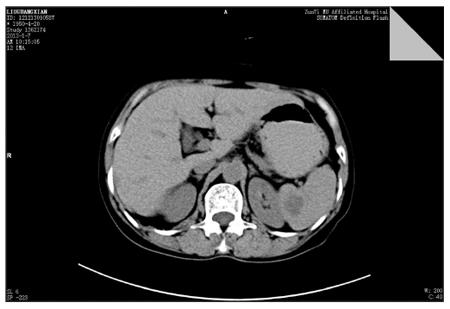
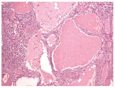
A 62-year-old female patient was admitted to our hospital with a history of ambiguous upper abdominal pain for the past two months. She did not have a history of abdominal distention, nausea, vomiting or fever. She gave no medical history of abdominal trauma or surgery, no family history of malignant tumor, and no weight loss. Physical examination revealed mild tenderness on the left upper abdominal quadrant. No abnormalities in laboratory test, including serum tumor markers, were observed. Ultrasonography failed to find a splenic lesion. Computed tomography (CT) demonstrated an 18 mm × 19 mm hypodense cyst occupying the spleen, with an irregular and rough edge, and an intracapsular area without enhancement (Figure 1). Diagnosis of the splenic occupying lesion was hemangioma or lymphangioma. The patient underwent total splenectomy. During surgery, the visible spleen size was approximately 11 cm × 6 cm × 3 cm, with normal color and smooth surface, and it was soft. A specimen section showed a gray and red region 2 cm in diameter, which was hard and without capsule. Microscopically, the tumor was mainly composed of lymphatic and blood vessels (Figure 2). The definitive histological diagnosis was hemolymphangioma of the spleen. No post-splenectomy infectious complications occurred. Her postoperative course was uneventful and she was discharged 16 d after surgery. Neither symptoms nor recurrence was found during the one-year follow-up period.
Splenic occupying lesions include splenic cyst, splenic abscess and splenic tumor. Primary splenic tumors are uncommon. Hemolymphangioma is a malformation of both lymphatic vessels and blood vessels[5]. Its incidence varies from 1.2 to 2.8 per 1000 newborns, and both genders are equally affected[6]. Splenic hemolymphangioma is extremely rare[1], and may pose a diagnostic challenge. The most common symptom of splenic hemolymphangioma is upper quadrant abdominal discomfort or pain, which depends on the size of the tumor, anatomical location and relationship to neighboring tissues. Physical examination may show mild tenderness, however, no obvious mass was detected in our case. Blood examination results, such as tumor markers, are often normal, which was also demonstrated in our case. CT findings of splenic hemolymphangioma include single or multiple hypodense cysts occupying the spleen, with an irregular and rough edge, an intracapsular area without enhancement, and are correlated with pathologic configuration. However, imaging examinations often fail to distinguish benign and malignant tumor, or misdiagnose it as other occupying lesions of the spleen, such as lymphoma or cyst, which was also seen in our case. Therefore, it is difficult to establish an accurate preoperative diagnosis according to radiological features. When a cystic tumor of the spleen is found, especially when there is no sufficient evidence to diagnose hemangioma, lymphoma or cyst, splenic hemolymphangioma should be considered in the differential diagnosis. The most effective therapy for splenic hemolymphangioma is complete resection of the tumor. In the past, the major treatment of choice for splenic tumors was total splenectomy. Currently, in order to avoid overwhelming post-splenectomy infection, partial splenectomy is increasingly advocated. Zhang et al[1] reported successfully that partial splenectomy by laparoscope was used for therapy of splenic hemolymphangioma. Microscopically, some dysplastic lymphatic vessels and blood vessels are found in the tumor, which is the gold standard for the diagnosis of splenic hemolymphangioma.
In conclusion, splenic hemolymphangioma lacks specific symptoms or signs. Pathologic examination is essential for diagnosis, and surgical treatment yields a good prognosis.
A 62-year-old woman with ambiguous upper quadrant abdominal pain for two months was found to have an occupying lesion in the spleen on computed tomography (CT).
Diagnosis of the splenic occupying lesion was hemangioma or lymphangioma.
Differentiatial diagnosis consisted of cystic hemangioma, cystic lymphangioma, and epidermoid or dermoid cyst of the spleen.
No abnormalities in laboratory test, including serum tumor markers, were observed.
CT findings of splenic hemolymphangioma include single or multiple hypodense cysts occupying the spleen, without enhancement, which are correlated with pathologic configuration.
Specimen histology revealed that the tumor was mainly composed of lymphatic and blood vessels.
The patient was treated with total splenectomy. According to the patient’s condition and location of the tumor, selective laparoscopic partial splenectomy can be considered.
In 2014, Zhang et al reported successfully that partial splenectomy by laparoscope was used for therapy of splenic hemolymphangioma.
Hemolymphangioma is a benign tumor, which consist of some dysplastic lymphatic vessels and blood vessels.
Splenic hemolymphangioma lacks specific symptoms or signs. Pathologic examination is essential for diagnosis, and surgical treatment yields a good prognosis.
This study introduces an interesting rare case of splenic hemolymphangioma, which has added evidence for the diagnosis of benign tumor of the spleen.
P- Reviewer: Lima M S- Editor: Ma YJ L- Editor: A E- Editor: Ma S
| 1. | Zhang Y, Chen XM, Sun DL, Yang C. Treatment of hemolymphangioma of the spleen by laparoscopic partial splenectomy: a case report. World J Surg Oncol. 2014;12:60. [RCA] [PubMed] [DOI] [Full Text] [Full Text (PDF)] [Cited by in Crossref: 16] [Cited by in RCA: 16] [Article Influence: 1.5] [Reference Citation Analysis (0)] |
| 2. | Scaltriti F, Manenti A. [Hemolymphangioma of the lower pole of the spleen (migrated into the pelvis minor)]. Chir Ital. 1967;19:543-554. [PubMed] |
| 3. | Bethouart M, Houcke M, Proye C, Linquette M. [Hepatosplenic hemolymphangioma]. Lille Med. 1980;25:288-290. [PubMed] |
| 4. | Chen G, Cui W, Ji XQ, Du JF. Diffuse hemolymphangioma of the rectum: a report of a rare case. World J Gastroenterol. 2013;19:1494-1497. [RCA] [PubMed] [DOI] [Full Text] [Full Text (PDF)] [Cited by in CrossRef: 18] [Cited by in RCA: 19] [Article Influence: 1.6] [Reference Citation Analysis (0)] |
| 5. | Dong F, Zheng Y, Wu JJ, Fu YB, Jin K, Chao M. Hemolymphangioma: a rare differential diagnosis of cystic-solid or cystic tumors of the pancreas. World J Gastroenterol. 2013;19:3520-3523. [RCA] [PubMed] [DOI] [Full Text] [Full Text (PDF)] [Cited by in CrossRef: 13] [Cited by in RCA: 15] [Article Influence: 1.3] [Reference Citation Analysis (0)] |
| 6. | Fang YF, Qiu LF, Du Y, Jiang ZN, Gao M. Small intestinal hemolymphangioma with bleeding: a case report. World J Gastroenterol. 2012;18:2145-2146. [RCA] [PubMed] [DOI] [Full Text] [Full Text (PDF)] [Cited by in CrossRef: 41] [Cited by in RCA: 45] [Article Influence: 3.5] [Reference Citation Analysis (0)] |


