Published online Mar 14, 2015. doi: 10.3748/wjg.v21.i10.3127
Peer-review started: July 16, 2014
First decision: August 6, 2014
Revised: December 8, 2014
Accepted: December 19, 2014
Article in press: December 22, 2014
Published online: March 14, 2015
Processing time: 244 Days and 7.8 Hours
A colonic lipoma is a very rare benign tumor that is usually asymptomatic and is found incidentally by colonoscopy. Patients with a large colonic lipoma may present with symptoms such as abdominal pain, bleeding, and colonic obstruction or intussusceptions. We report two patients with large colonic lipomas and symptoms. Standard endoscopic submucosal dissection (ESD) was performed to remove the lipomas instead of conventional surgical bowel resection. No complications were observed during or after the procedure. The tumors were resected en bloc, and the patients were discharged 2 d after ESD with a regular diet. The results indicate that ESD can be applied as safe and effective treatment for a large colonic lipoma.
Core tip: Endoscopic submucosal dissection (ESD) was a safe, effective, and suitable treatment modality to remove large submucosal colonic lipomas from patients with symptoms despite technical difficulties. In addition, using other equipment, including special knifes, a transparent cap, and an electrosurgical unit may help to remove a large colonic lipoma easier and safer by ESD. We report two patients with large colonic lipomas and symptoms, whose lipomas were successfully removed by ESD.
- Citation: Lee JM, Kim JH, Kim M, Kim JH, Lee YB, Lee JH, Lim CW. Endoscopic submucosal dissection of a large colonic lipoma: Report of two cases. World J Gastroenterol 2015; 21(10): 3127-3131
- URL: https://www.wjgnet.com/1007-9327/full/v21/i10/3127.htm
- DOI: https://dx.doi.org/10.3748/wjg.v21.i10.3127
Lipoma of the gastrointestinal tract is usually an asymptomatic, benign, and submucosal tumor[1]. It is most commonly found in the cecum and confined to the submucosal layer[2]. Colonic lipomas discovered incidentally during colonoscopy or surgery are commonly small and clinically negligible. However, a patient with a large colonic lipoma may present with various symptoms such as abdominal pain, bleeding, constipation, and colonic obstruction or intussusceptions[3,4].
Management of a patient with a large colonic lipoma and symptoms has typically included surgical treatment; however, endoscopic therapy has increased with the technological advances in endoscopic procedures and equipment. Herein, we report two patients with large colonic lipomas and symptoms, whose lipomas were successfully removed by endoscopic submucosal dissection (ESD).
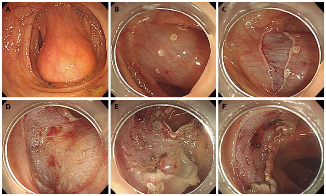
A 41-year-old man was admitted to our hospital because of repeated lower abdominal pain and constipation. He had no medical or family history. A physical examination showed no abnormalities, and initial laboratory data were within normal limits. A colonoscopy revealed a soft and yellowish submucosal tumor about 5 cm in diameter on the distal descending colon (Figure 1A). We diagnosed it as a colonic lipoma and decided to perform an ESD procedure. Markings were made around the base of the tumor on the distal side (anal side) after a transparent cap was equipped on the end of the scope. A lifting solution was injected to make a sufficient submucosal cushion from the muscle layer (Figure 1B). The solution was a mixture of 10% concentrated glycerin with 5% fructose and 0.9% sodium chloride and a small amount of epinephrine. The tumor located in the dependent position for an easy approach. Then, the precut was made just below the marking sites, and submucosal dissection was performed beneath the yellowish submucosal tumor using a dual knife (KD-650U, Olympus, Tokyo, Japan) (Figure 1C-E). The procedure time was approximately 28 min. No complications occurred during or after ESD. The 5.0 cm × 3.0 cm × 1.5 cm diameter tumor was resected en bloc (Figure 1F). A histopathological examination revealed a lipoma. The patient was discharged without complications and relieved of symptoms afterwards.
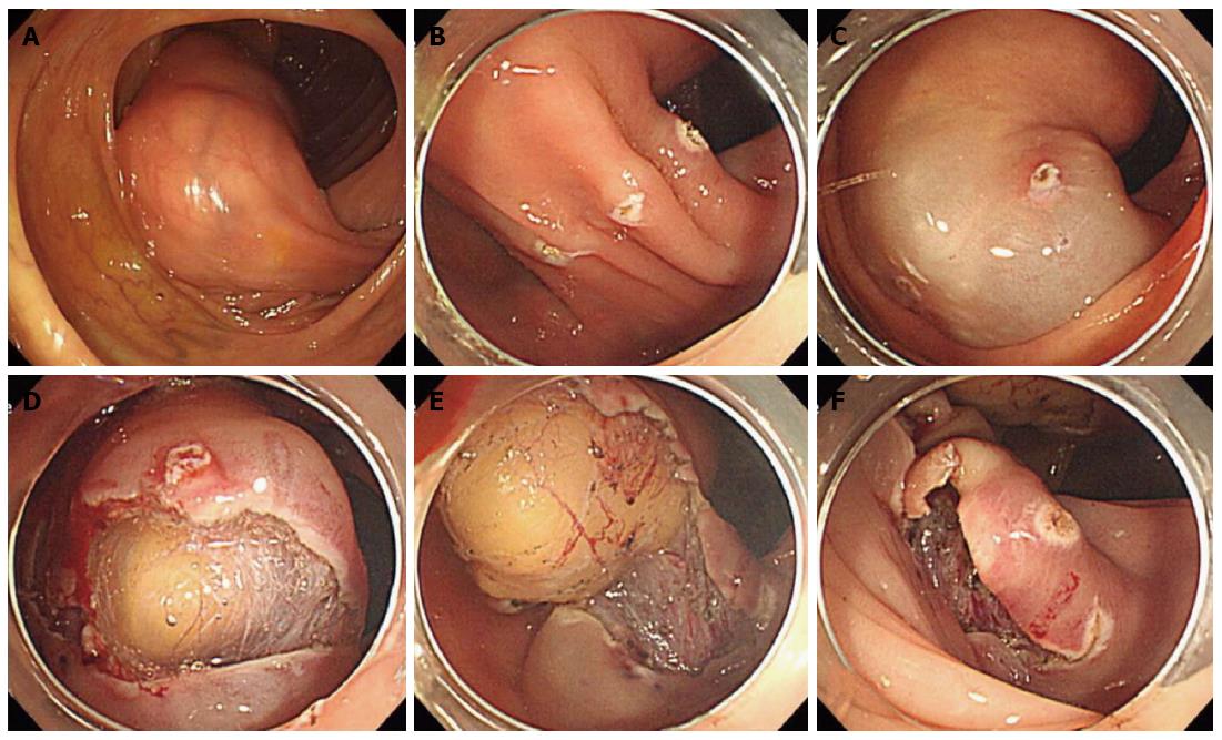
A 48-year-old man was referred to our hospital due to a large colonic mass detected during a colonoscopy. He had repeated lower abdominal pain for 1 year. He had no medical or family history. The physical examination at admission was unremarkable, and initial laboratory data were within normal limits. A colonoscopy revealed a yellowish submucosal mass approximately 7 cm in diameter on the proximal ascending colon (Figure 2A). The mass was soft and compressible, suggesting a colonic lipoma, and ESD was planned. Markings were made around the base of the tumor on the distal side (anal side) after a transparent cap was equipped at the end of the scope (Figure 2B). A lifting solution was injected to make a sufficient submucosal cushion (Figure 2C). The solution was a mixture of 10% concentrated glycerin with 5% fructose and 0.9% sodium chloride, with a small amount of epinephrine. The tumor was located in the dependent position for an easy approach. Next, a precut was made just below the marking sites, and the submucosal layer was dissected beneath the submucosal tumor using a dual knife (KD-650U, Olympus) and a hook knife (KD-620LR, Olympus) (Figure 2D). After submucosal dissection, the mucosal layer of the proximal side (oral side) was resected with the hook knife (Figure 2E). The procedure time was approximately 40 min. No complications occurred during or after ESD. The 7.5 cm × 4.5 cm × 4.0 cm diameter tumor was resected en bloc (Figure 2F). The histopathological examination revealed a lipoma. The patient was discharged without complications and relieved of symptoms afterwards.
A colonic lipoma is the second most common, but very rare, benign tumor after colon polyps[2]. The incidence of colonic lipoma is 0.15% at colonoscopy, and 0.4%-4.4% at autopsy[3,5]. Lipomas are confined to the submucosal layer in 90% of cases but are occasionally observed in the subserosal layer[6]. Although most cases are found incidentally in asymptomatic patients, some patients may present with symptoms such as abdominal pain, bleeding, constipation, and colonic obstruction or intussusceptions, particularly if the lipoma is > 3 cm in size[3,4].
A colonic lipoma can be diagnosed with barium enema, abdominal computed tomography (CT), colonoscopy, and endoscopic ultrasound (EUS). A colonic lipoma is observed as a round, smooth, and well demarcated filling defect on a barium series[2]. In addition, its shape and size can change according to colonic peristalsis (“squeeze sign”)[2]. Abdominal CT is only useful for diagnosing a large colonic lipoma and determining the extent of surgery[7]. It shows moderate signal intensity (-120 to -40 Hounsfield units) and can be distinguished from other tissue[7]. A colonic lipoma is usually observed as a yellowish, smooth, and round submucosal tumor without mucosal change during colonoscopy[6,8,9]. In particular, biopsy forceps may help to diagnose a lipoma; easily elevated mucosa over the lipoma with biopsy forceps (“tent sign”), recovery of the indented lipoma with biopsy forceps (“cushion or pillow sign”), or extrusion of fat tissue after biopsy (“naked fat sign”)[6,8,9]. A colonic lipoma on EUS shows typical features of a homogenous, high-echogenic tumor in the submucosal layer[5].
Treatment of a colonic lipoma is usually unnecessary because there is little possibility for malignant changes. However, treatment is required if it is difficult to distinguish the lipoma from other malignant tumors and it is associated with symptoms such as abdominal pain, bleeding, colonic obstruction, or intussusceptions[5,10]. Endoscopic removal of colonic lipomas has increased, and this has become a safe treatment modality with technological advances in colonoscopic procedures and equipment, particularly for small tumors[11]. However, because a colonic lipoma > 2 cm is associated with an increased risk of perforation during endoscopic removal, surgical removal is usually recommended[10,12]. Increasing the power of the electrosurgical device during endoscopic removal results in a high temperature, which may damage the colonic wall with subsequent perforation because fat tissue has low electric conductivity[13].
Thus, various endoscopic procedures, including snare resection, ESD, and unroofing or the Endo-loop technique have been introduced recently for safe resection[14-17]. Compared with other procedures, ESD is associated with a higher risk of complications such as perforation and bleeding[12]. However, ESD can be safe if performed while continuously observing the dissected layer. If submucosal dissection is performed using a knife in coagulation mode and a transparent cap is equipped at the end of the scope, it might be safer than other procedures. Submucosal dissection in coagulation mode is used to control the depth of the cut and reduce the risk of bleeding. In addition, because the exposed cutting surface is maintained using a transparent cap, submucosal dissection can be performed easily. In contrast, the proximal side (oral side) of the tumor margin cannot be seen during standard snare resection or the Endo-loop technique. Therefore, the risk of incomplete resection or perforation may increase. There are no reports demonstrating the indications and contraindications for ESD of a large colonic lipoma. Although surgery is not necessarily the first-line treatment option any more, the following circumstances are good candidates for operation; in case with wide-base sessile lipoma, unclear diagnosis, intussusceptions or obstruction, involvement of muscular and serosal layers[18].
In our cases, large colonic lipomas were successfully resected en bloc using the ESD technique without any complications. We created a sufficient submucosal cushion for safer dissection and changed the location of the colonic lipomas to the dependent position for easier dissection. In particular, changing the location of the lesion made the submucosal layer dissection easier, and the exposed cutting surface increased. Although colonic lipomas are confined to the submucosal layer, they have typical yellow color, which distinguishes them from surrounding tissues. Therefore, we could safely dissect the submucosal layer beneath the lipomas by coagulating exposed vessels.
In conclusion, ESD was a safe, effective, and suitable treatment modality to remove large submucosal colonic lipomas from patients with symptoms despite technical difficulties. However, this technique should be needed the special tool and experienced endoscopist.
A 41-years-old male and a 48-years-old male presented with repeated lower abdominal pain or constipation.
The physical examination at admission was unremarkable.
Colonic malignancy, irritable bowel syndrome, functional constipation, etc.
Initial laboratory data were within normal limits.
A colonoscopy revealed a soft and yellowish submucosal tumor about 5 cm and 7 cm in diameter on the distal descending colon and proximal ascending colon, respectively.
A histopathological examination revealed a lipoma.
Two patients were successfully treated with endoscopic submucosal dissection (ESD).
Using other equipment, including special knifes, a transparent cap, and an electrosurgical unit may help to remove a large colonic lipoma easier and safer by ESD.
ESD was a safe, effective, and suitable treatment modality to remove large submucosal colonic lipomas from patients with symptoms despite technical difficulties.
This article evaluated the safety and efficacy of ESD to remove a large submucosal colonic lipoma. Although endoscopic ultrasonography (EUS) was not performed to confirm the depth of lesion in the current cases, we think that the use of EUS can render the procedure safer.
P- Reviewer: Amornyotin S, Jonaitis L, Kapetanos D, Yamamoto S S- Editor: Qi Y L- Editor: A E- Editor: Liu XM
| 1. | Chung YF, Ho YH, Nyam DC, Leong AF, Seow-Choen F. Management of colonic lipomas. Aust N Z J Surg. 1998;68:133-135. [PubMed] |
| 2. | Michowitz M, Lazebnik N, Noy S, Lazebnik R. Lipoma of the colon. A report of 22 cases. Am Surg. 1985;51:449-454. [PubMed] |
| 3. | Ladurner R, Mussack T, Hohenbleicher F, Folwaczny C, Siebeck M, Hallfeld K. Laparoscopic-assisted resection of giant sigmoid lipoma under colonoscopic guidance. Surg Endosc. 2003;17:160. [RCA] [PubMed] [DOI] [Full Text] [Cited by in Crossref: 6] [Cited by in RCA: 16] [Article Influence: 0.7] [Reference Citation Analysis (0)] |
| 4. | Yu HG, Ding YM, Tan S, Luo HS, Yu JP. A safe and efficient strategy for endoscopic resection of large, gastrointestinal lipoma. Surg Endosc. 2007;21:265-269. [RCA] [PubMed] [DOI] [Full Text] [Cited by in Crossref: 42] [Cited by in RCA: 44] [Article Influence: 2.4] [Reference Citation Analysis (0)] |
| 5. | Murray MA, Kwan V, Williams SJ, Bourke MJ. Detachable nylon loop assisted removal of large clinically significant colonic lipomas. Gastrointest Endosc. 2005;61:756-759. [PubMed] |
| 6. | Bardají M, Roset F, Camps R, Sant F, Fernández-Layos MJ. Symptomatic colonic lipoma: differential diagnosis of large bowel tumors. Int J Colorectal Dis. 1998;13:1-2. [PubMed] |
| 7. | Liessi G, Pavanello M, Cesari S, Dell’Antonio C, Avventi P. Large lipomas of the colon: CT and MR findings in three symptomatic cases. Abdom Imaging. 1996;21:150-152. [PubMed] |
| 8. | Pfeil SA, Weaver MG, Abdul-Karim FW, Yang P. Colonic lipomas: outcome of endoscopic removal. Gastrointest Endosc. 1990;36:435-438. [PubMed] |
| 9. | Tascilar O, Cakmak GK, Gün BD, Uçan BH, Balbaloglu H, Cesur A, Emre AU, Comert M, Erdem LO, Aydemir S. Clinical evaluation of submucosal colonic lipomas: decision making. World J Gastroenterol. 2006;12:5075-5077. [PubMed] |
| 10. | El-Khalil T, Mourad FH, Uthman S. Sigmoid lipoma mimicking carcinoma: case report with review of diagnosis and management. Gastrointest Endosc. 2000;51:495-496. [PubMed] |
| 11. | Kim CY, Bandres D, Tio TL, Benjamin SB, Al-Kawas FH. Endoscopic removal of large colonic lipomas. Gastrointest Endosc. 2002;55:929-931. [PubMed] |
| 12. | Raju GS, Gomez G. Endoloop ligation of a large colonic lipoma: a novel technique. Gastrointest Endosc. 2005;62:988-990. [RCA] [PubMed] [DOI] [Full Text] [Cited by in Crossref: 40] [Cited by in RCA: 43] [Article Influence: 2.2] [Reference Citation Analysis (0)] |
| 13. | Bahadursingh AM, Robbins PL, Longo WE. Giant submucosal sigmoid colon lipoma. Am J Surg. 2003;186:81-82. [PubMed] |
| 14. | Geraci G, Pisello F, Arnone E, Sciuto A, Modica G, Sciumè C. Endoscopic Resection of a Large Colonic Lipoma: Case Report and Review of Literature. Case Rep Gastroenterol. 2010;4:6-11. [RCA] [PubMed] [DOI] [Full Text] [Full Text (PDF)] [Cited by in Crossref: 24] [Cited by in RCA: 26] [Article Influence: 1.7] [Reference Citation Analysis (0)] |
| 15. | Khorashad AK, Hosseini SM, Gaffarzadegan K, Farzanehfar MR, Zivarifar HR. Endoscopic resection of large colonic lipomas assisted by a prototype single-use endoloop device. J Res Med Sci. 2011;16:1511-1515. [PubMed] |
| 16. | Okada K, Shatari T, Suzuki K, Tamada T, Sasaki T, Suwa T, Hori M, Sakuma M. Is endoscopic submucosal dissection really contraindicated for a large submucosal lipoma of the colon? Endoscopy. 2008;40 Suppl 2:E227. [RCA] [PubMed] [DOI] [Full Text] [Cited by in Crossref: 8] [Cited by in RCA: 9] [Article Influence: 0.5] [Reference Citation Analysis (0)] |
| 17. | Sugimoto K, Sato K, Maekawa H, Sakurada M, Orita H, Ito T, Saita M, Ikota M, Yoshida Y, Yamano M. Unroofing technique for endoscopic resection of a large colonic lipoma. Case Rep Gastroenterol. 2012;6:557-562. [RCA] [PubMed] [DOI] [Full Text] [Full Text (PDF)] [Cited by in Crossref: 11] [Cited by in RCA: 12] [Article Influence: 0.9] [Reference Citation Analysis (0)] |
| 18. | Aydin HN, Bertin P, Singh K, Arregui M. Safe techniques for endoscopic resection of gastrointestinal lipomas. Surg Laparosc Endosc Percutan Tech. 2011;21:218-222. [RCA] [PubMed] [DOI] [Full Text] [Cited by in Crossref: 18] [Cited by in RCA: 19] [Article Influence: 1.5] [Reference Citation Analysis (0)] |


