Published online Feb 28, 2014. doi: 10.3748/wjg.v20.i8.2117
Revised: November 7, 2013
Accepted: December 12, 2013
Published online: February 28, 2014
Processing time: 151 Days and 8.3 Hours
Intestinal lipomatosis is a rare disease with an incidence at autopsy ranging from 0.04% to 4.5%. Because the lipomas are diffusely distributed in the intestine, most patients are symptom-free, and invasive intervention is not advised by most doctors. Here, we describe a case with intussusception due to small-bowel lipomatosis. Partial small bowel resection and anastomosis were performed because the intestinal wall was on the verge of perforation. This case indicates that regular follow-up is necessary and endoscopic treatment should be considered to avoid surgical procedures if the lipoma is large enough to cause intestinal obstruction.
Core tip: Intestinal lipomatosis is a rare disease, obstruction due to a large lipoma may result in acute abdomen, and surgical intervention is applied infrequently. When patients are presented with symptoms, endoscopic treatment should be considered.
- Citation: Gao PJ, Chen L, Wang FS, Zhu JY. Ileo-colonic intussusception secondary to small-bowel lipomatosis: A case report. World J Gastroenterol 2014; 20(8): 2117-2119
- URL: https://www.wjgnet.com/1007-9327/full/v20/i8/2117.htm
- DOI: https://dx.doi.org/10.3748/wjg.v20.i8.2117
Intestinal lipomatosis is a rare disease with an incidence at autopsy ranging from 0.04% to 4.5% and most patients are symptom-free[1]. Symptomatic cases usually present as paroxysmal abdominal pain, hemorrhage of digestive tract, or abdominal masses. We here describe a case with ileo-colonic intussusception secondary to small-bowel lipomatosis.
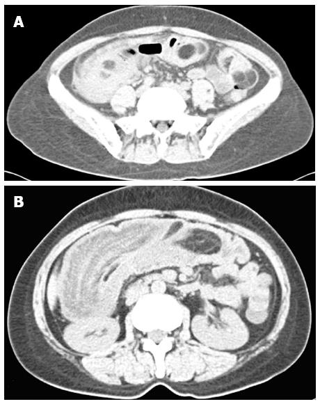
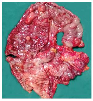
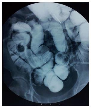
A 52-year-old woman presented to our outpatient clinic in May 2013 with a history of abdominal pain for 21 d. The patient suffered from persistent abdominal cramps, accompanied by vomiting. The upright plain abdominal film revealed dilated small bowel loops with air-fluid levels. Intussusception was diagnosed by colonoscopy. Computed tomography (CT) scan showed multiple lipomas in small intestine (Figure 1A) and intussusception (Figure 1B). The patient was admitted to the hospital and emergency exploratory laparotomy was performed. During the operation, numerous lipomas 0.3-5 cm in diameter were found diffusely distributed in the intestine and ileo-colonic intussusception due to a large lipoma was confirmed (Figure 2). Partial small bowel resection and anastomosis were carried out because the intestinal wall was on the verge of perforation. Pathological examination confirmed a diagnosis of lipomatosis. Postoperative wound infection and cholecystitis occurred. After dressing change and anti-infection treatment, the patient was discharged in good condition one month later. The patient has remained symptom-free for 3 mo, although a small intestinal double contrast radiography revealed a filling defect in the duodenum and multiple submucosal masses in the small intestine (Figure 3).
Primary small intestinal tumors are uncommon, which account for about 1% of all gastrointestinal tumors, and primary lipomatosis of the small intestine is rare[2]. Diffuse and multiple lipomas in the intestine were first reported by Hellstrom[3] in 1906. CT and barium examination are useful techniques to confirm the diagnosis. The typical lipomas are usually seen as smooth, nonulcerated filling defects. Most lipomas are small and display no symptoms, and invasive treatment is unnecessary. When patients are presented with paroxysmal abdominal pain, hemorrhage of digestive tract and other symptoms, endoscopic treatment including endoscopic submucosal dissection, endoscopic snare resection and endoscopic unroofing techniques should be considered[4]. If the lipoma is large enough to cause intestinal obstruction or intestinal obstruction secondary to intussusception as shown in our patient, surgical intervention may be inevitable[5]. Because the lipomas are diffusely distributed in the intestine, the object of the surgery is not to remove all the lipomas but to relieve the obstruction. During the operation, large lipoma can be dislodged by local resection.
This condition may be asymptomatic and abdomimal pain is the most common symptom.
Symptomatic cases may present as abdominal pain, intestinal obstruction, intussusception, volvulus, or bleeding due to mucosal ulceration.
With aid of computed tomography (CT), intestinal lipomatosis can be distinguished from liposarcoma by the homogeneity of its fatty content and absence of areas of increased density.
Laboratory testing was not used for this condition.
Barium studies showed multiple submucosal masses in the small intestine and abdominal CT revealed multiple intramural fat density masses in the small intestine.
Under microscopy, the specimen showed multiple submucosal lipomatous nodules composed of abnormal collections of mature adipose tissue.
If intestinal obstruction secondary to intussusception or volvulus occur, surgical intervention should be considered.
Endoscopic treatment is recommended to remove the large lipomas to prevent obstruction.
Intestinal lipomatosis is characterized by diffuse and multiple lipomas in the intestine.
Most patients with lipomatosis may be asymptomatic, but when intestinal obstruction secondary to intussusception or volvulus occur, surgical intervention may be inevitable. Endoscopic treatment is ideal in theory, but it is too difficult to perform in most hospitals.
Lipomatosis is a rare disease, and intussusception secondary to lipomatosis is even rare. There are few literatures about this disease.
P- Reviewers: Akyuz F, Akyuz U, Reddy DN S- Editor: Gou SX L- Editor: A E- Editor: Wu HL
| 1. | Suárez Moreno RM, Hernández Ramírez DA, Madrazo Navarro M, Salazar Lozano CR, Martínez Gen R. Multiple intestinal lipomatosis. Case report. Cir Cir. 2010;78:163-165. [PubMed] |
| 2. | Fang SH, Dong DJ, Chen FH, Jin M, Zhong BS. Small intestinal lipomas: diagnostic value of multi-slice CT enterography. World J Gastroenterol. 2010;16:2677-2681. [RCA] [PubMed] [DOI] [Full Text] [Full Text (PDF)] [Cited by in CrossRef: 26] [Cited by in RCA: 24] [Article Influence: 1.6] [Reference Citation Analysis (0)] |
| 3. | Hellstrom N. Kasuistische Beitrage zur Kenntis des Intestinallipoms. Dtsch Z Chir. 1906;84:488–511 Available from: http//link.springer.com/article/10.1007%2FBF02790963#page-1. |
| 4. | Fu CK, Hsieh TY, Huang TY. Lipomatosis of the small intestine: detection and endoscopic unroofing by single-balloon enteroscopy. Dig Endosc. 2013;25:85-86. [RCA] [PubMed] [DOI] [Full Text] [Cited by in RCA: 1] [Reference Citation Analysis (0)] |
| 5. | Tzeng YD, Liu SI, Yang MC, Mok KT. Bowel obstruction with intestinal lipomatosis. Dig Liver Dis. 2012;44:e4. [RCA] [PubMed] [DOI] [Full Text] [Cited by in Crossref: 2] [Cited by in RCA: 3] [Article Influence: 0.2] [Reference Citation Analysis (0)] |


