Case Report Open Access
Copyright ©2014 Baishideng Publishing Group Co., Limited. All rights reserved.
World J Gastroenterol. Feb 28, 2014; 20(8): 2113-2116
Published online Feb 28, 2014. doi: 10.3748/wjg.v20.i8.2113
Pleomorphic hepatocellular carcinoma following consumption of hypericum perforatum in alcoholic cirrhosis
Evangeli S Lampri, Vasiliki Malamou-Mitsi, Department of Pathology, Ioannina University Medical School, 45110 Ioannina, Greece
Elli Ioachim, Eufemia Balasi, Department of Pathology, G. Hatzikosta General Hospital, 45001 Ioannina, Greece
Haralampos Harissis, Department of Surgery, Ioannina University Medical School, 45110 Ioannina, Greece
Antigoni Mitselou, Department of Forensic Pathology, Ioannina University Medical School, 45110 Ioannina, Greece
Author contributions: Lampri ES and Ioachim E designed the study; Harisis H was the attending doctor and performed the surgical operation; Malamou-Mitsi V performed the cytological examination; Lampri ES, Ioachim E, Malamou-Mitsi V, Balasi E and Mitselou A performed the pathological examinations; Ioachim E organized the case report; Lampri E wrote the manuscript.
Correspondence to: Evangeli S Lampri, MD, Department of Pathology, Ioannina University Medical School, Leoforos Stavrou Niarxou, PO Box 1186, 45110 Ioannina, Greece. evangeli.lampri@gmail.com
Telephone: +30-265-1080428 Fax: +30-265-1080428
Received: April 23, 2013
Revised: June 6, 2013
Accepted: July 17, 2013
Published online: February 28, 2014
Processing time: 309 Days and 6.9 Hours

Abstract

Hepatocellular carcinoma (HCC) often develops in patients with underlying liver disease, yet HCC with syncytial giant cells (SGCs) is extremely rare. Herein, we report a 55-year-old man with a 6-year history of alcoholic cirrhosis who during his regular checkup presented with marked elevation of alpha-fetoprotein. Clinical examination and imaging analyses revealed a tumor-like lesion in segment 4 of the liver, which was removed by limited wedge resection. Histological analysis by hematoxylin and eosin staining indicated pleomorphic and atypical nodules, with some SGCs, embedded within the boundaries of the neoplastic lesion. The adjacent liver parenchyma showed microvesicular steatosis, pericellular fibrosis, and moderate hemosiderin accumulation (grade 2, as determined by Prussian blue iron stain) in hepatocytes and Kupffer cells but no copper accumulation (as determined by orcein stain). Immunohistochemical analysis showed hepatocyte antigen-positive staining for the neoplastic cells and SGCs. The diagnosis was made for cirrhosis-related HCC with SGCs. The previous reports of pleomorphic HCC have featured osteoclast-like (i.e., mesenchymal type) giant cells, making this case of epithelial type giant cells very rare. The patient’s 6-month history of hypericum perforatum/St John’s wort self-medication may have prompted the cirrhosis or HCC progression or the unusual SGC manifestation.

Key Words: Hepatocellular carcinoma; Giant cell carcinoma; Hypericum perforatum; St John’s wort; Syncytial giant cell; Pleomorphic liver tumor; Alcoholic cirrhosis

Core tip: This case report describes the clinicopathological-based finding of an unusual hepatocellular carcinoma (HCC) with syncytial giant cells (SGCs) in a 55-year-old man with a 6-year history of alcoholic cirrhosis. Unlike the previous case reports of these rare tumor types that have demonstrated the mesenchymal and non-neoplastic nature of the giant cells, the current case showed an epithelial and hepatocyte-originated neoplasmic nature. An intriguing feature of case is the patient’s 6-month history of hypericum perforatum/St John’s wort self-medication, which may have prompted the cirrhosis or HCC progression or the unusual SGC manifestation.


INTRODUCTION

Hepatocellular carcinoma (HCC) often develops in patients with underlying liver disease, yet the HCC type with syncytial giant cells (SGC) is extremely rare. Multinucleated giant cells have been reported in a variety of neoplasms from many organs, including the liver, but these cells have featured an osteoclast phenotype[1-5]. The cellular origin of these osteoclast-like giant cells remains controversial, and various studies have provided evidence suggesting mesenchymal derivation as well as epithelial derivation[4-6].

Herein, we report the clinicopathological-based finding of cirrhosis-related HCC with SGC in an adult male with a long-term history of alcoholism and a short-term history of hypericum perforatum [commonly known as St John’s wort (SJW)]. Immunohistochemical evidence indicated that the SGCs are of epithelial and not mesenchymal origin. Moreover, the patient’s ingestion of SJW raises the intriguing possibility of its involvment with this unusual manifestation.

CASE REPORT

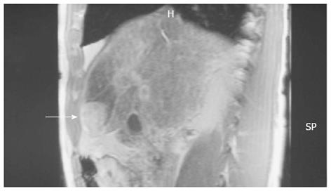
A 55-year-old man with a 6-year history of alcoholic cirrhosis diagnosis presented with marked elevation of alpha-fetoprotein (a-FP; 14000 ng/mL, reference range: 0-40 ng/mL) during his regular checkup. Magnetic resonance imaging revealed a tumor-like lesion on segment 4 of the liver (Figure 1). The patient was admitted to the Department of Surgery for treatment on 1 April 2008. During the initial interview, the patient self-described a long-term history of large-quantity daily alcohol intake (10 L of wine/d for 10 years) and a short-term history of unknown quantity daily intake of SJW (for the last few months). Hepatomegaly was found upon physical examination, but further serologic analyses revealed normal levels of ferritin (120 g/L, reference range: 24-248 g/L) and ceruloplasmin (32 g/L, reference range: 20-50 g/L) and negative results for autoimmune markers and hepatitis B- and C-specific antigens.

Figure 1
Figure 1 Magnetic resonance imaging showed a tumor-like lesion (arrow) and ischemia in segment 4 of the liver.

Fine needle aspiration of the tumor performed on 2 April 2008 revealed normal and atypical hepatocytes, nodular regenerative atypia, and transitional and differentiated bile duct epithelial cells. In addition, three distinct hepatocyte aggregations were observed, which were characterized by pleomorphism, non-uniform nuclear size, increased nucleocytoplasmic ratio, diffuse chromatin distribution, and large, prominent nucleoli. While the presence of these aggregates suggested a neoplastic character for the tumor-like lesion, atypia could not be excluded due to the presence of the regenerative nodules and cirrhotic features.

The patient underwent segment 4 resection on 4 April 2008. The procedure was modified perioperatively to a limited wedge resection with ethanol injection of the cut liver surface according to the observations of substantial ischemia in the left liver lobe, gross pathological features of cirrhosis in the right lobe, and presence of three small (about 2 mm each) satellite nodules. The resected specimen measured 3 cm × 2.5 cm × 1.5 cm. Sectioning revealed multiple white nodules throughout the tumor tissue, which itself was surrounded by normal liver tissue with a micro-nodular cirrhotic appearance.

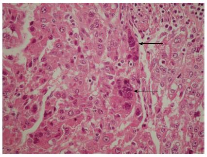
Subsequent microscopic analysis of the nodules revealed disordered architecture of the surrounding liver cell. The cells comprising the nodules showed pleomorphism and atypia, with some syncytial type multinuclear giant cells that had features similar to infantile giant cell hepatitis (Figure 2). Moreover, these giant cells also displayed the same nuclear atypia as the other HCC cells, which suggested that the cells were part of the tumor itself and not merely entrapped non-neoplastic cells; the marked lack of any giant cell changes in the adjacent liver tissue further reinforced this hypothesis.

Figure 2
Figure 2 Hematoxylin and eosin-staining of a representative hepatocellular carcinoma section showing the multinuclear giant syncytial cells (arrows).

A small number of mitotic figures, both typical and atypical and with or without apoptotic bodies, were observed in the tumor-like lesion, including the nodules, as well as beyond the lesion boundary, in the adjacent liver parenchyma. Inflammatory cells, such as lymphocytes and plasmatocytes, were present throughout the HCC and adjacent liver tissue, with greater incidence in the septa. Hyperplasia was observed in the marginal area of the nodules and small bile ducts. The adjacent liver parenchyma showed microvesicular steatosis, pericellular fibrosis, and moderate hemosiderin accumulation (grade 2, as determined by Prussian blue iron stain) in hepatocytes and Kupffer cells but no copper accumulation (as determined by orcein stain).

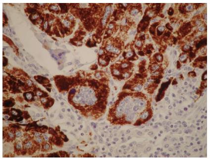
Immunohistochemical analysis showed hepatocyte antigen-positive staining for the neoplastic cells and SGCs (Figure 3). No cells showed immunoreactivity for macrophage-specific markers, such as CD68, but occasional cells showed immunoreactivity for the proliferation marker Ki-67/MIB-1 or the oncoprotein p53. The giant cells’ nuclei showed a remarkable lack of immunoreactivity for all of the markers detected.

Figure 3
Figure 3 Mononuclear and multinuclear carcinoma cells showed reactivity for the hepatocyte antigen (brown). Magnification × 40.

According to the collective histological findings, the diagnosis was made for cirrhosis-related HCC with SGCs. Up to the last follow-up examination, performed on 10 April 2013, the patient had maintained a-FP levels within the normal range and showed no signs of tumor recurrence by liver imaging.

DISCUSSION

While adult cases of giant cell HCC are relatively common, and its clinical and histologic features are well characterized, the case of giant cell HCC presented herein highlights the unusual histological manifestation of syncytial type cells[1-5]. Previous case reports of these rare tumor types have demonstrated the mesenchymal and non-neoplastic nature of the giant cells, leading to the suggested nomenclature of liver cell carcinoma with osteoclast-like giant cells[4-6]. However, the giant cells of the current case showed an epithelial and hepatocyte-originated neoplasmic nature, leading to the diagnosis of HCC with syncytial giant cells.

Another distinctive feature of this HCC case is its manifestation in cirrhotic liver. While cirrhosis was a likely etiology of the HCC, as suggested by the presence of nodular cirrhosis and microvesicular steatosis, the etiology of the cirrhosis was not obvious, and could have been long-term alcoholism or hemosiderosis. The patient’s chronic consumption of SJW represents another potential complicating factor. The known hepatoxic properties of this herbal medicine may have contributed to or been the primary inducing factor of the syncytial phenotype of the giant cells.

The liver is among the first organs to experience toxic injury, as it is the main site of endo- and xenobiotic bio-transformation. As such, a primary focus of drug development and clinical trials is determining the liver-related side effects of pharmaceutical and herbal medicines. Among the most common liver injuries are acute liver necrosis, hepatitis, cholestasis, fibrosis and cirrhosis; in addition, many of the medicines show either direct or indirect carcinogenic potential. Over the past few decades, the traditional herbal preparation of SJW extract has risen tremendously in popularity in the Western world, largely due to its observed benefits in treating mild to moderate depression[7-9], and bladder cancer[10,11].

Recent studies have indicated a potential clinical benefit of SJW for treating alcoholism, and implicated the bioactive phytochemical hyperforin as the constituent responsible for this response[12]. Interestingly, hyperforin has also been shown to have anti-microbial activity against Gram-positive bacteria and numerous viruses[13,14]. Studies of the molecular mechanisms underlying the bioactive activities of hyperforin have revealed a complex functional interaction with the other SJW constituents of essential oils, phloroglucinols, and flavonoids to induce a photoactivation process, possibly leading to production of singlet oxygen molecules[13,14]. Photoactivated-free radicals may exert the protective anti-infective or anti-tumor effects-for the latter, possibly through activation of apoptosis-related pathways[15,16].

In general, SJW is well tolerated as both topical and oral preparations. The most commonly reported adverse effects are gastrointestinal-related (i.e., nausea, hypogastric pain, loss of appetite, diarrhea, gastric complaints[17] and sedative-related (i.e., dizziness, confusion, and fatigue)[18]. Rare cases of photosensitivity development have also been reported, most often associated with systemic administration[19], and possible fertility risks have been suggested by some studies as well[20].

Another unknown mechanism of SJW is its ability to interact, either beneficially or detrimentally, with conventional drugs concomitantly-administered[21]. This possibly was highlighted by the observation of SJW-associated enhancement of P-glycoprotein, an intestinal multidrug transporter, and the cytochrome P450 (CYP) oxidative enzyme superfamily members CYP3A4 and CYP2C9. The induction of CYP3A4 represents a particularly troubling (and unresolved) clinical concern because of its abundance in the liver and intestine, both important sites of drug bio-transformation[22]. In addition, high-dosage SJW has been shown to activate the detoxification pathway mediated by the pregnane X receptor (PXR), a particularly promiscuous receptor; PXR overstimulation is detrimental and may result from SJW interactions with other drugs[23]. Finally, the non-standardized compositions of SJW extract preparations, available as over-the-counter medicines and often taken without clinical oversight, further complicate our ability to understand the benefits and risks of SJW, such as which may have contributed to the current case[24].

Despite these unknown features of SJW bioactivity, the significant anti-tumor effects of hyperforin in cell culture and animal systems have prompted continued efforts to investigate its potential as an efficacious cancer therapy that is less toxic than the current chemo- and radio-therapies. For example, in vitro studies of various rat and human cancers (e.g., mammary carcinoma, squamous cell carcinoma, malignant melanoma, and lymphoma) showed equal or greater anti-tumor effects of hyperforin compared to the common cytostatic drugs camptothecin, paclitaxel, and vincristine[25]. Moreover, the molecular mechanism of hyperforin-mediated tumor cell apoptosis was shown to involve, at least partially, mitochondrial release of cytochrome c and caspase activation, thereby triggering cell death pathways[25,26]. However, as with any pharmacologic or herbal medicine, absolute anti-tumor activity is not guaranteed and inappropriate doses or concomitant drug administrations may create a pro-tumor situation.

For the case of cirrhosis-related HCC with SGCs described herein, we cannot conclude the role played by SJW with any certainty. The patient’s ingestion of SJW may have created a hepatotoxic condition that promoted the progression of cirrhosis or HCC. Nonetheless, this case highlights the need for further studies of SJW to determine its side effects profile and safe dosages under various physiologic and pathogenic conditions; such knowledge is particularly important as the manufacture (i.e., compositions) and use (i.e., dose, duration, and in conjunction with other drugs) of SJW is non-uniform and widespread.

Footnotes

P- Reviewers: Sazci A, Sanai FM S- Editor: Wen LL L- Editor: A E- Editor: Wang CH

References
1.  Atra A, Al-Asiri R, Wali S, Al-Husseini H, Al-Bassas A, Zimmermann A. Hepatocellular carcinoma, syncytial giant cell: a novel variant in children: a case report. Ann Diagn Pathol. 2007;11:61-63.  [PubMed]  [DOI]  [Cited in This Article: ]  [Cited by in Crossref: 8]  [Cited by in F6Publishing: 10]  [Article Influence: 0.6]  [Reference Citation Analysis (0)]
2.  Kuwano H, Sonoda T, Hashimoto H, Enjoji M. Hepatocellular carcinoma with osteoclast-like giant cells. Cancer. 1984;54:837-842.  [PubMed]  [DOI]  [Cited in This Article: ]
3.  Hood DL, Bauer TW, Leibel SA, McMahon JT. Hepatic giant cell carcinoma. An ultrastructural and immunohistochemical study. Am J Clin Pathol. 1990;93:111-116.  [PubMed]  [DOI]  [Cited in This Article: ]
4.  Sasaki A, Yokoyama S, Nakayama I, Nakashima K, Kim YI, Kitano S. Sarcomatoid hepatocellular carcinoma with osteoclast-like giant cells: case report and immunohistochemical observations. Pathol Int. 1997;47:318-324.  [PubMed]  [DOI]  [Cited in This Article: ]
5.  Bauditz J, Rudolph B, Wermke W. Osteoclast-like giant cell tumors of the pancreas and liver. World J Gastroenterol. 2006;12:7878-7883.  [PubMed]  [DOI]  [Cited in This Article: ]
6.  Rosai J. Liver cell carcinoma with osteoclast-like giant cells: nonepitheliogenic giant cells in diverse malignancies. Hepatology. 1990;12:782-783.  [PubMed]  [DOI]  [Cited in This Article: ]
7.  Gaster B, Holroyd J. St John’s wort for depression: a systematic review. Arch Intern Med. 2000;160:152-156.  [PubMed]  [DOI]  [Cited in This Article: ]
8.  Laakmann G, Jahn G, Schüle C. Hypericum perforatum extract in treatment of mild to moderate depression. Clinical and pharmacological aspects. Nervenarzt. 2002;73:600-612.  [PubMed]  [DOI]  [Cited in This Article: ]
9.  Linde K, Ramirez G, Mulrow CD, Pauls A, Weidenhammer W, Melchart D. St John’s wort for depression--an overview and meta-analysis of randomised clinical trials. BMJ. 1996;313:253-258.  [PubMed]  [DOI]  [Cited in This Article: ]
10.  Orellane C. St John’s wort helps to fight bladder cancer. Lancet Oncol. 2001;2:399.  [PubMed]  [DOI]  [Cited in This Article: ]
11.  Skalkos D, Stavropoulos NE, Tsimaris I, Gioti E, Stalikas CD, Nseyo UO, Ioachim E, Agnantis NJ. The lipophilic extract of Hypericum perforatum exerts significant cytotoxic activity against T24 and NBT-II urinary bladder tumor cells. Planta Med. 2005;71:1030-1035.  [PubMed]  [DOI]  [Cited in This Article: ]  [Cited by in Crossref: 15]  [Cited by in F6Publishing: 10]  [Article Influence: 0.5]  [Reference Citation Analysis (0)]
12.  Perfumi M, Mattioli L, Cucculelli M, Massi M. Reduction of ethanol intake by chronic treatment with Hypericum perforatum, alone or combined with naltrexone in rats. J Psychopharmacol. 2005;19:448-454.  [PubMed]  [DOI]  [Cited in This Article: ]  [Cited by in Crossref: 16]  [Cited by in F6Publishing: 17]  [Article Influence: 0.9]  [Reference Citation Analysis (0)]
13.  Weber ND, Murray BK, North JA, Wood SG. The antiviral agent hypericin has in vitro activity against HSV-1 through non-specific association with viral and cellular membranes. Antivir Chem Chemother. 1994;5:83-90.  [PubMed]  [DOI]  [Cited in This Article: ]
14.  Lenard J, Rabson A, Vanderoef R. Photodynamic inactivation of infectivity of human immunodeficiency virus and other enveloped viruses using hypericin and rose bengal: inhibition of fusion and syncytia formation. Proc Natl Acad Sci USA. 1993;90:158-162.  [PubMed]  [DOI]  [Cited in This Article: ]
15.  Thomas C, MacGill RS, Miller GC, Pardini RS. Photoactivation of hypericin generates singlet oxygen in mitochondria and inhibits succinoxidase. Photochem Photobiol. 1992;55:47-53.  [PubMed]  [DOI]  [Cited in This Article: ]
16.  Hadjur C, Richard MJ, Parat MO, Favier A, Jardon P. Photodynamically induced cytotoxicity of hypericin dye on human fibroblast cell line MRC5. J Photochem Photobiol B. 1995;27:139-146.  [PubMed]  [DOI]  [Cited in This Article: ]
17.  Schulz V, John A. Side effects and drug interactions. St. John’s Wort and its Active Principles in Depression and Anxiety - Milestones in Drug Therapy MDT. New York: Springer 2005; 145-160.  [PubMed]  [DOI]  [Cited in This Article: ]
18.  Barnes J, Anderson LA, Phillipson JD.  Herbal Medicines: A guide for healthcare professionals. 2nd ed. London: Pharmaceutical Press 2002; .  [PubMed]  [DOI]  [Cited in This Article: ]
19.  Ernst E, Rand JI, Barnes J, Stevinson C. Adverse effects profile of the herbal antidepressant St. John’s wort (Hypericum perforatum L.). Eur J Clin Pharmacol. 1998;54:589-594.  [PubMed]  [DOI]  [Cited in This Article: ]
20.  Stahl S Essential psychopharmacology: neuroscientific basis and practical applications. 2nd ed. United Kingdom: Cambridge University Press 2000; .  [PubMed]  [DOI]  [Cited in This Article: ]
21.  Ioannides C. Pharmacokinetic interactions between herbal remedies and medicinal drugs. Xenobiotica. 2002;32:451-478.  [PubMed]  [DOI]  [Cited in This Article: ]  [Cited by in Crossref: 167]  [Cited by in F6Publishing: 171]  [Article Influence: 7.8]  [Reference Citation Analysis (0)]
22.  Maurel P The CYP3A family. In Cytochrome P450: Metabolic and Toxicological. Aspects Boca Raton, FL: CRC Press 1996; 241-270.  [PubMed]  [DOI]  [Cited in This Article: ]
23.  Moore LB, Goodwin B, Jones SA, Wisely GB, Serabjit-Singh CJ, Willson TM, Collins JL, Kliewer SA. St. John’s wort induces hepatic drug metabolism through activation of the pregnane X receptor. Proc Natl Acad Sci USA. 2000;97:7500-7502.  [PubMed]  [DOI]  [Cited in This Article: ]  [Cited by in Crossref: 677]  [Cited by in F6Publishing: 607]  [Article Influence: 25.3]  [Reference Citation Analysis (0)]
24.  Cantoni L, Rozio M, Mangolini A, Hauri L, Caccia S. Hyperforin contributes to the hepatic CYP3A-inducing effect of Hypericum perforatum extract in the mouse. Toxicol Sci. 2003;75:25-30.  [PubMed]  [DOI]  [Cited in This Article: ]  [Cited by in Crossref: 41]  [Cited by in F6Publishing: 42]  [Article Influence: 2.0]  [Reference Citation Analysis (0)]
25.  Schempp CM, Kirkin V, Simon-Haarhaus B, Kersten A, Kiss J, Termeer CC, Gilb B, Kaufmann T, Borner C, Sleeman JP. Inhibition of tumour cell growth by hyperforin, a novel anticancer drug from St. John’s wort that acts by induction of apoptosis. Oncogene. 2002;21:1242-1250.  [PubMed]  [DOI]  [Cited in This Article: ]  [Cited by in Crossref: 185]  [Cited by in F6Publishing: 161]  [Article Influence: 7.3]  [Reference Citation Analysis (0)]
26.  Schempp CM; Monograph. Hypericum perforatum. Altern Med Rev. 2004;9:318-325.  [PubMed]  [DOI]  [Cited in This Article: ]