Published online Dec 7, 2014. doi: 10.3748/wjg.v20.i45.17092
Revised: May 29, 2014
Accepted: July 11, 2014
Published online: December 7, 2014
Processing time: 289 Days and 3.6 Hours
AIM: To explore the inhibitory effects of dobutamine on gastric adenocarcinoma cells.
METHODS: Dobutamine was used to treat gastric adenocarcinoma cells (SGC-7901) and cell viability was determined by the 3-(4,5-dimethylthiazol-2-yl)-2,5-diphenyltetrazolium bromide (MTT) assay. The effects of dobutamine combined with cisplatin on cell viability were also analyzed. Cell migration was studied using the wound healing assay, and cell proliferation was analyzed using the colony formation assay. A cell invasion assay was carried out using Transwell cell culture chambers. The cell cycle and cell apoptosis were analyzed by flow cytometry. Western blot and immunocytochemistry were performed to determine the expression of Yes-associated protein (YAP) in treated cells.
RESULTS: Dobutamine significantly inhibited cell growth, migration, cell colony formation, and cell invasion into Matrigel. Dobutamine also arrested the cell cycle at G1/S phase, and increased the rate of apoptosis of gastric adenocarcinoma cells. The expression of YAP was detected mainly in the nucleus in the absence of dobutamine. However, reduced expression of phosphorylated YAP was mainly found in the cytosol following treatment with dobutamine.
CONCLUSION: Dobutamine has significant inhibitory effects on gastric adenocarcinoma cells and may be used in neoadjuvant therapy not only for gastric cancer, but also for other tumors.
Core tip: Dobutamine inhibited the viability, migration, proliferation and invasion of SGC-7901 cells. Dobutamine arrested the cell cycle at G1/S phase, and increased the rate of apoptosis of gastric adenocarcinoma cells. Phosphorylated Yes-associated protein was found mainly in the cytosol after treatment with dobutamine.
- Citation: Zheng HX, Wu LN, Xiao H, Du Q, Liang JF. Inhibitory effects of dobutamine on human gastric adenocarcinoma. World J Gastroenterol 2014; 20(45): 17092-17099
- URL: https://www.wjgnet.com/1007-9327/full/v20/i45/17092.htm
- DOI: https://dx.doi.org/10.3748/wjg.v20.i45.17092
Gastric carcinoma is now one of the most common malignancies with a relatively high incidence and mortality rate. It was estimated that in 2012 there were 21320 new cases and 10540 deaths in the United States[1]. However, traditional chemotherapy for the treatment of gastric carcinoma is still not very effective. Therefore, new therapies and drugs are required.
Dobutamine is a synthetic sympathomimetic amine, and is widely used in patients with congestive heart failure. Dobutamine is a β1-adrenoceptor agonist, but it can also stimulate β2- and α1-adrenoceptors in the cardiovascular system[2,3]. In addition, dobutamine has been used in cardiovascular testing, including dobutamine stress cardiovascular magnetic resonance (DCMR)[4]. Recently, dobutamine was shown to significantly affect human osteosarcoma cells. Researchers confirmed that dobutamine attenuated Yes-associated protein (YAP)-dependent transcription by inhibiting YAP nuclear translocation[5]. This may result in a new application for dobutamine in cancer treatment.
YAP, which is present in chromosome 11q22, is highly conserved in mammalian systems, and is considered to be the nuclear effector of the Hippo pathway. The Hippo pathway is very important in the regulation of cell growth, division, and apoptosis[6]. This pathway was first identified by mosaic screens in Drosophila melanogaster, as a pathway that determined organ size[7]. It was found that the Hippo pathway is commonly altered during carcinogenesis. Recently, high-expression of YAP was observed in many types of tumors, including hepatocellular, colorectal, gastric, ovarian, breast, and lung cancers, and was correlated with poor prognosis[8-14]. These observations suggest that YAP may contribute to a malignant cellular phenotype[15,16]. During the process of carcinogenesis, the effects of YAP are not unique, but are universal. Therefore, it is important to identify reagents which can inhibit YAP in clinical medicine, which will be useful in tumor treatment.
YAP is a carcinogenic factor which is over-expressed in gastric carcinoma. Dobutamine may reduce the expression of YAP. In this study, dobutamine was used to treat gastric adenocarcinoma cells, and its therapeutic value in these cells was assessed. The combined effects of dobutamine and cisplatin were also determined. We expect that dobutamine will be approved as an adjuvant therapy in gastric cancer.
Human gastric adenocarcinoma SGC-7901 cells (Institute of Biochemistry and Cell Biology, Shanghai, PRC) were cultured in RPMI-1640 containing 10% fetal bovine serum, 100 U/mL penicillin and 100 μg/mL streptomycin. The cells were maintained at 37 °C in a humidified atmosphere containing 5% CO2 at 37 °C.
The influence of dobutamine (Sigma-Aldrich, MO, United States) on cell viability was determined using the 3-(4,5-dimethylthiazol-2-yl)-2,5-diphenyltetrazolium bromide (MTT) assay. Dobutamine was diluted in 0.9% saline solution and stored at 37 °C, and freshly diluted in culture medium before each experiment. SGC-7901 cells (5 × 10⁴/mL, 200 μL) were cultured in a 96-well plate overnight and stimulated with various concentrations of dobutamine (1 μmol/L, 3 μmol/L and 10 μmol/L) for 24, 48 and 72 h, respectively. Twenty μL MTT was added to each well, and incubated for a further 4 h. After careful removal of the medium, 150 μL dimethyl sulfoxide (DMSO) was added to each well, and the plate was then shaken until the crystals were solubilized. Finally, the absorbance was measured at 490 nm. Cell survival was calculated using the mean of pooled data from three separate experiments with five wells. The inhibitory rate was calculated using the following equation: Inhibitory rate (IR) (%) = (1 - ODtreatment/ODcontrol) × 100%.
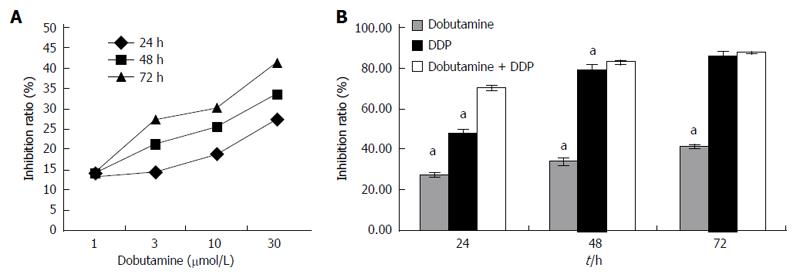
In addition, the effect of dobutamine (30 μmol/L) combined with cisplatin (DDP, 8 μg/mL) on cell viability was also analyzed by MTT.
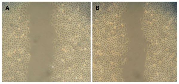
The effect of dobutamine on the migration of SGC-7901 cells was examined using the wound healing assay. Cells were cultured in 6-well plates and allowed to grow to 90% confluence. A wound track (approximately 5 mm in size) was scored in each dish with a plastic scraper. The debris was removed by washing with PBS. After incubation with 30 μmol/L dobutamine for 24 h, the cells which migrated into the wound area were visualized and photographed. Experiments were performed in triplicate.
The effect of dobutamine on the proliferation of SGC-7901 cells was analyzed by the colony formation assay. Cells were firstly trypsinized, and 1 × 10³ cells were mixed with a 0.3% agar solution in RPMI-1640 containing 10% FBS and layered on top of a 0.6% agar layer in plates. Three, 10 and 30 μmol/L dobutamine were used in this experiment. The plates were then incubated for 2 wk at 37 °C in 5% CO2. Only colonies containing more than 50 cells were counted. The cells were fixed with 10% acetic acid and 10% methanol, and then the colonies were stained with 1% crystal violet. The results were recorded as the mean number of colonies observed in five wells. Experiments were repeated three times.
The invasion assay was carried out using Transwell cell culture chambers. SGC-7901 cells were treated with different concentrations of dobutamine (1, 3, 10 and 30 μmol/L) for 48 h. The cells were later harvested with 0.02% EDTA and suspended in RPMI-1640 serum-free medium. A cell suspension of 100 μL (2 × 10⁵/mL) was added to the upper compartment of the chamber, and RPMI-1640 containing 20% FBS was placed in the lower compartment of the chemotaxis chamber as a chemoattractant source. After incubation at 37 °C for 4 h, cells on the upper surface of the filter were removed with cotton swabs, and those on the lower side were fixed in methanol and then stained with hematoxylin. The stained cells were observed under an inverted microscope. More than 10 fields of view at 200 × magnification were counted randomly. The invading cells were expressed as the mean number of invaded cells located on the lower side of the chambers. The experiments were performed three times.
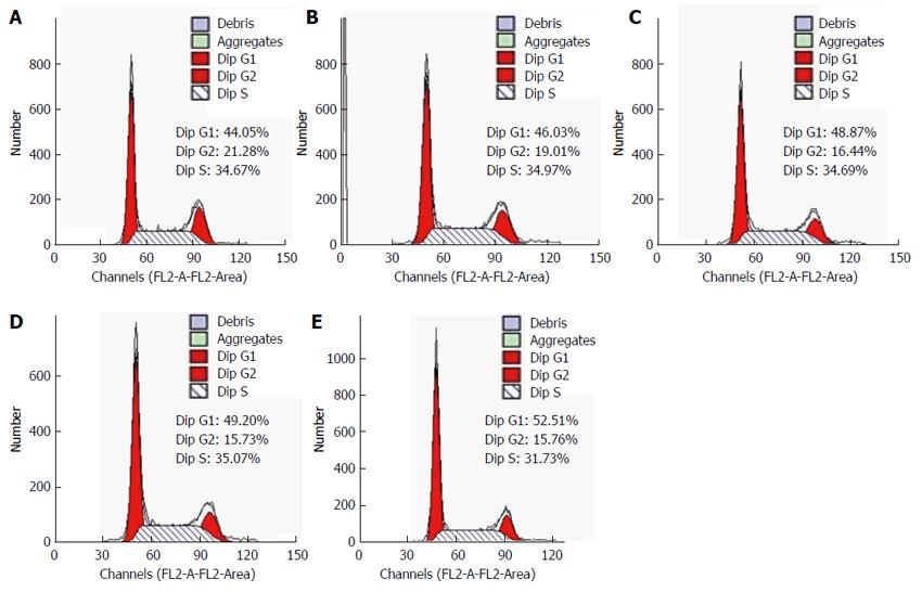
SGC-7901 (2 × 10⁵) cells were treated with different concentrations of dobutamine (1, 3, 10 and 30 μmol/L) for 48 h, and then fixed in 70% ethanol and stained with propidium iodide (PI), and the DNA content was analyzed by flow cytometry.
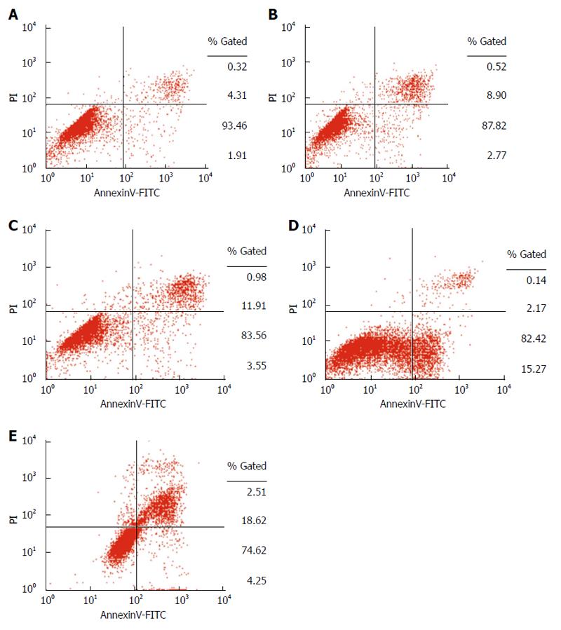
SGC-7901 cells were treated with different concentrations of dobutamine (1, 3, 10 and 30 μmol/L) for 72 h, and then the cells were washed twice with cold PBS and resuspended in 1 × binding buffer (BD Biosciences, San Jose, CA, United States). Apoptosis of SGC-7901 cells was quantified by staining with 5 μL annexin V-fluorescein isothiocyanate (FITC) and 2.5 μL PI (Beckman Coulter, Inc., CA, United States). The cells were then incubated on ice for 10 min in the dark, followed by the addition of 400 μL 1 × binding buffer. Samples for analysis of apoptosis were analyzed using flow cytometry within 30 min.
After treatment with dobutamine for 72 h, the cells were first washed in ice-cold phosphate buffered saline (PBS), lysed in whole-cell extraction buffer, and centrifuged at 14000 g for 20 min. Approximately 30 μL of proteins was isolated by 8% SDS-PAGE and transferred into a polyvinylidene fluoride membrane. The membrane was blocked with 5% skim milk and then probed with antibodies specific to human YAP (Santa Cruz Biotechnology, CA, United States; 1/1000), Phospho-YAP (Cell Signaling Technology Inc., Boston, MA, United States; 1/500) and GAPDH (Santa Cruz Biotechnology, CA, United States; 1/500) overnight at 4 °C. An appropriate secondary antibody was chosen and the sample was incubated at room temperature for 2 h.
Immunodetected protein YAP was visualized by a DAB system according to the manufacturer’s instructions. The corresponding immunocytochemistry was carried out according to the manufacturer’s instructions.
Statistical analyses were performed using the SPSS 15.0 software package (SPSS Inc. Chicago, IL, United States). Comparisons between two samples were performed using the Student’s t test. P < 0.05 was considered statistically significant.
Cell viability was determined by the MTT assay after treatment with dobutamine. The viability of SGC-7901 cells was significantly inhibited by dobutamine in a time- and dose-dependent manner (Figure 1A). The median effective concentrations (IC50) of dobutamine for inhibition of SGC-7901 cell viability were 27.16, 21.25, and 16.84 μmol/L at 24, 48 and 72 h, respectively. The inhibition ratio was significantly increased after DDP was added (Figure 1B).
An in vitro wound healing assay was performed to determine whether dobutamine affected SGC-7901 cell migration. As shown in Figure 2, the number of migrated cells increased in the control group. However, the number of migrated cells was significantly reduced following the addition of dobutamine for 24 h.
The effect of dobutamine on the proliferation of SGC-7901 cells was analyzed using the colony formation assay. The colonies formed in the dobutamine groups were smaller and fewer than those formed in the control group. The number of colonies formed in the dobutamine groups was significantly reduced (3 μmol/L: 17.8 ± 1.21, 10 μmol/L: 14.96 ± 1.36, and 30 μmol/L: 12.43 ± 1.09 vs control: 22.20 ± 1.49, P < 0.05). This indicated that dobutamine inhibited colony formation in SGC-7901 cells.
The in vitro invasion analysis was employed to determine whether dobutamine affected SGC-7901 cell invasion into matrigel. The number of invaded cells was 122.67 ± 5.51 (1 μmol/L), 116.00 ± 5.29 (3 μmol/L), 97.00 ± 6.56 (10 μmol/L), and 67.33 ± 5.69 (30 μmol/L) in the dobutamine groups, and 56.67 ± 3.06 in the control group after 48 h, respectively. These findings indicated that dobutamine significantly inhibited Matrigel invasion (P < 0.01). A dose response was also observed.
To elucidate the inhibitory mechanism of dobutamine on cell growth, flow cytometry was used to compare distribution of the cell cycle between dobutamine-treated and control cells. As shown in Figure 3, the percentage of SGC-7901 cells in the G0/G1 phase was significantly increased (3 μmol/L: 47.89% ± 1.01%, 10 μmol/L: 9.46% ± 0.66%, and 30 μmol/L: 51.77% ± 0.77% vs control: 45.36% ± 1.14%, P < 0.01), whereas the percentage in the S-phase was significantly decreased (3 μmol/L: 33.89% ± 0.74%, 10 μmol/L: 33.70% ± 1.30%, and 30 μmol/L: 31.55% ± 1.19% vs control: 34.23% ± 1.90%, P < 0.05). However, no difference was found between the 1 μmol/L group and the control group. These results suggested that dobutamine arrested the cell cycle at G1/S phase.
Cell apoptosis was also influenced by dobutamine. After 72 h treatment, dobutamine significantly increased the rate of apoptosis at 1 μmol/L (9.96% ± 2.12%), 3 μmol/L (15.14% ± 1.06%), 10 μmol/L (18.50% ± 0.93%) and 30 μmol/L (23.85% ± 1.09%) compared with the rate of apoptosis in the control group (6.41% ± 0.64%) (P < 0.05) (Figure 4). The rate of apoptosis was dose-dependent.
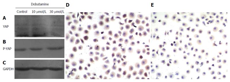
The expression of YAP was detected in cells after treatment with dobutamine. The expression of YAP was decreased, but phosphorylated YAP was increased after treatment with dobutamine for 72 h (Figure 5A). Immunocytochemistry showed that the expression of YAP was mainly found in the nucleus in the absence of dobutamine. However, phosphorylated YAP was mainly found in the cytosol following treatment with dobutamine (Figure 5B and C). Therefore, we suspect that YAP was recruited from the nucleus to the cytosol after treatment with dobutamine.
Gastric carcinoma is one of the most common malignancies worldwide. Researchers recently found that dobutamine inhibited YAP nuclear translocation, which is very important in the regulation of cell growth, division, and apoptosis[5]. Therefore, we suspect that dobutamine will be accepted as an adjuvant therapy in gastric cancer. In this study, we found that dobutamine significantly inhibited cell growth, migration, cell colony formation, and invasion into Matrigel. In addition, dobutamine arrested the cell cycle at G1/S phase, and increased the rate of apoptosis of gastric adenocarcinoma cells. The expression of YAP was mainly found in the nucleus in the absence of dobutamine. However, the expression of phosphorylated YAP was reduced and was mainly found in the cytosol following treatment with dobutamine.
YAP is a proline-rich phosphoprotein, which acts as a transcriptional co-activator in the regulation of cellular processes, including cell proliferation and apoptosis. Many genes related to cell growth and apoptosis are influenced by YAP, such as Ki67, C-mye, Sox4, H19 and AFP[17]. YAP is the nuclear effector of the Hippo pathway, and will normally flow to the cytoplasm[18]. When the Hippo pathway is activated, YAP is phosphorylated and recruited from the nucleus to the cytosol, then YAP-dependent transcription is turned off. In contrast, when the Hippo pathway is impaired, YAP remains in the nucleus, consequently resulting in tissue over-growth and tumorigenesis[19]. Recently, emerging evidence suggests that YAP may be a candidate oncogene. As reported, high-expression of YAP has been found in many types of tumors, including gastric carcinoma[8]. YAP may play an important role as a carcinogenic factor in gastric carcinoma, and eliminating YAP may inhibit gastric carcinoma cell proliferation and metastasis[14].
As reported, dobutamine can lead to altered expression of YAP[5]. Although the effect of dobutamine is independent of the Hippo pathway, it inhibits YAP-dependent gene transcription. It is believed that phosphorylation of LATS1 and LATS2 is a hallmark of Hippo pathway activation, and is the classical Hippo pathway. However, different from the classical pathway, dobutamine may enhance the phosphorylation of Akt and activate the Hippo pathway independently. Akt is known to phosphorylate Ser127 of YAP and recruit YAP to the cytoplasm[20]. Ser127 of YAP is the most important phosphorylation site and is a determinant of the sub-cellular localization of YAP. When YAP locates in the cytoplasm, YAP-dependent transcription of the cell proliferation-promoting and anti-apoptotic gene is shut off. In the present study, we determined the effects of dobutamine on SGC-7901 cells. It was shown that the influence of dobutamine on SGC-7901 cells was extensive. Dobutamine may inhibit the biological behavior of SGC-7901 cells, including cell viability, migration, proliferation and invasion. These data are similar to the results obtained when the YAP gene was silenced using RNA interference[21]. In addition, research demonstrated that YAP could activate apoptosis in response to DNA damage by interacting with p73 in several cancer cell lines[22]. In this study, dobutamine arrested the cell cycle at G1/S transition and augmented cell apoptosis.
Nowadays, the mainly obstacle in the successful management of patients with gastric carcinoma is intrinsic or acquired drug resistance[23]. Cisplatin is widely used as a primary anti-tumor chemotherapeutic agent to treat various cancers. However, resistance to cisplatin has been investigated, but is not fully understood[24]. In this study, we found that the combined effects of dobutamine and cisplatin were favorable in gastric adenocarcinoma cells. Cell growth and viability were inhibited more greatly following treatment with both drugs. Dobutamine may ameliorate resistance to cisplatin.
In conclusion, as YAP is shown to play an important role as a carcinogenic factor in many malignant tumors, YAP represents an important therapeutic target in human cancer. Following treatment with dobutamine, YAP was recruited from the nucleus to the cytosol. The inhibitory effect of dobutamine as seen in SGC-7901 cells may be used in neoadjuvant therapy not only for gastric cancer, but also for other tumors.
Gastric carcinoma is one of the most common malignancies with a relatively high incidence and mortality rate. However, traditional chemotherapy in the treatment of gastric carcinoma is still uneffective. Therefore, new therapies and drugs are required.
High-expression of Yes-associated protein (YAP) has been found in many types of tumors, including gastric carcinoma, and is correlated with poor prognosis. The identification of new reagents which can inhibit YAP in clinical medicine will be useful in the treatment of tumors.
Dobutamine is a synthetic sympathomimetic amine, and is widely used in patients with congestive heart failure and in cardiovascular testing. Recently, researchers confirmed that dobutamine attenuated YAP-dependent transcription by inhibiting YAP nuclear translocation. This may result in a new application for dobutamine in cancer treatment. In this study, dobutamine was used to treat gastric adenocarcinoma cells, and the therapeutic value of dobutamine in these cells was assessed. In addition, the combined effects of dobutamine and cisplatin were also determined. The authors expect that dobutamine will be approved as an adjuvant therapy for gastric cancer.
The study results suggest that YAP is a potential therapeutic target, and the inhibitory effect of dobutamine on SGC-7901 cells may be useful in the neoadjuvant therapy for not only gastric cancer, but also for other tumors.
This is a good exploratory study in which authors investigated the effect of dobutamine on gastric adenocarcinoma cells. The results are interesting and suggest that dobutamine has a significant inhibitory effect on gastric adenocarcinoma cells and is a potential therapeutic reagent for treatment of tumors.
P- Reviewer: Kouraklis G S- Editor: Ding Y L- Editor: O’Neill M E- Editor: Ma S
| 1. | Siegel R, Naishadham D, Jemal A. Cancer statistics, 2012. CA Cancer J Clin. 2012;62:10-29. [RCA] [PubMed] [DOI] [Full Text] [Cited by in Crossref: 8406] [Cited by in RCA: 8970] [Article Influence: 690.0] [Reference Citation Analysis (0)] |
| 2. | Ruffolo RR. The pharmacology of dobutamine. Am J Med Sci. 1987;294:244-248. [RCA] [PubMed] [DOI] [Full Text] [Cited by in Crossref: 242] [Cited by in RCA: 218] [Article Influence: 5.7] [Reference Citation Analysis (0)] |
| 3. | Vallet B, Dupuis B, Chopin C. [Dobutamine: mechanisms of action and use in acute cardiovascular pathology]. Ann Cardiol Angeiol (Paris). 1991;40:397-402. [PubMed] |
| 4. | Robbers-Visser D, Luijnenburg SE, van den Berg J, Roos-Hesselink JW, Strengers JL, Kapusta L, Moelker A, Helbing WA. Safety and observer variability of cardiac magnetic resonance imaging combined with low-dose dobutamine stress-testing in patients with complex congenital heart disease. Int J Cardiol. 2011;147:214-218. [RCA] [PubMed] [DOI] [Full Text] [Cited by in Crossref: 12] [Cited by in RCA: 9] [Article Influence: 0.6] [Reference Citation Analysis (0)] |
| 5. | Bao Y, Nakagawa K, Yang Z, Ikeda M, Withanage K, Ishigami-Yuasa M, Okuno Y, Hata S, Nishina H, Hata Y. A cell-based assay to screen stimulators of the Hippo pathway reveals the inhibitory effect of dobutamine on the YAP-dependent gene transcription. J Biochem. 2011;150:199-208. [RCA] [PubMed] [DOI] [Full Text] [Cited by in Crossref: 133] [Cited by in RCA: 148] [Article Influence: 10.6] [Reference Citation Analysis (0)] |
| 6. | Edgar BA. From cell structure to transcription: Hippo forges a new path. Cell. 2006;124:267-273. [RCA] [PubMed] [DOI] [Full Text] [Cited by in Crossref: 262] [Cited by in RCA: 276] [Article Influence: 14.5] [Reference Citation Analysis (0)] |
| 7. | Pan D. Hippo signaling in organ size control. Genes Dev. 2007;21:886-897. [RCA] [PubMed] [DOI] [Full Text] [Cited by in Crossref: 487] [Cited by in RCA: 533] [Article Influence: 29.6] [Reference Citation Analysis (0)] |
| 8. | Steinhardt AA, Gayyed MF, Klein AP, Dong J, Maitra A, Pan D, Montgomery EA, Anders RA. Expression of Yes-associated protein in common solid tumors. Hum Pathol. 2008;39:1582-1589. [RCA] [PubMed] [DOI] [Full Text] [Full Text (PDF)] [Cited by in Crossref: 449] [Cited by in RCA: 463] [Article Influence: 27.2] [Reference Citation Analysis (0)] |
| 9. | Zender L, Spector MS, Xue W, Flemming P, Cordon-Cardo C, Silke J, Fan ST, Luk JM, Wigler M, Hannon GJ. Identification and validation of oncogenes in liver cancer using an integrative oncogenomic approach. Cell. 2006;125:1253-1267. [RCA] [PubMed] [DOI] [Full Text] [Full Text (PDF)] [Cited by in Crossref: 904] [Cited by in RCA: 910] [Article Influence: 47.9] [Reference Citation Analysis (0)] |
| 10. | Overholtzer M, Zhang J, Smolen GA, Muir B, Li W, Sgroi DC, Deng CX, Brugge JS, Haber DA. Transforming properties of YAP, a candidate oncogene on the chromosome 11q22 amplicon. Proc Natl Acad Sci USA. 2006;103:12405-12410. [RCA] [PubMed] [DOI] [Full Text] [Cited by in Crossref: 673] [Cited by in RCA: 762] [Article Influence: 40.1] [Reference Citation Analysis (0)] |
| 11. | Roden R, Wu TC. How will HPV vaccines affect cervical cancer? Nat Rev Cancer. 2006;6:753-763. [PubMed] |
| 12. | Castle PE, Dockter J, Giachetti C, Garcia FA, McCormick MK, Mitchell AL, Holladay EB, Kolk DP. A cross-sectional study of a prototype carcinogenic human papillomavirus E6/E7 messenger RNA assay for detection of cervical precancer and cancer. Clin Cancer Res. 2007;13:2599-2605. [RCA] [PubMed] [DOI] [Full Text] [Cited by in Crossref: 109] [Cited by in RCA: 107] [Article Influence: 5.9] [Reference Citation Analysis (0)] |
| 13. | Cuschieri KS, Whitley MJ, Cubie HA. Human papillomavirus type specific DNA and RNA persistence--implications for cervical disease progression and monitoring. J Med Virol. 2004;73:65-70. [PubMed] |
| 14. | Da CL, Xin Y, Zhao J, Luo XD. Significance and relationship between Yes-associated protein and survivin expression in gastric carcinoma and precancerous lesions. World J Gastroenterol. 2009;15:4055-4061. [RCA] [PubMed] [DOI] [Full Text] [Full Text (PDF)] [Cited by in CrossRef: 50] [Cited by in RCA: 63] [Article Influence: 3.9] [Reference Citation Analysis (0)] |
| 15. | Yuan M, Tomlinson V, Lara R, Holliday D, Chelala C, Harada T, Gangeswaran R, Manson-Bishop C, Smith P, Danovi SA. Yes-associated protein (YAP) functions as a tumor suppressor in breast. Cell Death Differ. 2008;15:1752-1759. [RCA] [PubMed] [DOI] [Full Text] [Cited by in Crossref: 225] [Cited by in RCA: 259] [Article Influence: 15.2] [Reference Citation Analysis (0)] |
| 16. | Molden T, Kraus I, Karlsen F, Skomedal H, Hagmar B. Human papillomavirus E6/E7 mRNA expression in women younger than 30 years of age. Gynecol Oncol. 2006;100:95-100. [RCA] [PubMed] [DOI] [Full Text] [Cited by in Crossref: 51] [Cited by in RCA: 49] [Article Influence: 2.5] [Reference Citation Analysis (0)] |
| 17. | Zeng Q, Hong W. The emerging role of the hippo pathway in cell contact inhibition, organ size control, and cancer development in mammals. Cancer Cell. 2008;13:188-192. [RCA] [PubMed] [DOI] [Full Text] [Cited by in Crossref: 356] [Cited by in RCA: 379] [Article Influence: 22.3] [Reference Citation Analysis (0)] |
| 18. | Oka T, Remue E, Meerschaert K, Vanloo B, Boucherie C, Gfeller D, Bader GD, Sidhu SS, Vandekerckhove J, Gettemans J. Functional complexes between YAP2 and ZO-2 are PDZ domain-dependent, and regulate YAP2 nuclear localization and signalling. Biochem J. 2010;432:461-472. [RCA] [PubMed] [DOI] [Full Text] [Cited by in Crossref: 149] [Cited by in RCA: 160] [Article Influence: 11.4] [Reference Citation Analysis (0)] |
| 19. | Zhao B, Lei QY, Guan KL. The Hippo-YAP pathway: new connections between regulation of organ size and cancer. Curr Opin Cell Biol. 2008;20:638-646. [RCA] [PubMed] [DOI] [Full Text] [Full Text (PDF)] [Cited by in Crossref: 375] [Cited by in RCA: 376] [Article Influence: 22.1] [Reference Citation Analysis (0)] |
| 20. | Zhang H, Wu S, Xing D. Inhibition of Aβ(25-35)-induced cell apoptosis by low-power-laser-irradiation (LPLI) through promoting Akt-dependent YAP cytoplasmic translocation. Cell Signal. 2012;24:224-232. [RCA] [PubMed] [DOI] [Full Text] [Cited by in Crossref: 43] [Cited by in RCA: 50] [Article Influence: 3.6] [Reference Citation Analysis (0)] |
| 21. | Zhou Z, Zhu JS, Xu ZP. RNA interference mediated YAP gene silencing inhibits invasion and metastasis of human gastric cancer cell line SGC-7901. Hepatogastroenterology. 2011;58:2156-2161. [RCA] [PubMed] [DOI] [Full Text] [Cited by in Crossref: 10] [Cited by in RCA: 15] [Article Influence: 1.2] [Reference Citation Analysis (0)] |
| 22. | Lapi E, Di Agostino S, Donzelli S, Gal H, Domany E, Rechavi G, Pandolfi PP, Givol D, Strano S, Lu X. PML, YAP, and p73 are components of a proapoptotic autoregulatory feedback loop. Mol Cell. 2008;32:803-814. [RCA] [PubMed] [DOI] [Full Text] [Cited by in Crossref: 178] [Cited by in RCA: 206] [Article Influence: 12.9] [Reference Citation Analysis (0)] |
| 23. | Liu FS. Mechanisms of chemotherapeutic drug resistance in cancer therapy--a quick review. Taiwan J Obstet Gynecol. 2009;48:239-244. [RCA] [PubMed] [DOI] [Full Text] [Cited by in Crossref: 136] [Cited by in RCA: 144] [Article Influence: 9.0] [Reference Citation Analysis (0)] |
| 24. | Cho HJ, Baek KE, Nam IK, Park SM, Kim IK, Park SH, Im MJ, Ryu KJ, Yoo JM, Hong SC. PLCγ is required for RhoGDI2-mediated cisplatin resistance in gastric cancer. Biochem Biophys Res Commun. 2011;414:575-580. [RCA] [PubMed] [DOI] [Full Text] [Cited by in Crossref: 16] [Cited by in RCA: 18] [Article Influence: 1.3] [Reference Citation Analysis (0)] |


