Published online Nov 28, 2014. doi: 10.3748/wjg.v20.i44.16779
Revised: July 10, 2014
Accepted: August 13, 2014
Published online: November 28, 2014
Processing time: 259 Days and 10.9 Hours
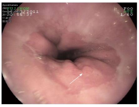
The heterotopic pancreas, which is usually described as an untypical presence of pancreatic tissue without any anatomic or vascular continuity with the pancreas, is relatively rare. Clinical manifestations may include bleeding, inflammation, pain and obstruction; however, in most cases it remains silent and is diagnosed during autopsy. Here, we report a case of ectopic pancreatic lesion located in the gastric cardia. The patient was a 73-year-old woman who had a history (over four months) of chronic epigastric pain accompanied by heartburn. Esophagogastroduodenoscopy revealed inflammatory changes throughout the stomach and lower esophagus, as well as a flat polypoid mass with benign features located in the gastric cardia, approx. 10 mm below the “Z” line, measuring approx. 7 mm in diameter. Endoscopic biopsy forceps were used to remove the lesion. Histological examination of the lesion revealed the presence of heterotopic pancreatic tissue in the gastric mucosa. On the basis of the presented case, we suggest that pancreatic ectopia should be a part of differential diagnosis, not only when dealing with submucosal gastric lesions, but also with those that are small, flat and/or untypically located.
Core tip: The heterotopic pancreas, which is usually described as an untypical presence of pancreatic tissue without any anatomic or vascular continuity with the pancreas, is relatively rare. As an intra- or submucosal lesion, is usually found incidentally, most often in the gastric antrum. However, it may also be found anywhere in the digestive tract. We report an ectopic pancreatic lesion atypically located in the gastric cardia in a 73-year-old woman with chronic epigastric pain accompanied by heartburn.
- Citation: Filip R, Walczak E, Huk J, Radzki RP, Bieńko M. Heterotopic pancreatic tissue in the gastric cardia: A case report and literature review. World J Gastroenterol 2014; 20(44): 16779-16781
- URL: https://www.wjgnet.com/1007-9327/full/v20/i44/16779.htm
- DOI: https://dx.doi.org/10.3748/wjg.v20.i44.16779
The heterotopic pancreas, which is usually described as an untypical presence of pancreatic tissue without any anatomic or vascular continuity with the pancreas, was probably firstly described in the 18th century when it was found in an ileal diverticulum[1]. It may be found at different sites in the gastrointestinal tract, with a propensity to affect the small intestine and stomach. Although a heterotopic pancreas can occur at any age, it is most common, after 50 years of age[2,3]. Clinical manifestations may include bleeding, inflammation, pain and obstruction; however, in most cases it remains silent and is diagnosed during autopsy[1,4]. On the other hand, in such tissue there is also an increased risk of malignant transformation[5].
We report the case of a 73-year-old female with an ectopic pancreatic lesion in the gastric cardia.
In March 2011, a 73-year-old woman was admitted to hospital because of pain located in the left upper- and middle-abdomen, accompanied by heartburn, nausea and vomiting. The history of the above symptoms lasted for about four months with remarkable aggravation a few days before being admitted to hospital. Her past medical history, apart from colon diverticulosis, also contained arterial hypertension, nodular goiter and diabetes type 2. Physical examination was unremarkable, as were the results of complete blood count and routine biochemical investigations. Esophagogastroduodenoscopy revealed inflammatory changes throughout the stomach and lower esophagus, as well as a flat polypoid mass with benign features located in the gastric cardia, approx. 10 mm below the “Z” line, measuring approx. 7 mm in diameter (Figure 1). The duodenum was unremarkable. Endoscopic biopsy forceps removed the lesion, which submitted for histopathological assessment. The endoscopic diagnosis was superficial gastritis, gastroesophageal reflux disease (GERD) with the presence of a gastroesophageal junction hyperplastic polyp. Histological examination of the lesion revealed the presence of heterotopic pancreatic tissue in the cardiac mucosa with fully developed acini (Figure 2). An abdominal ultrasound was normal, apart from the presence of nephrolithiasis. After discharge from hospital, she was under the supervision of the gastroenterology outpatients clinic, where she passed colonoscopy with the polypectomy of the two tubular adenomas of approx. 6 mm and 8 mm in diameter, as well as whole gastrointestinal tract radiography with barium contrast, which was normal. Two control esophagogastroduodenoscopical examinations with random biopsies were performed, the last in June 2012, and no abnormalities in the location of the previously described lesion were found, nor in any other location within the upper gastrointestinal tract (GIT).
The origins of pancreatic ectopia are not fully elucidated; however, the most probable theories are based on the fetal migration of pancreatic cells and on the penetration of immature gastric mucosa inside the submucosa, followed by its pancreatic metaplasia[6]. The possible localizations within the GIT are the esophagus, stomach, small intestine, common bile duct and gallbladder, papilla of Vater, Meckel’s diverticulum and mesocolon[6,7]. However, most commonly, an ectopic pancreas is seen in the stomach - up to 38%, of which 95% are located in the greater curvature in the antrum[8]. The occurrence in particular layers of the stomach wall is as follows: 73% in the submucosal layer, 17% in the muscularis propria layer and 10% in the subserosal layer[7]. Notably, we could not find any information in the literature about the occurrence of ectopic pancreatic tissue on or within the epithelial layer of the gastric mucosa.
The clinical symptoms of pancreatic ectopia depend of location, size and other pathological features that may occasionally coexist, e.g., secretion of pancreatic enzymes that can result in local inflammation or/and secreting the hormones that may exert a whole body effect. Lesions smaller than 15 mm in diameter remain asymptomatic until they cause local inflammation or obstruction, and are usually detected accidentally. Last, but not least, pancreatic ectopia may occasionally turn into adenocarcinoma or a neuroendocrine neoplasm[1,2,7]. In pediatric patients, the clinical picture of pancreatic ectopia can be different. Most characteristic are GIT obstructions and intrasuspection that can also be associated with some congenital abnormalities, including granular pancreas, esophageal atresia, Meckel’s diverticulum, malrotation, choledochal cyst and extrahepatic biliary atresia[9].
Endoscopical examination usually shows a well-circumscribed submucosal tumor, sometimes with central “umbilication”, covered with normal mucosa. Therefore, the surface biopsy results are usually inconclusive, and the final diagnosis is based on histological verification after surgery or endoscopic submucosal resection/dissection[2]. Lymphoma, carcinoid, gastrointestinal stromal tumors, as well as some other abnormalities within the GIT, should be a part of the differential diagnosis, and available imaging techniques, including CT or MR scanning, and EUS enhanced with fine needle aspiration, allow the selection of the best resection technique[10]. It is clear that a symptomatic heterotopic pancreas should be resected, irrespective of the method chosen; however, clear recommendations for the management of asymptomatic and histologically verified lesions have not been established to date.
In the presented case, the first diagnosis based on the endoscopic view was typical for patients with GERD-“gastroesophageal junction hyperplastic (inflammatory) polyp”; however, the histology result was very surprising, since data on the occurrence of pancreatic ectopia on the surface of the gastric mucosa in gastric cardia are lacking. In such a case, one could expect pancreatic metaplasia in the gastric mucosa, which is sometimes seen in, e.g., the gastric patch[11]; however, the histological examination revealed pancreatic ectopia. The polyp was completely and safely removed, and several further endoscopical controls did not show any relapse.
In summary, although an ectopic pancreas is relatively rare, it is always a part of differential diagnosis when dealing with submucosal or polyp-like gastric lesions. On the basis of the presented case, we suggest, that even small, flat and/or untypically located lesions (e.g., gastric cardia) should also be considered to be formed from the ectopic pancreatic mass.
The authors would like to acknowledge the expert assistance in the field of pathology and for sharing Figure 2 of Dr. Jaroslaw Swatek, Chair and Department of Clinical Pathomorphology, Lublin Medical University, Lublin, Poland.
A 73-year-old female with a history of recurrent epigastric pain and heartburn.
Ectopic pancreatic lesion located in the gastric cardia.
Hyperplastic polyp, gastric adenoma, fundic gland polyp, gastrointestinal stromal tumor (GIST), lymphoma.
Blood morphology, metabolic panel and liver function tests were within normal limits.
Esophagogastroduodenoscopy revealed inflammatory changes throughout the stomach and lower esophagus, as well as a flat polypoid mass with benign features located in the gastric cardia.
Histology revealed heterotopic pancreatic tissue in the cardiac mucosa with fully developed acini.
Endoscopic biopsy forceps removed the lesion.
The diagnosis may be complex because of the gross similarity of pancreatic heterotopia with GIST, carcinoid, lymphoma, adenoma or even gastric carcinoma.
The presented case report suggests that even small, flat and/or untypically located lesions (e.g., cardia) should also be considered to be formed from the pancreatic ectopic mass.
This article describes a pancreatic ectopia mimicking a flat polyp, atypically located in the gastric cardia.
P- Reviewer: Eysselein VE, Xu HX, Wu SL S- Editor: Qi Y L- Editor: Stewart G E- Editor: Wang CH
| 1. | Christodoulidis G, Zacharoulis D, Barbanis S, Katsogridakis E, Hatzitheofilou K. Heterotopic pancreas in the stomach: a case report and literature review. World J Gastroenterol. 2007;13:6098-6100. [RCA] [PubMed] [DOI] [Full Text] [Cited by in CrossRef: 107] [Cited by in RCA: 31] [Article Influence: 1.7] [Reference Citation Analysis (0)] |
| 2. | Agale SV, Agale VG, Zode RR, Grover S, Joshi S. Heterotopic pancreas involving stomach and duodenum. J Assoc Physicians India. 2009;57:653-654. [PubMed] |
| 3. | Yuan Z, Chen J, Zheng Q, Huang XY, Yang Z, Tang J. Heterotopic pancreas in the gastrointestinal tract. World J Gastroenterol. 2009;15:3701-3703. [RCA] [PubMed] [DOI] [Full Text] [Full Text (PDF)] [Cited by in CrossRef: 35] [Cited by in RCA: 39] [Article Influence: 2.4] [Reference Citation Analysis (0)] |
| 4. | Jiang LX, Xu J, Wang XW, Zhou FR, Gao W, Yu GH, Lv ZC, Zheng HT. Gastric outlet obstruction caused by heterotopic pancreas: A case report and a quick review. World J Gastroenterol. 2008;14:6757-6759. [RCA] [PubMed] [DOI] [Full Text] [Full Text (PDF)] [Cited by in CrossRef: 38] [Cited by in RCA: 42] [Article Influence: 2.5] [Reference Citation Analysis (0)] |
| 5. | Cullen JJ, Weydert C, Hinkhouse MM, Ritchie J, Domann FE, Spitz D, Oberley LW. The role of manganese superoxide dismutase in the growth of pancreatic adenocarcinoma. Cancer Res. 2003;63:1297-1303. [PubMed] |
| 6. | Sadeghi NR, Godambe A, Shienbaum AJ, Alloy A. Premalignant gastric heterotopic pancreas. Gastroenterol Hepatol (N Y). 2008;4:218-221. [PubMed] |
| 7. | Gurocak B, Gokturk HS, Kayacetin S, Bakdik S. A rare case of heterotopic pancreas in the stomach which caused closed perforation. Neth J Med. 2009;67:285-287. [PubMed] |
| 8. | Seneviratne SA, Ramanayaka IT, Samarasekera DN. Heterotopic pancreas in the body of the stomach. Ceylon Med J. 2009;54:57-58. [RCA] [PubMed] [DOI] [Full Text] [Cited by in Crossref: 6] [Cited by in RCA: 7] [Article Influence: 0.5] [Reference Citation Analysis (0)] |
| 9. | Ogata H, Oshio T, Ishibashi H, Takano S, Yagi M. Heterotopic pancreas in children: review of the literature and report of 12 cases. Pediatr Surg Int. 2008;24:271-275. [RCA] [PubMed] [DOI] [Full Text] [Cited by in Crossref: 40] [Cited by in RCA: 34] [Article Influence: 2.0] [Reference Citation Analysis (0)] |
| 10. | Catalano F, Rodella L, Lombardo F, Silano M, Tomezzoli A, Fuini A, Di Cosmo MA, de Manzoni G, Trecca A. Endoscopic submucosal dissection in the treatment of gastric submucosal tumors: results from a retrospective cohort study. Gastric Cancer. 2013;16:563-570. [RCA] [PubMed] [DOI] [Full Text] [Cited by in Crossref: 48] [Cited by in RCA: 55] [Article Influence: 4.6] [Reference Citation Analysis (0)] |
| 11. | Tang P, McKinley MJ, Sporrer M, Kahn E. Inlet patch: prevalence, histologic type, and association with esophagitis, Barrett esophagus, and antritis. Arch Pathol Lab Med. 2004;128:444-447. [PubMed] |


