Published online Nov 21, 2014. doi: 10.3748/wjg.v20.i43.16377
Revised: April 22, 2014
Accepted: July 22, 2014
Published online: November 21, 2014
Processing time: 253 Days and 21.4 Hours
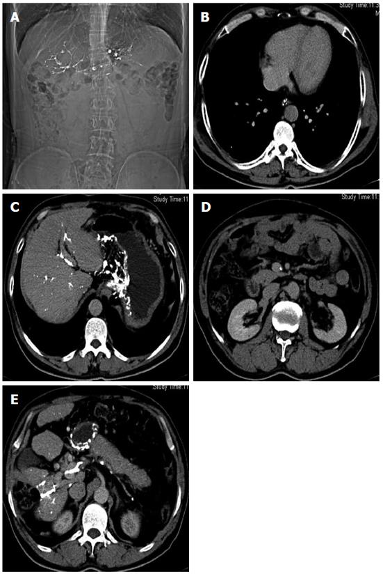
Calcification of the portal venous system is a rare entity that can be incidentally discovered during computed tomography (CT). We describe a case of extensive calcifications in the portal venous system in a middle-aged male patient with hepatocellular carcinoma (HCC). This patient presented with epigastric pain that had no obvious origin prior to admission. Laboratory examinations were positive for hepatitis B surface antigen and α-fetoprotein, and severe esophageal and gastric varices were detected during gastroscopy. Abdominal X-ray plain film showed well-defined linear and track-like calcification, with irregular margins directed along the course of the portal venous system. CT revealed extensive calcifications along the course of the portal, splenic, superior mesenteric and gastroesophageal veins. He underwent splenectomy 22 years ago due to splenomegaly and partial hepatectomy seven months before because of HCC of low-grade differentiation, confirmed by pathology. Finally, the patient was diagnosed with postoperative recurrent HCC and extensive portal venous system calcification after selective hepatic angiography under digital subtraction angiography.
Core tip: We reported a case of extensive calcifications in portal venous system combined with hepatocellular carcinoma (HCC) in a middle-aged man. He underwent splenectomy 22 years ago for splenomegaly and partial hepatectomy 7 mo before due to HCC. Extensive calcifications along the course of the portal vein were found on computed tomography.
- Citation: Wang CE, Sun CJ, Huang S, Wang YH, Xie LL. Extensive calcifications in portal venous system in a patient with hepatocarcinoma. World J Gastroenterol 2014; 20(43): 16377-16380
- URL: https://www.wjgnet.com/1007-9327/full/v20/i43/16377.htm
- DOI: https://dx.doi.org/10.3748/wjg.v20.i43.16377
Portal vein calcification seen on abdominal radiography is rare, and occurs even more rarely in the portal trunk and its branches, such as the splenic and superior mesenteric veins. To the best of our knowledge, such extensive calcifications in the portal venous system combined with hepatocelluar carcinoma (HCC) have not been previously reported. Here, we report a recent case with evidence of calcifications in the portal venous system confirmed by computed tomography (CT).
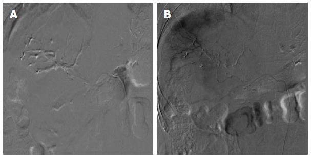
A 46-year-old man was admitted for upper abdominal pain of 2 mo duration on February 25, 2014. He underwent splenectomy 22 years ago due to splenomegaly, pancytopenia, and portal hypertension caused by hypertension-related liver cirrhosis, and partial hepatectomy seven months before due to HCC of low-grade differentiation, confirmed by surgery and pathology. He denied drug and alcohol abuse. Physical examination was essentially negative except for mild epigastric tenderness and two scars below the bilateral costal arch. Hemoglobin (144 g/L), white blood cell count (8.04 × 10⁹/L) and platelet count (290 × 109/L) were within normal ranges. Liver function tests were unremarkable except for a total bilirubin level of 22.5 μmol/L, with a direct bilirubin level of 4.8 μmol/L. Serum levels of α-fetoprotein and hepatitis B surface antigen were 235.6 ng/mL and 3.15 IU/mL, respectively. Endoscopic examination of the esophagus revealed severe gastroesophageal varices that did not receive any therapy before admission. Plain abdominal radiography revealed linear and track-like calcification with irregular margins directed along the course of the portal vein (Figure 1A). Calcifications in the portal, splenic, superior mesenteric and gastroesophageal veins were detected on abdominal CT (Figure 1B-E). On digital subtraction angiography, the tree structure of the calcified portal vein was clearly demonstrated (Figure 2A). On the arterial phase of selective celiac arteriography, multiple tumor staining in the right upper quadrant was detected (Figure 2B).
Calcification in the portal vein or its tributaries visible on abdominal radiography is a rare radiological finding, and almost always occurs in patients with long-standing portal hypertension, regardless of underlying etiology[1,2]. Sclerosis and calcification within the thickened intima and media of the vein may result from mechanical stress[3,4]. Since Moberg[5] first described in 1943 a case in which the diagnosis of portal vein thrombosis could be made from a plain abdominal roentgenograph, the clinical presentation has generally been that of recurrent hemorrhage, and less than 50 well-documented cases of portal vein calcification have been reported in the English-language literature[4-18]. A review of 21 cases of portal vein calcification reported by Kawasaki et al[2] revealed that the average age was 53.7 ± 10.2 years and the male-to-female ratio was 17:4, reflecting the higher morbidity in adult men. All the reported cases of portal vein calcification were associated with portal hypertension, and there was histological evidence of cirrhosis in the majority of cases. Most patients had esophageal varices and a clinical history of hematemesis[2].
The portal vein arises at the junction of the superior mesenteric and splenic veins and extends for about 8 cm superiorly and laterally before dividing into left and right branches that enter the liver parenchyma[19]. The calcified lesions were located in the portal vein in all the patients, splenic vein in 62%, superior mesenteric vein in 33%, and inferior mesenteric vein in none. The precise cause was not elucidated in any of the previous cases reviewed. Calcium may be deposited either in a thrombus or, as in the present case, in the vessel wall[19,20]. A distinctive radiographic feature of portal venous calcification is the presence of radiodensity that corresponds to the course of the vein. Calcification of the portal vein should be considered if elongated opacity is seen corresponding to the location and direction of the vein. Minimal calcification can be frequently missed on plain film radiography and even on pathological examination. The widespread use of CT is of value in improving the positive rate of calcification. Ayuso et al[20] reviewed patients with portal vein calcification who were examined with abdominal plain film (n = 10) and CT (n = 9). Calcium was seen on CT scans in nine cases and on abdominal plain films in only five. Abdominal CT showed this abnormality with increasing frequency because of its ability to demonstrate small amounts of calcium not visible on plain film radiography. Furthermore, the characteristic position, location and pattern of portal vein wall calcification permit its diagnosis from abdominal radiographs.
Verma et al[21] found a high operative mortality associated with calcifications in portal venous system in patients undergoing liver transplantation. In that study, two of seven patients with portal and mesenteric calcification present on CT died during liver transplantation as a result of portal venous thrombosis[21]. None of the patients without venous calcification on abdominal CT and receiving liver transplantation were found to have portal vein thrombosis during surgery. This finding indicates that the detectable portal and mesenteric calcifications on CT may be a sign of portal vein thrombosis at surgery, which may result in difficult transplantation or even failure[21]. In addition, the presence of portal vein calcification is of clinical significance, as calcified portal vein is a difficult vessel with which to perform a transjugular intrahepatic portosystemic stent shunt[2].
A 46-year-old man with a history of splenectomy due to splenomegaly presented with epigastric pain.
Upper abdominal pain and mild epigastric tenderness were detected in the patient with long-standing portal hypertension.
The differential diagnosis included hepatolithiasis, cholelithiasis and schistosomiasis.
Hemoglobin (144 g/L), white blood cell count (8.04 × 10⁹/L), platelet count (290 × 109/L) and liver enzymes were within normal ranges except for a total bilirubin level of 22.5 μmol/L, direct bilirubin level of 4.8 μmol/L, α- fetoprotein level of 235.6 ng/mL and hepatitis B surface antigen level of 3.15 IU/mL.
Computed tomography scan showed extensive calcifications along the course of the portal vein.
Pathology revealed hepatocellular carcinoma of low-grade differentiation combined with portal vein calcification.
The patient was treated with splenectomy and partial hepatectomy.
Fewer than 50 cases of portal vein calcification have been reported, and the mechanism of calcification is unclear.
Calcification of the portal venous system is defined as calcium deposited either in a thrombus or in the vessel wall of the portal venous system.
Calcification of the portal venous system can be diagnosed by radiographic examination and can offer some assistance to patients undergoing liver transplantation.
This article reports an extremely rare case of extensive calcifications in the portal venous system in a patient with hepatocellular carcinoma, which has not been documented previously.
P- Reviewer: Komatsu H, Kamiyama T, Lau WY, Jin B S- Editor: Ma N L- Editor: A E- Editor: Zhang DN
| 1. | Pickhardt PJ, Balfe DM. Portal vein calcification and associated biliary stricture in idiopathic portal hypertension (Banti’s syndrome). Abdom Imaging. 1998;23:180-182. [RCA] [PubMed] [DOI] [Full Text] [Cited by in Crossref: 7] [Cited by in RCA: 8] [Article Influence: 0.3] [Reference Citation Analysis (0)] |
| 2. | Kawasaki T, Kambayashi J, Sakon M, Shiba E, Yokota M, Doki Y, Suehisa E, Nishimura K, Amino N, Miyai K. Portal vein calcification: a clinical review of the last 50 years and report of a case associated with dysplasminogenemia. Surg Today. 1993;23:176-181. [RCA] [PubMed] [DOI] [Full Text] [Cited by in Crossref: 14] [Cited by in RCA: 14] [Article Influence: 0.4] [Reference Citation Analysis (0)] |
| 3. | Smallwood RA, Davidson JS. Calcification in the portal system. Gastroenterology. 1968;54:265-270. [PubMed] |
| 4. | Araki T, Kachi K, Monzawa S, Matsusako M, Hihara T, Ogata H. Computed tomographic detection of intestinal calcification of Schistosomiasis japonica. Gastrointest Radiol. 1989;14:360-362. [PubMed] |
| 5. | Moberg G. Calcified thrombosis in the portal system diagnosed by roentgen examination. Acta Radiologica. 1943;24:374-383. [RCA] [DOI] [Full Text] [Cited by in Crossref: 10] [Cited by in RCA: 10] [Article Influence: 0.7] [Reference Citation Analysis (0)] |
| 6. | Magovern GJ, Muehsam GE. Calcification of the portal and splenic veins. Am J Roentgenol Radium Ther Nucl Med. 1954;71:84-88. [PubMed] |
| 7. | Mackenzie RL, Tubbs HR, Laws JW, Dawson JL, Williams R. Obstructive jaundice and portal vein calcification. Br J Radiol. 1978;51:953-955. [RCA] [PubMed] [DOI] [Full Text] [Cited by in Crossref: 16] [Cited by in RCA: 15] [Article Influence: 0.3] [Reference Citation Analysis (0)] |
| 8. | Schneider K, Hartl M, Fendel H. Umbilical and portal vein calcification following umbilical vein catheterization. Pediatr Radiol. 1989;19:468-470. [RCA] [PubMed] [DOI] [Full Text] [Cited by in Crossref: 9] [Cited by in RCA: 10] [Article Influence: 0.3] [Reference Citation Analysis (0)] |
| 9. | Friedman AP, Haller JO, Boyer B, Cooper R. Calcified portal vein thromboemboli in infants: radiography and ultrasonography. Radiology. 1981;140:381-382. [PubMed] |
| 10. | Broker MH, Baker SR. Calcification in the portal vein wall demonstrated by computed tomography. J Comput Assist Tomogr. 1985;9:444-446. [RCA] [PubMed] [DOI] [Full Text] [Cited by in Crossref: 9] [Cited by in RCA: 9] [Article Influence: 0.2] [Reference Citation Analysis (0)] |
| 11. | Blanc WA, Berdon WE, Baker DH, Wigger HJ. Calcified portal vein thromboemboli in newborn and stillborn infants. Radiology. 1967;88:287-292. [PubMed] |
| 12. | Perlemuter G, Béjanin H, Fritsch J, Prat F, Gaudric M, Chaussade S, Buffet C. Biliary obstruction caused by portal cavernoma: a study of 8 cases. J Hepatol. 1996;25:58-63. [RCA] [PubMed] [DOI] [Full Text] [Cited by in Crossref: 62] [Cited by in RCA: 58] [Article Influence: 2.0] [Reference Citation Analysis (0)] |
| 13. | Mata JM, Alegret X, Martínez A. Calcification in the portal and collateral veins wall: CT findings. Gastrointest Radiol. 1987;12:206-208. [PubMed] |
| 14. | Sherrick DW, Kincaid OW, Gambill EE. Calcification in the portal venous system. unusual radiologic sign of portal venous thrombosis. JAMA. 1964;187:861-862. [RCA] [PubMed] [DOI] [Full Text] [Cited by in Crossref: 7] [Cited by in RCA: 7] [Article Influence: 0.1] [Reference Citation Analysis (0)] |
| 15. | Kocakoc E, Kiris A, Bozgeyik Z, Uysal H, Artas H. Splenic vein aneurysm with calcification of splenic and portal veins. J Clin Ultrasound. 2005;33:251-253. [RCA] [PubMed] [DOI] [Full Text] [Cited by in Crossref: 9] [Cited by in RCA: 6] [Article Influence: 0.3] [Reference Citation Analysis (0)] |
| 16. | Hadar H, Sommer R. Calcification in the portal venous system demonstrated by computed tomography. Eur J Radiol. 1983;3:187-188. [PubMed] |
| 17. | Haddow RA, Kemp-Harper RA. Calcification in the liver and portal system. Clin Radiol. 1967;18:225-236. [RCA] [PubMed] [DOI] [Full Text] [Cited by in Crossref: 23] [Cited by in RCA: 21] [Article Influence: 0.4] [Reference Citation Analysis (0)] |
| 18. | Freund M, Heller M. Portal venous calcifications 20 years after portosystemic shunting: demonstration by spiral CT with CT angiography and 3D reconstructions. Eur J Radiol. 2000;36:165-169. [PubMed] |
| 19. | Baker SR, Broker MH, Charnsangavej C, Sitron AP. Calcification in the portal vein wall. Radiology. 1984;152:18. [PubMed] |
| 20. | Ayuso C, Luburich P, Vilana R, Bru C, Bruix J. Calcifications in the portal venous system: comparison of plain films, sonography, and CT. AJR Am J Roentgenol. 1992;159:321-323. [RCA] [PubMed] [DOI] [Full Text] [Cited by in Crossref: 24] [Cited by in RCA: 23] [Article Influence: 0.7] [Reference Citation Analysis (0)] |
| 21. | Verma V, Cronin DC, Dachman AH. Portal and mesenteric venous calcification in patients with advanced cirrhosis. AJR Am J Roentgenol. 2001;176:489-492. [RCA] [PubMed] [DOI] [Full Text] [Cited by in Crossref: 26] [Cited by in RCA: 28] [Article Influence: 1.2] [Reference Citation Analysis (0)] |


