Published online Jun 14, 2014. doi: 10.3748/wjg.v20.i22.7075
Revised: January 22, 2014
Accepted: March 8, 2014
Published online: June 14, 2014
Processing time: 241 Days and 18.8 Hours
Fish bones are the most common foreign objects leading to bowel perforation. Most cases are confined to the extraluminal space without penetration of an adjacent organ. However, abscess formation due to the perforation of the rectosigmoid colon by a fish bone can lead to the penetration of the urinary bladder and may subsequently cause the fish bone to migrate into the urinary bladder. In the presented case, a 42-year-old female was admitted for lower abdominal pain. The computed tomography (CT) demonstrated a 5cm pelvic abscess containing a thin and curvilinear foreign body. After conservative management, the patient was discharged. After 1 mo, the subject developed a mechanical ileus. Surgery had to be delayed due to her hyperthyroidism. Migration of the foreign body to the urinary bladder was shown on additional CT. A Yellowish fish bone 3.5 cm in size was removed through intra-operative cystoscopy. The patient was discharged 8 d after the operation without any unexpected event.
Core tip: Fish bones are the most commonly observed objects leading to bowel perforation. In this case report, abscess formation due to perforation of rectosigmoid colon by a fish bone can lead to its migration to the urinary bladder. The operation timing for removal of foreign body should be tactically considered if the vital signs are stable, but we should always keep in mind that delayed complications such as migration of foreign body to adjacent organ could happen.
- Citation: Cho MK, Lee MS, Han HY, Woo SH. Fish bone migration to the urinary bladder after rectosigmoid colon perforation. World J Gastroenterol 2014; 20(22): 7075-7078
- URL: https://www.wjgnet.com/1007-9327/full/v20/i22/7075.htm
- DOI: https://dx.doi.org/10.3748/wjg.v20.i22.7075
The ingestion of a foreign body (FB) is not uncommon and often goes unnoticed. The majority of FBs that pass into the stomach traverse the gastrointestinal tract without complication[1]. Less than 1% of FBs cause perforation, depending on the size and type of the FB[2]. The most common perforation site is the terminal ileum, followed by the rectosigmoid junction. Fish bones are the most commonly observed objects that result in bowel perforation[3], and these FBs may occasionally lead to penetration injuries, resulting in complications, such as the formation of an abscess. In most reported cases, foreign bodies that have perforated the gastrointestinal tract were confined to the extraluminal space without penetrating an adjacent organ. However, in this case, the ingested FB, a fish bone, caused the formation of an abscess formation after its perforation of the rectosigmoid colon and lead to the penetration of the urinary bladder, resulting in its migration into the urinary bladder.
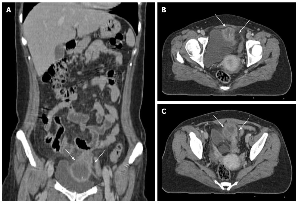
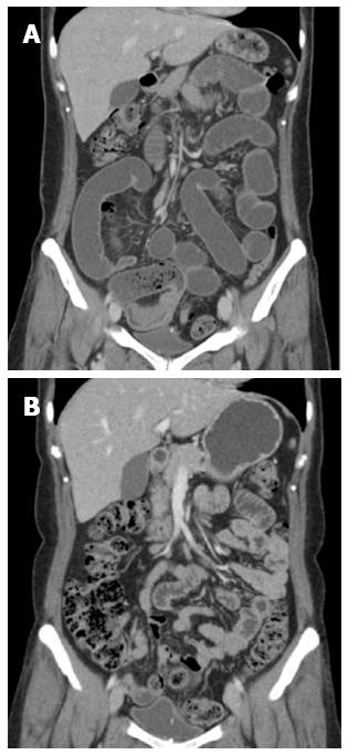
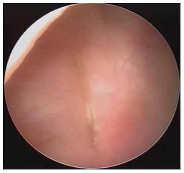
A 42-year-old female was admitted for lower abdominal pain and fever that lasted for 1 wk. The physical examination revealed mild abdominal tenderness without rebound tenderness in the supra-pubic area. The patient’s vital signs were recorded: body temperature was 37.4 °C, heart frequency was 108 beats/min and blood pressure was 100/63 mmHg. No mass or hernia was identified. Laboratory tests showed an elevated white blood cell count (15870 cells/L) and increased C-reactive protein level (3.74 mg/dL). The patient had no history of abdominal surgery, suffered from uncontrolled hyperthyroidism, which was not treated with medication. The thyroid function test showed elevated T3 levels of 281 ng/dL (60-181), elevated free T4 levels of 4.47 ng/dL (0.7-1.4) and a low thyroid stimulating hormone (TSH) level, which was shown to be less than 0.01 mIU/L (0.4-4.0). These laboratory results demonstrated an active hyperthyroidism state, which required medication. Computed tomography (CT) demonstrated a pelvic abscess, 5 cm in size, in the lower abdomen, and this abscess contained a thin and curvilinear hyperdense foreign body (Figure 1). However, the patient had no memory of swallowing any foreign body, such as a fish bone. After conservative management including antibiotics, the symptoms and laboratory data were returned to normal, and the subject was discharged. After 1 mo, the patient returned to the hospital and presented with vomiting and the absence of gas passage. In the follow-up CT, a mechanical ileus was observed due to a transitional point, with the presence of a 2 cm pelvic abscess in the right lower abdomen, which had decreased compared to the previous study (Figure 2A). The follow-up thyroid function test showed T3 levels of > 800 ng/dL, free T4 levels of 7.89 ng/dL, and TSH < 0.01 mIU/L, which may suggest poor medication compliance. The surgery had to be delayed to properly manage the patient’s hyperthyroidism. An additional CT was performed 7 d before the operation, and it revealed the migration of the foreign body into the urinary bladder (Figure 2B). An adhesion of sigmoid colon was found near the urinary bladder in the pelvic area, and a small abscess was observed adjacent to the bladder wall during laparoscopic exploration. However, there was no bowel perforation or fistula. Using intra-operative cystoscopy, a yellowish foreign body was observed between the dome and lateral wall of the urinary bladder, and this foreign body was removed using forceps (Figure 3). The surgical specimen consisted of a thin, curvilinear fish bone, 3.5 cm in length. There were no other specific intra-operative events. The patient was discharged 8 d after the operation, without any unexpected event.
Most ingested foreign bodies uneventfully pass through the gastrointestinal tract within 1 wk[4,5]. However, the perforation rate can be as high as 15%-35% when sharp foreign objects are ingested[6,7]. Fish bones are the most commonly observed objects that lead to bowel perforations. The most common intestinal perforation site is the terminal ileum, followed by the rectosigmoid colon, due to its narrow and angulating anatomical nature[8]. Intestinal perforation by a foreign body may result in the development of an abscess, enteric fistula, or intestinal obstruction, as well as peritonitis with or without pneumoperitoneum. In most cases, the reports of foreign bodies are confined to the extraluminal space, without any penetration of an adjacent organ. However, there are reported cases of the migration of a needle into the liver or the migration of a fish bone into the urinary bladder, similar to this case[9,10]. Therefore, the migration of a foreign body toward an adjacent organ, as well as the intestinal perforation, should be considered if a sharp foreign body is ingested.
Because a foreign body may perforate any part of the gastrointestinal tract, the clinical presentations may vary and mimic diverse surgical conditions such as diverticulitis, appendicitis, and peptic ulcer perforation, which makes the diagnosis of intestinal perforation due to the ingestion of an unrecognized foreign body very difficult[11]. Moreover, fish bones are invisible on plain film. A CT scan is helpful in recognizing the intestinal perforation caused by the fish bone, which usually presents as a curvilinear foreign body[12]. Moreover, CT scans allow the differential diagnosis of other causes of abdominal pain, and the scans also show the surrounding anatomical connections. The possibility of the migration of a sharp foreign body, such as a fish bone, into adjacent organs should always be considered. If the operation should be delayed, additional CT should be performed prior to the operation.
The standard management for foreign body-induced bowel perforation is emergency surgery. However, there are reported cases in which the foreign body-induced bowel perforation as successfully treated using only conservative management. A non-operative management could be employed if there is a lack of peritoneal irritation, as assessed during physical examination, or if the patient has a relative contraindication for surgery. In that case, close observation is required in order to detect possible delayed complications, such as mechanical ileus caused by inflammation or the penetration of an adjacent organ, as was the case in this study. It may be difficult to remove the foreign body due to inflammation during the acute period. If the operation is delayed for too long, the extent of bowel resection and the rate of complication may increase as the time until the operation increases. However, delaying the emergency operation may, paradoxically, downscale the range of operation in some cases. There are reported cases in which the foreign body was successfully removed after the resolution of the initial inflammation[10]. Therefore, the operation timing for the removal of a foreign body should be tactically considered if the patient’s vital signs are stable, but we should always keep in mind that delayed complications, such as the migration of the foreign body into an adjacent organ, could occur.
A 42-year-old female was admitted for lower abdominal pain and fever that lasted for 1 wk.
Clinical findings suggested localized peritonitis.
The symptoms were also similar to pelvic inflammatory disease and acute appendicitis; further evaluation was required.
Laboratory tests showed elevated white blood cell count (15870/L) with increased C-reactive protein level (3.74 mg/dL) suggesting acute inflammation.
Computed tomography (CT) demonstrated 5 cm-sized pelvic abscess in lower abdomen containing thin and curvilinear hyperdense foreign body.
On intra-operative cystoscopy, yellowish foreign body was observed in the urinary bladder and was removed.
The patient initially received conservative management using only antibiotics, and was discharged, but she developed mechanical ileus and follow up CT finding showed foreign body in the urinary bladder; surgery had to be performed to remove the foreign body.
There are reported cases of migration of needle to liver or the migration of fish bone to urinary bladder similar to this case[9,10].
The operation timing for removal of foreign body should be tactically considered if the vital signs are stable, but the authors should always keep in mind that delayed complications such as migration of foreign body to adjacent organ could happen.
This is a well written and interesting case report on a rare case of a gastrointestinal foreign body penetrating the intestinal wall and migrating into the lumen of the bladder.
P- Reviewers: Luk WH, Woo HH S- Editor: Ma YJ L- Editor: A E- Editor: Ma S
| 1. | Abel RM, Fischer JE, Hendren WH. Penetration of the alimentary tract by a foreign body with migration to the liver. Arch Surg. 1971;102:227-228. [RCA] [PubMed] [DOI] [Full Text] [Cited by in Crossref: 37] [Cited by in RCA: 39] [Article Influence: 0.7] [Reference Citation Analysis (0)] |
| 2. | Mapelli P, Head LH, Conner WE, Ferrante WE, Ray JE. Perforation of colon by ingested chicken bone diagnosed by colonoscope. Gastrointest Endosc. 1980;26:20-21. [RCA] [PubMed] [DOI] [Full Text] [Cited by in Crossref: 9] [Cited by in RCA: 11] [Article Influence: 0.2] [Reference Citation Analysis (0)] |
| 3. | Leijonmarck CE, Fenyö G, Räf L. Nontraumatic perforation of the small intestine. Acta Chir Scand. 1984;150:405-411. [PubMed] |
| 4. | Strauss JE, Balthazar EJ, Naidich DP. Jejunal perforation by a toothpick: CT demonstration. J Comput Assist Tomogr. 1985;9:812-814. [RCA] [PubMed] [DOI] [Full Text] [Cited by in Crossref: 21] [Cited by in RCA: 22] [Article Influence: 0.6] [Reference Citation Analysis (0)] |
| 5. | McCanse DE, Kurchin A, Hinshaw JR. Gastrointestinal foreign bodies. Am J Surg. 1981;142:335-337. [RCA] [PubMed] [DOI] [Full Text] [Cited by in Crossref: 93] [Cited by in RCA: 106] [Article Influence: 2.4] [Reference Citation Analysis (0)] |
| 6. | Henderson CT, Engel J, Schlesinger P. Foreign body ingestion: review and suggested guidelines for management. Endoscopy. 1987;19:68-71. [RCA] [PubMed] [DOI] [Full Text] [Cited by in Crossref: 105] [Cited by in RCA: 92] [Article Influence: 2.4] [Reference Citation Analysis (1)] |
| 7. | Vizcarrondo FJ, Brady PG, Nord HJ. Foreign bodies of the upper gastrointestinal tract. Gastrointest Endosc. 1983;29:208-210. [RCA] [PubMed] [DOI] [Full Text] [Cited by in Crossref: 134] [Cited by in RCA: 120] [Article Influence: 2.9] [Reference Citation Analysis (0)] |
| 8. | Singh RP, Gardner JA. Perforation of the sigmoid colon by swallowed chicken bone: case reports and review of literature. Int Surg. 1981;66:181-183. [PubMed] |
| 9. | Kodama K, Ofude M, Motoi I, Hinoue Y, Saito K. Successful endoscopic management of fish bone embedded into the bladder wall. Case Rep Med. 2010;2010:578058. [PubMed] |
| 10. | Avcu S, Unal O, Ozen O, Bora A, Dülger AC. A swallowed sewing needle migrating to the liver. N Am J Med Sci. 2009;1:193-195. [PubMed] |
| 11. | Chiu JJ, Chen TL, Zhan YL. Perforation of the transverse colon by a fish bone: a case report. J Emerg Med. 2009;36:345-347. [RCA] [PubMed] [DOI] [Full Text] [Cited by in Crossref: 14] [Cited by in RCA: 16] [Article Influence: 0.9] [Reference Citation Analysis (0)] |
| 12. | Ngan JH, Fok PJ, Lai EC, Branicki FJ, Wong J. A prospective study on fish bone ingestion. Experience of 358 patients. Ann Surg. 1990;211:459-462. [RCA] [PubMed] [DOI] [Full Text] [Cited by in Crossref: 163] [Cited by in RCA: 167] [Article Influence: 4.8] [Reference Citation Analysis (0)] |


