Published online Jun 7, 2014. doi: 10.3748/wjg.v20.i21.6698
Revised: February 14, 2014
Accepted: March 5, 2014
Published online: June 7, 2014
Processing time: 156 Days and 15.8 Hours
We performed endoscopic submucosal dissection of a gastric fundus tumor. It was difficult to strip the tumor completely due to space limitation, and we used blunt dissection to remove the tumor quickly and safely. Firstly, the basal area of the 2.5 cm submucosal tumor located in the gastric fundus was cut open, and the mucosa was dissected. The tumor was difficult to peel, therefore, a snare was used and the tumor was pulled and tightened slightly. Short electronic coagulation was used during the procedure. The tumor was then bluntly dissected. This method ensured rapid and complete removal of the tumor.
Core tip: We performed endoscopic submucosal dissection (ESD) of a gastric fundus tumor. It was difficult to strip the tumor completely due to space limitation, therefore, we used blunt dissection and removed the tumor quickly and safely. This is a new method based on traditional ESD, and ensured quick and safe removal of the tumor in this patient.
- Citation: Wen ZQ, Wu GY, Yu SP, Lin XD, Li SH, Huang XG, Zhang F, Zeng XY, Huang HY, Li AM. Application of blunt dissection in ESD of a gastric submucosal tumor. World J Gastroenterol 2014; 20(21): 6698-6700
- URL: https://www.wjgnet.com/1007-9327/full/v20/i21/6698.htm
- DOI: https://dx.doi.org/10.3748/wjg.v20.i21.6698
Endoscopic submucosal dissection of a submucosal tumor located in the gastric fundus adjacent to the gastric cardiac region or which has extended into the cardioesphageal junction, is difficult to perform due to space limitation. Recently, we performed endoscopic submucosal dissection (ESD) of a 2.5 cm gastric fundus submucosal tumor adjacent to the gastric cardiac region. The tumor was removed safely and quickly using blunt dissection.
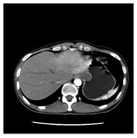
A 30-year-old female was admitted due to recurrent nausea of 3 years, which had worsened in the previous 2 wk. A neoplasm was found in the stomach. Computed tomography of the epigastric zone showed irregular thickening of the gastric fundus wall adjacent to the gastric cardiac region, local nodosity, and the tumor was found to be approximately 2.4 cm in size (Figure 1). The first therapeutic choice was ESD.
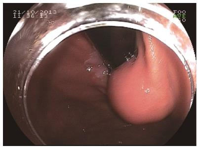
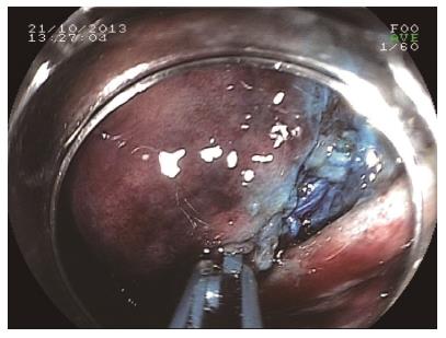
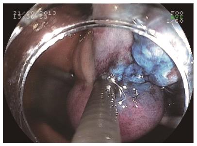
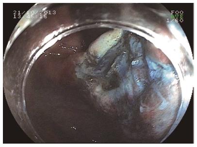
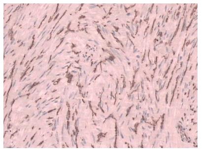
A hyaline cap was placed in front of the gastroscope and a hemispheroid submucosal tumor approximately 2.0 cm × 2.5 cm was seen in the gastric fundus (Figure 2). The range was marked, and a mixture of methylene blue, epinephrine, and physiological saline was injected. The mucosa was incised, the tumor was removed from the basal area using an IT knife, and the submucosa was dissected using a cut knife (Figure 3). The patient’s tumor extended to the gastric cardiac region, and it was difficult to peel from the submucosa due to space limitation. The submucosa around the tumor was injected with the above-mentioned solution, the tumor was removed using a snare which was pulled and tightened slightly around the tumor and short electronic coagulation was used during the procedure. This process was carefully repeated to avoid excessive traction. The tumor was bluntly dissected cautiously to avoid bleeding caused by mechanical cutting (Figure 4). The tumor was completely removed, the raw surface was clean with no residual tumor, bleeding or perforation (Figure 5). Pathology and immunohistochemistry results confirmed that the tumor was a gastrointestinal stromal tumor, with a low risk of malignancy (Figure 6). The patient was discharged after 5 d of observation without bleeding, perforation or other complications.
Gastrointestinal stromal tumors are the most common mesenchymal tissue tumors in the gastrointestinal tract, and arise from the Cajal mesenchymal cells or their co-stem cells[1]. The standard treatment is laparoscopy or laparotomy. Endoscopic therapy has been gradually developed in recent years, and the main endoscopic techniques include endoscopic band ligation, endoscopic submucosal dissection (ESD), endoscopic mucosal resection and endoscopic full-thickness resection. En bloc dissection of stromal tumors, whatever the size or shape of the tumor, is an advantage of ESD. During electromagnetic resonance, the neoplastic tissue is resected rapidly, and the endoscopist has little or no control in adjusting the plane or the margin of resection; during ESD, the endoscopist deliberately and diligently creates a plane of dissection through the submucosa, while attempting to achieve a margin that is free of neoplastic tissue[2]. Endoscopic therapy has developed rapidly in recent years, and several methods had been modified based on ESD. BR Liu et al[3] reported an endoscopic technique known as endoscopic muscularis dissection for removing lesions located in upper gastrointestinal subepithelium. Takizawa et al[4] reported a technique to remove lesions in the colon using blunt balloon dissection.
In our patient, we examined the status of the tumor and the gastric fundus wall using computed tomography, and initially determined the possibility and risk of endoscopic therapy in this patient. ESD was the first choice in the treatment of this tumor. When dissected, the tumor body was exposed during the submucosa dissection, and it was difficult to peel the tumor tissue accurately and effectively using the cut knife due to space limitation. Following submucosa injection around the tumor, the tumor was removed at the basal area using a snare, which was slightly pulled and tightened around the tumor. Short electronic coagulation was used during the procedure. This process was carefully repeated to avoid excessive traction. The tumor was bluntly dissected cautiously, as mechanical cutting needs to be well managed to avoid excessive traction, and the tumor was completely removed. It is impossible to bluntly dissect the tumor with a snare directly without ESD. The snare may enclose too much or too little tissue, which makes it impossible to manage the range of dissection and the tissue can easily be perforated. The repair of perforated tissue is complex. In this case, the use of blunt dissection allowed peeling of the tumor, resulting in a good outcome.
Recurrent nausea of 3 years which worsened in the previous 2 wk. A neoplasm was found in the stomach.
Submucosal tumor of the gastric fundus.
The tumor seemed to be benign from gastroscopy and computed tomography.
Blood tests for tumor markers were all negative.
A hemispheroid submucosal tumor approximately 2.0 cm × 2.5 cm was seen in the gastric fundus by gastroscopy and computed tomography of the epigastric zone revealed irregular thickening of the gastric fundus wall adjacent to the gastric cardiac region.
Pathology and immunohistochemistry results confirmed that the tumor was a gastrointestinal stromal tumor, with a low risk of malignancy.
The first treatment choice was endoscopic submucosal dissection (ESD).
ESD of a tumor located in the gastric fundus requires considerable practice, skill, and is a difficult operation. The authors peformed ESD of a gastric fundus tumor using blunt dissection, which allowed quick and safe removal of the tumor.
This method applyed blund dissection in ESD which removed the tumor quick and safe, it still needs to be tested to proove its safety and time saving, application of gastrointestinal stromal tumor still need to expolre.
P- Reviewer: Huang MT S- Editor: Qi Y L- Editor: Webster JR E- Editor: Wang CH
| 1. | Kang YK, Kim KM, Sohn T, Choi D, Kang HJ, Ryu MH, Kim WH, Yang HK. Clinical practice guideline for accurate diagnosis and effective treatment of gastrointestinal stromal tumor in Korea. J Korean Med Sci. 2010;25:1543-1552. [RCA] [PubMed] [DOI] [Full Text] [Full Text (PDF)] [Cited by in Crossref: 21] [Cited by in RCA: 21] [Article Influence: 1.4] [Reference Citation Analysis (0)] |
| 2. | Das A. Endoscopic submucosal dissection--cure in one piece. Endoscopy. 2006;38:1044-1046. [PubMed] |
| 3. | Liu BR, Song JT, Qu B, Wen JF, Yin JB, Liu W. Endoscopic muscularis dissection for upper gastrointestinal subepithelial tumors originating from the muscularis propria. Surg Endosc. 2012;26:3141-3148. [RCA] [PubMed] [DOI] [Full Text] [Cited by in Crossref: 67] [Cited by in RCA: 73] [Article Influence: 5.6] [Reference Citation Analysis (0)] |
| 4. | Takizawa K, Knipschield MA, Gostout CJ. Submucosal endoscopy with mucosal resection (SEMR): a new hybrid technique of endoscopic submucosal balloon dissection in the porcine rectosigmoid colon. Surg Endosc. 2013;27:4457-4462. [RCA] [PubMed] [DOI] [Full Text] [Cited by in Crossref: 8] [Cited by in RCA: 11] [Article Influence: 0.9] [Reference Citation Analysis (0)] |


