Case Report Open Access
Copyright ©2014 Baishideng Publishing Group Co., Limited. All rights reserved.
World J Gastroenterol. Mar 28, 2014; 20(12): 3376-3382
Published online Mar 28, 2014. doi: 10.3748/wjg.v20.i12.3376
Helicobacter heilmannii sensu stricto-related gastric ulcers: A case report
Takehisa Matsumoto, Masatomo Kawakubo, Naoko Ogiwara, Seiko Kubota, Mitsutoshi Sugano, Tsutomu Katsuyama, Departments of Laboratory Medicine, Shinshu University Hospital, 3-1-1 Asahi, Matsumoto 390-8621, Japan
Tsutomu Katsuyama, Maruko Central Hospital, 1771-1 Maruko, Ueda 386-0405, Japan
Taiji Akamatsu, Endoscopy Center, Suzaka Hospital, Nagano Prefectural Hospital Organization, 1332 Suzaka, Suzaka, Nagano 382-0091, Japan
Naohiko Koide, Department of Surgery, Nagano Prefectural Kiso Hospital, Kiso-Fukushima 397-0001, Japan
Yoshiyuki Kawakami, Hiroyoshi Ota, Department of Health and Medical Sciences, Shinshu University Graduate School of Medicine, 3-1-1 Asahi, Matsumoto, Nagano 390-8621, Japan
Author contributions: Matsumoto T designed and conducted research, wrote the manuscript and had primary responsibility for final content; Kawakubo M, Kubota S and Sugano M performed the research; Ogiwara N performed transmission electron microscopy analysis; Akamatsu T and Koide N performed endoscopic procedures and treatment; Kawakami Y proofread and revised the manuscript; Katsuyama T made the pathological diagnosis, and Ota H designed research, made the pathological diagnosis and wrote the manuscript.
Supported by A Grant-in-Aid for Scientific Research C-20590564 from the Ministry of Education, Culture, Sports, Science and Technology, Japan, to Ota H
Correspondence to: Takehisa Matsumoto, PhD, Departments of Laboratory Medicine, Shinshu University Hospital, 3-1-1 Asahi, Matsumoto 390-8621, Japan. ggatcc@shinshu-u.ac.jp
Telephone: +81-263-373493 Fax: +81-263-373492
Received: November 27, 2013
Revised: January 10, 2014
Accepted: January 20, 2014
Published online: March 28, 2014
Processing time: 119 Days and 15.7 Hours

Abstract

A spiral bacterium (SH9), morphologically different from Helicobacter pylori (H. pylori), was found in a 62-year-old woman’s gastric mucosa. Gastroscopic examination revealed multiple gastric ulcers near the pyloric ring; mapping gastric biopsy showed mild mononuclear infiltration with large lymphoid follicles in the antrum, without corpus atrophy. Urea breath test and H. pylori culture were negative, but Giemsa staining of biopsies revealed tightly coiled bacteria that immunostained with anti-H. pylori antibody. Sequencing of SH9 16S rRNA and the partial urease A and B subunit genes showed that the former sequence had highest similarity (99%; 1302/1315 bp) to Helicobacter heilmannii (H. heilmannii) sensu stricto (H. heilmannii s.s.) BC1 obtained from a bobcat, while the latter sequence confirmed highest similarity (98.3%; 1467/1493 bp) to H. heilmannii s.s. HU2 obtained from a human. The patient was diagnosed with multiple gastric ulcers associated with H. heilmannii s.s. infection. After triple therapy (amoxicillin, clarithromycin, and lansoprazole) with regimen for eradicating H. pylori, gastroscopy showed ulcer improvement and no H. heilmannii s.s. upon biopsy.

Key Words: Helicobacter heilmannii sensu stricto; Helicobacter heilmannii” type 2; Gastric ulcers; Urease gene; Non-Helicobacter pylori helicobacters

Core tip: Herein we report a case of Helicobacter heilmannii sensu stricto (H. heilmannii s.s.)-related gastric ulcers. The organism was identified as H. heilmannii s.s which is a recently identified species of the former “H. heilmannii” type 2 group by transmission electron microscopy, 16S rRNA gene and the partial urease gene sequences. After triple therapy with regimen for eradicating Helicobacter pylori, gastroscopy showed ulcer improvement and no H. heilmannii s.s. upon biopsy.


INTRODUCTION

Helicobacter spp. have been isolated from an ever-expanding range of host species[1].

Although gastric non-Helicobacter pylori (H. pylori.) helicobacters (NHPH)[2-5] are ubiquitous in animals, including primates, dogs, cats and pigs, their occurrence in humans is rather low compared to the occurrence of H. pylori. NHPH infection in human gastric mucosa has been suggested to occur through zoonotic transmission[6]. Sequence analysis of 16S rRNA genes detected in NHPH-positive gastric biopsies revealed the presence of 2 sequence types. This has led to the sub-classification of gastric NHPH as “Helicobacter heilmannii (H. heilmannii)” type 1 and “H. heilmannii” type 2. “H. heilmannii” type 1 is identical to Helicobacter suis, which colonizes the stomachs of pigs[7]. “H. heilmannii” type 2 represents a group of species, including Helicobacter felis, Helicobacter bizzozeronii, Helicobacter salomonis, and the previously named “Candidatus H. heilmannii” or “H. heilmannii”[8].

Furthermore, to avoid confusion with the name H. heilmannii, Haesebrouck et al[9] have proposed that H. heilmannii sensu lato (H. heilmannii s.l.) be used to refer to the whole group of non-H. pylori helicobacters detected in the human or animal stomach, for instance, when only histopathology, electron microscopy, or crude taxonomic data are available. The name H. heilmannii sensu stricto (H. heilmannii s.s.) or other species names should be used whenever bacteria are really identified to the species level.

In line with this concept, Smet et al[10] reported the successful isolation of H. heilmannii s.s. (formerly “Candidatus H. heilmannii”) from the gastric mucosa of cats. The bacterium was commonly found in feline rather than canine hosts, and sequence analysis of the urease gene of the bacterium showed high similarity to that of the originally named “Candidatus H. heilmannii”[10,11].

To date, few studies have identified gastric NHPH to the species level by genetic analysis[5,11-14]. Thus far, the differences in the pathogenicity and eradication therapy response between Helicobacter suis (H. suis) and H. heilmannii s.s. infection in humans have not been understood.

We describe here the case of a patient with multiple gastric ulcers infected with H. heilmannii s.s., as defined in a report by Smet et al[10].

CASE REPORT

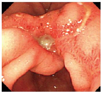
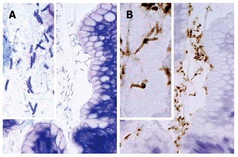
A 62-year-old woman underwent gastrointestinal endoscopy because of the sudden onset of epigastralgia and multiple gastric ulcers were found near the pyloric ring (Figure 1). Gastric tissue biopsies were taken from the ulcer edge and revealed regenerative changes with active inflammation. For the treatment of gastric ulcers, lansoprazole (30 mg/d) was administered once daily to the patient for 4 wk, which resulted in the resolution of her symptoms. Gastrointestinal endoscopic examination after lansoprazole therapy revealed multiple gastric scars near the pyloric ring. Gastric tissue biopsies were taken from the scars and revealed the regenerative changes with active inflammation and the presence of H. heilmannii s.l. on hematoxylin and eosin (H and E) and Giemsa staining in the mucus covering the gastric mucosa; these bacteria were larger and more tightly coiled than H. pylori (Figure 2A). The spiral bacteria were immunostained with a rabbit polyclonal antibody against H. pylori (Dako, Carpinteria, CA) (Figure 2B); this antibody is generated using the whole H. pylori bacterial extract as the antigen, and it exhibits cross-reactivity with “Candidatus H. heilmannii”[15] and H. suis[16]. The cross-reactivity of anti-H. pylori antibodies from different sources with “Candidatus H. heilmannii”[17] and H. suis[18] have been reported to be similar.

Figure 1
Figure 1 Initial gastrointestinal endoscopy shows gastric ulcers in the antrum.
Figure 2
Figure 2 Histological findings in non-Helicobacter pylori helicobacter SH9 in gastric mucosa. The organisms are present in small groups in gastric pits (A, B). The organisms display a tightly coiled spiral configuration and (B) are positive for the anti-Helicobacter pylori (H. pylori) antibody. (A) HE, original magnification, × 1000; (B) Immunostaining with anti-H. pylori antibody, original magnification, × 1000.

The patient was referred to our hospital for verification of the diagnosis of H. heilmannii s.l.. The patient’s laboratory data were within the normal range. Serum anti-H. pylori immunoglobulin G antibody, measured with an enzyme-linked immunosorbent assay (ELISA) kit using the E plate test (Eiken Kagaku, Inc., Tokyo, Japan), was 2.1 IU/mL (cut-off value: 10 U/mL ). The 13C-urea breath test result (UBiT, Otsuka Pharmaceutical Co., Tokushima, Japan) was negative. The patient was not administered any non-steroidal anti-inflammatory drugs. The patient had 3 household cats.

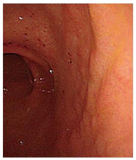
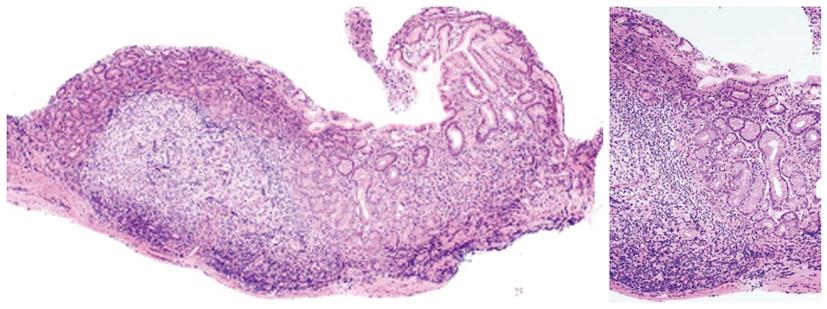
Gastrointestinal endoscopy and mapping gastric biopsy according to the updated Sydney system[19] was performed in our hospital for the histological evaluation of gastritis. In addition, antral biopsy specimens were taken for the culture of H. pylori and for the inoculation of mice by peroral administration of gastric mucosal homogenates. On gastrointestinal endoscopic examination in our hospital, background gastric mucosa of the present patient showed erosive and a slightly coarse appearance in the antrum (Figure 3), and normal appearing mucosa, without atrophy, in the corpus. Histopathological examination of gastric biopsy specimens showed mild mononuclear infiltration with large lymphoid follicles, and without neutrophil infiltration and epithelial degeneration in the antrum (Figure 4) and normal morphology in the corpus. Tightly coiled spiral bacteria were identified in the antrum, but not in the corpus, by histological examination; however, H. pylori was not isolated by microbiological culture. Based on these clinical, endoscopic, and histological findings, a diagnosis of multiple gastric ulcers associated with H. heilmannii s.l. (designated as SH9) infection was made.

Figure 3
Figure 3 Gastrointestinal endoscopy after lansoprazole therapy shows erosion and a slightly coarse granular appearance in the antrum.
Figure 4
Figure 4 Histological findings of the lesser curvature of gastric antrum from a SH9 (Helicobacter heilmannii sensu stricto)-infected patient after the administration of lansoprazole for the treatment of gastric ulcers. The gastric mucosa shows large lymphoid follicles with germinal centers without epithelial degeneration.

After confirmation of the H. heilmannii s.l. infection, the patient was treated with 1-wk triple therapy consisting of lansoprazole (30 mg/d), amoxicillin (1500 mg/d), and clarithromycin (1200 mg/d) according to the standard protocol for eradicating H. pylori infection. Twelve wks after the cessation of treatment, gastrointestinal endoscopy showed gastric scars. Further, gastric biopsy mapping showed smaller lymphoid follicles in a biopsy obtained from the greater curvature of the antrum as well as the normal appearance of the corpus, without the presence of these organisms, as evaluated by histology and immunostaining with anti-H. pylori antibody. The patient showed complete remission at the follow-up endoscopy performed 7 years after the completion of eradication therapy, and currently remains healthy.

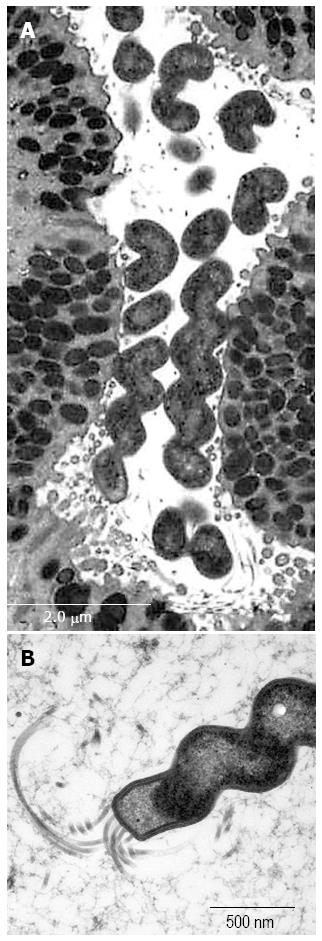
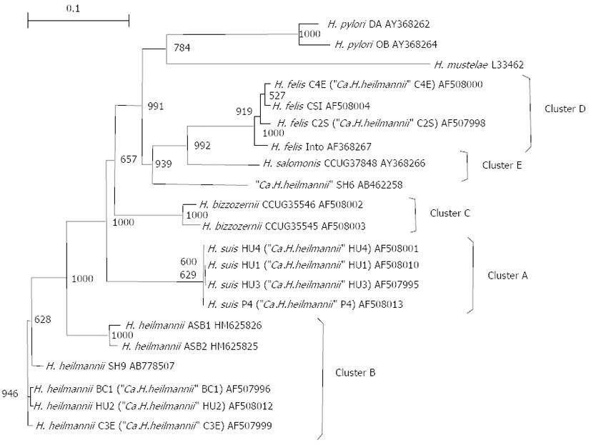
An attempt was made to maintain and propagate SH9 in vivo by inoculating homogenized whole-antral biopsy specimens from the patient into the stomachs of specific pathogen-free mice[20,21]. Transmission electron microscopy of the mouse gastric mucosa colonized with SH9 revealed that the bacteria showed 5-9 coils per cell with flagella at each pole, and were 4-6 μm long and 1 μm wide, which is larger and more coiled than H. pylori (Figure 5). In order to characterize SH9, the 16S rRNA and urease genes were sequenced. Polymerase chain reaction (PCR) amplification of the 16S rRNA gene from SH9-derived DNA isolated from the mouse was performed using 2 sets of primers: 79F (5’-GTGAGTAATGCATAGATGACATGCCC, originally designed in this study) and H679R (5’-ATTCCACCTACCTCTCCCA), and H279F (5’-CTATGACGGGTATCCGGC) and 1494R (5’-TACGGCTACCTTGTTACGAC)[22-24] The urease gene was amplified by PCR using the primers U430F (5’-GCKGAWTTGATGCAAGAAGG)[11] and UH6R2 (5’-TTATCCAAGTGGTGGCACACC)[13] and sequenced. The sequence obtained represented the partial 16S rRNA gene with a readable sequence of 1323 bp (GenBank/EMBL/DDBJ accession no. AB778508) determined from the DNA. The closest match of the 16S rRNA sequence with H. heilmannii s.s BC1 obtained from a bobcat was obtained with 99% (1302/1315 bp) identity (GenBank/EMBL/DDBJ accession no. AF506772). The urease gene from SH9 was amplified by PCR using the primer U430F together with H6-R2. The sequencing represented the partial urease gene with a readable sequence of 1493 bp (GenBank/EMBL/DDBJ accession no. AB778507). The comparatively high similarities of the DNA sequence to that of H. heilmannii s.s HU2 (AF508012) obtained from a human, BC1 (AF507996), ASB2 (HM625825) isolated from an European feline, and ASB1 (HM625826) isolated from an European feline were 98.3% (1467/1493 bp), 98.3% (1427/1451 bp), 90.5% (964/1065 bp), and 90.4% (958/1060 bp), respectively. The DNA sequences of the urease genes from SH9 and from reference organisms were aligned using GENETYX ver. 9 (GENETYX Corp., Tokyo, Japan), and the resulting alignment was modified to remove regions containing gaps. A phylogenetic tree was constructed according to the neighbor-joining method[25,26] and the Kimura model. The stabilities of the relationships were assessed by bootstrap analysis comprising 1000 data settings. Figure 6 demonstrates a phylogenetic tree constructed on the basis of urease gene analysis, and grouping of clusters as shown by O’Rourke et al[11]. The results demonstrated high levels of homology with cluster B.

Figure 5
Figure 5 Ultrastructure of SH9 (Helicobacter heilmannii s. s.) infected mouse gastric mucosa. The organisms showing tightly coiled spiral configuration (A) with flagella (B) are positioned on the apical surface of the surface mucous cells.
Figure 6
Figure 6 Phylogenetic tree based on partial ureA and ureB gene sequences, demonstrating the relationship between Helicobacter spp. from animals and humans. Clusters A through E correspond to Helicobacter suis (H.suis), Helicobacter heilmannii sensu stricto (H. heilmannii s.s.), Helicobacter bizzozerronii (H. bizzozerronii), Helicobacter felis (H. felis), and Helicobacter salomonis (H. salomonis), as described by O'Rourke et al[11]. Ca.H.heilmannii: Candidatus H. heilmannii; H. pylori: Helicobacter pylori.
DISCUSSION

We investigated the H. heilmannii s.s., designated as SH9, of human origin, both histologically and genetically by analysis of the sequences of both the 16S rRNA and urease genes.

We were unable to detect H. pylori, but detected a population of corkscrew-shaped organisms in gastric tissue biopsy samples taken from the marginal region of the gastric ulcers and from the antrum. The organisms were found in the mucous gel or on the surface of mucous cells without adhesion to gastric epithelial cells. Background gastric mucosa of the patient showed chronic gastritis with prominent lymphoid follicles without atrophy, although the mapping biopsy was performed after administration of lansoprazole for the treatment of gastric ulcers. These findings are similar to those obtained in previous studies of H. heilmannii s.l.[15,27-29]. The negative antibody test and urea breath-test results of the present patient were also consistent with previous results of H. heilmannii s.l. infection[15]. The sensitivity of urea breath-test for detection of H. heilmannii s.l. was reported to be low[30] and in this study, urea breath-test was negative. A false-negative urea breath-test test may occur due to the lower activity of H. heilmannii s.l. urease, as suggested by previous studies[30-32].

The 16S rRNA gene sequence of SH9 exhibited 99.1% (1302/1314 bp) similarity to H. heilmannii s.s. BC1; this was higher than that to H. suis. In the subsequent comparison of the urease gene sequence, SH9 revealed apparently higher levels of urease gene sequence similarities to H. heilmannii s.s. HU2 and H. heilmannii s.s. BC1. In the phylogenetic analysis following sequencing of the urease genes, SH9 was classified as belonging to Cluster B, proposed by O’Rourke et al[11] (Figure 6). Consequently, we identified SH9 as H. heilmannii s.s.. Interestingly, homologies to the urease gene sequences of H. heilmannii s.s. ASB1 and ASB2 isolated from European felines were approximately 90%. The slightly differences between the urease genes of SH9 and the European isolates may suggest geographical differences.

H. heilmannii s.l. infection have been reported in patients with acute[29] and chronic gastritis[15,27,28], duodenal ulcers[33], gastric mucosa-associated lymphoid tissue (MALT) lymphoma[15,27,34], or gastric carcinoma[35]. However, since identification at the species level of H. heilmannii s.l. is often lacking, the pathogenicity of H. suis and H. heilmannii s.s. has not been clear. H. suis infection has, however, been reported to cause gastric MALT lymphoma in experimental animals[16,18,36]. In contrast to H. suis, H. heilmannii s.s. infection in humans has rarely been reported. Interestingly, Dieterich et al[12] reported multiple H. heilmannii s.l. strains infection in a gastric ulcer patient and in his 2 cats after partially analyzing the urease B gene and suggested transmission of H. heilmannii s.l. from the patient’s household cats. Based on partial urease B gene sequences of H. heilmannii s.l. strains in their report, all of their H. heilmannii s.l. strains can be defined as H. heilmannii s.s. as the partial urease B gene sequences they reported demonstrated the highest similarity 98% with H. heilmannii s.s (GenBank/EMBL/DDBJ accession no. L25079). In addition, the reported H. heilmannii s.s. isolates have commonly been observed in feline rather than canine hosts[11]. The present patient also had 3 cats, again suggesting transmission of H. heilmannii s.s from the patient’s household cats.

H. heilmannii s.l. eradication by antimicrobial treatment regimens that are used for eradicating H. pylori results in clinical and histological improvement in H. heilmannii s.l.-associated gastroduodenal diseases[15,29,33,34,37]. Furthermore, in the present case, 1-wk triple therapy (amoxicillin, clarithromycin, and lansoprazole) for the eradication of H. pylori was effective. Thus, the regimen for H. pylori eradication may also be effective for H. heilmannii s.s. PCR analysis is desirable for the precise evaluation of H. heilmannii s.s infection status after eradication therapy. In the present case, H. heilmannii s.s. was considered to be successfully eradicated, because the patient showed complete remission at the follow-up endoscopy performed 7 years after the completion of eradication therapy. Currently, the patient is healthy.

In conclusion, we presented a case of H. heilmannii s.s.-associated multiple gastric ulcers. Further studies are needed to clarify the pathogenicity of H. heilmannii s.s. and to verify effective eradication strategies for H. heilmannii s.s..

COMMENTS
Case characteristics

A 62-year-old female with Helicobacter heilmannii sensu stricto (H. heilmannii s.s.)-related gastric ulcers.

Clinical diagnosis

Sudden onset of epigastralgia.

Differential diagnosis

Gastroesophageal reflux disease, Gastric cancer, nonsteroidal anti-inflammatory drug-related gastric uler, Helicobacter pylori (H. pylori)-related gastric ulcer.

Laboratory diagnosis

Laboratory data were within the normal range. Serum anti-H. pylori immunoglobulin G antibody and the 13C-urea breath test result were negative.

Imaging diagnosis

Gastroscopic examination revealed multiple gastric ulcers near the pyloric ring.

Pathological diagnosis

Pathological and molecular biological examinations revealed the gastric ulcer with H. heilmannii s.s. infection.

Treatment

The patient was treated with 1-wk triple therapy consisting of lansoprazole (30 mg/d), amoxicillin (1500 mg/d), and clarithromycin (1200 mg/d) according to the standard protocol for eradicating H. pylori infection.

Related reports

H. heilmannii s.s. infection in humans is not very well reported.

Term explanation

The name Helicobacter heilmannii sensu stricto is H. heilmannii identified to the species level.

Experiences and lessons

H. heilmannii s.s. is also related to a human gastric ulcer.

Peer review

This article shows a rare case of H. heilmannii s.s. infection in a patient with multiple gastric ulcers.

Footnotes

P- Reviewers: Daphan CE, Ulasoglu C S- Editor: Wen LL L- Editor: A E- Editor: Wu HL

References
1.  Solnick JV, Schauer DB. Emergence of diverse Helicobacter species in the pathogenesis of gastric and enterohepatic diseases. Clin Microbiol Rev. 2001;14:59-97.  [PubMed]  [DOI]  [Cited in This Article: ]  [Cited by in Crossref: 273]  [Cited by in F6Publishing: 255]  [Article Influence: 11.1]  [Reference Citation Analysis (0)]
2.  Solnick JV, O’Rourke J, Lee A, Paster BJ, Dewhirst FE, Tompkins LS. An uncultured gastric spiral organism is a newly identified Helicobacter in humans. J Infect Dis. 1993;168:379-385.  [PubMed]  [DOI]  [Cited in This Article: ]  [Cited by in Crossref: 162]  [Cited by in F6Publishing: 170]  [Article Influence: 5.5]  [Reference Citation Analysis (0)]
3.  Jalava K, On SL, Harrington CS, Andersen LP, Hänninen ML, Vandamme P. A cultured strain of “Helicobacter heilmannii,” a human gastric pathogen, identified as H. bizzozeronii: evidence for zoonotic potential of Helicobacter. Emerg Infect Dis. 2001;7:1036-1038.  [PubMed]  [DOI]  [Cited in This Article: ]  [Cited by in Crossref: 57]  [Cited by in F6Publishing: 60]  [Article Influence: 2.6]  [Reference Citation Analysis (0)]
4.  De Bock M, Van den Bulck K, Hellemans A, Daminet S, Coche JC, Debongnie JC, Decostere A, Haesebrouck F, Ducatelle R. Peptic ulcer disease associated with Helicobacter felis in a dog owner. Eur J Gastroenterol Hepatol. 2007;19:79-82.  [PubMed]  [DOI]  [Cited in This Article: ]  [Cited by in Crossref: 18]  [Cited by in F6Publishing: 19]  [Article Influence: 1.1]  [Reference Citation Analysis (0)]
5.  Wüppenhorst N, von Loewenich F, Hobmaier B, Vetter-Knoll M, Mohadjer S, Kist M. Culture of a gastric non-Helicobacter pylori Helicobacter from the stomach of a 14-year-old girl. Helicobacter. 2013;18:1-5.  [PubMed]  [DOI]  [Cited in This Article: ]  [Cited by in Crossref: 10]  [Cited by in F6Publishing: 11]  [Article Influence: 1.0]  [Reference Citation Analysis (0)]
6.  Stolte M, Wellens E, Bethke B, Ritter M, Eidt H. Helicobacter heilmannii (formerly Gastrospirillum hominis) gastritis: an infection transmitted by animals? Scand J Gastroenterol. 1994;29:1061-1064.  [PubMed]  [DOI]  [Cited in This Article: ]  [Cited by in Crossref: 79]  [Cited by in F6Publishing: 84]  [Article Influence: 2.8]  [Reference Citation Analysis (0)]
7.  Baele M, Decostere A, Vandamme P, Ceelen L, Hellemans A, Mast J, Chiers K, Ducatelle R, Haesebrouck F. Isolation and characterization of Helicobacter suis sp. nov. from pig stomachs. Int J Syst Evol Microbiol. 2008;58:1350-1358.  [PubMed]  [DOI]  [Cited in This Article: ]  [Cited by in Crossref: 117]  [Cited by in F6Publishing: 121]  [Article Influence: 7.6]  [Reference Citation Analysis (0)]
8.  Haesebrouck F, Pasmans F, Flahou B, Chiers K, Baele M, Meyns T, Decostere A, Ducatelle R. Gastric helicobacters in domestic animals and nonhuman primates and their significance for human health. Clin Microbiol Rev. 2009;22:202-203, Table of Contents.  [PubMed]  [DOI]  [Cited in This Article: ]  [Cited by in Crossref: 214]  [Cited by in F6Publishing: 198]  [Article Influence: 13.2]  [Reference Citation Analysis (0)]
9.  Haesebrouck F, Pasmans F, Flahou B, Smet A, Vandamme P, Ducatelle R. Non-Helicobacter pylori Helicobacter species in the human gastric mucosa: a proposal to introduce the terms H. heilmannii sensu lato and sensu stricto. Helicobacter. 2011;16:339-340.  [PubMed]  [DOI]  [Cited in This Article: ]  [Cited by in Crossref: 48]  [Cited by in F6Publishing: 43]  [Article Influence: 3.3]  [Reference Citation Analysis (0)]
10.  Smet A, Flahou B, D’Herde K, Vandamme P, Cleenwerck I, Ducatelle R, Pasmans F, Haesebrouck F. Helicobacter heilmannii sp. nov., isolated from feline gastric mucosa. Int J Syst Evol Microbiol. 2012;62:299-306.  [PubMed]  [DOI]  [Cited in This Article: ]  [Cited by in Crossref: 43]  [Cited by in F6Publishing: 39]  [Article Influence: 3.0]  [Reference Citation Analysis (0)]
11.  O’Rourke JL, Solnick JV, Neilan BA, Seidel K, Hayter R, Hansen LM, Lee A. Description of ‘Candidatus Helicobacter heilmannii’ based on DNA sequence analysis of 16S rRNA and urease genes. Int J Syst Evol Microbiol. 2004;54:2203-2211.  [PubMed]  [DOI]  [Cited in This Article: ]  [Cited by in Crossref: 104]  [Cited by in F6Publishing: 101]  [Article Influence: 5.3]  [Reference Citation Analysis (0)]
12.  Dieterich C, Wiesel P, Neiger R, Blum A, Corthésy-Theulaz I. Presence of multiple “Helicobacter heilmannii” strains in an individual suffering from ulcers and in his two cats. J Clin Microbiol. 1998;36:1366-1370.  [PubMed]  [DOI]  [Cited in This Article: ]
13.  Matsumoto T, Kawakubo M, Shiohara M, Kumagai T, Hidaka E, Yamauchi K, Oana K, Matsuzawa K, Ota H, Kawakami Y. Phylogeny of a novel “Helicobacter heilmannii” organism from a Japanese patient with chronic gastritis based on DNA sequence analysis of 16S rRNA and urease genes. J Microbiol. 2009;47:201-207.  [PubMed]  [DOI]  [Cited in This Article: ]  [Cited by in Crossref: 14]  [Cited by in F6Publishing: 11]  [Article Influence: 0.7]  [Reference Citation Analysis (0)]
14.  Kivistö R, Linros J, Rossi M, Rautelin H, Hänninen ML. Characterization of multiple Helicobacter bizzozeronii isolates from a Finnish patient with severe dyspeptic symptoms and chronic active gastritis. Helicobacter. 2010;15:58-66.  [PubMed]  [DOI]  [Cited in This Article: ]  [Cited by in Crossref: 32]  [Cited by in F6Publishing: 29]  [Article Influence: 2.1]  [Reference Citation Analysis (0)]
15.  Okiyama Y, Matsuzawa K, Hidaka E, Sano K, Akamatsu T, Ota H. Helicobacter heilmannii infection: clinical, endoscopic and histopathological features in Japanese patients. Pathol Int. 2005;55:398-404.  [PubMed]  [DOI]  [Cited in This Article: ]  [Cited by in Crossref: 58]  [Cited by in F6Publishing: 62]  [Article Influence: 3.3]  [Reference Citation Analysis (0)]
16.  Suzuki A, Kobayashi M, Matsuda K, Matsumoto T, Kawakubo M, Kumazawa S, Koide N, Miyagawa S, Ota H. Induction of high endothelial venule-like vessels expressing GlcNAc6ST-1-mediated L-selectin ligand carbohydrate and mucosal addressin cell adhesion molecule 1 (MAdCAM-1) in a mouse model of “Candidatus Helicobacter heilmannii”-induced gastritis and gastric mucosa-associated lymphoid tissue (MALT) lymphoma. Helicobacter. 2010;15:538-548.  [PubMed]  [DOI]  [Cited in This Article: ]  [Cited by in Crossref: 13]  [Cited by in F6Publishing: 12]  [Article Influence: 0.9]  [Reference Citation Analysis (0)]
17.  Singhal AV, Sepulveda AR. Helicobacter heilmannii gastritis: a case study with review of literature. Am J Surg Pathol. 2005;29:1537-1539.  [PubMed]  [DOI]  [Cited in This Article: ]  [Cited by in Crossref: 33]  [Cited by in F6Publishing: 22]  [Article Influence: 1.2]  [Reference Citation Analysis (0)]
18.  Nakamura M, Murayama SY, Serizawa H, Sekiya Y, Eguchi M, Takahashi S, Nishikawa K, Takahashi T, Matsumoto T, Yamada H. “Candidatus Helicobacter heilmannii” from a cynomolgus monkey induces gastric mucosa-associated lymphoid tissue lymphomas in C57BL/6 mice. Infect Immun. 2007;75:1214-1222.  [PubMed]  [DOI]  [Cited in This Article: ]
19.  Dixon MF, Genta RM, Yardley JH, Correa P. Classification and grading of gastritis. The updated Sydney System. International Workshop on the Histopathology of Gastritis, Houston 1994. Am J Surg Pathol. 1996;20:1161-1181.  [PubMed]  [DOI]  [Cited in This Article: ]  [Cited by in Crossref: 3221]  [Cited by in F6Publishing: 3427]  [Article Influence: 122.4]  [Reference Citation Analysis (3)]
20.  Dick E, Lee A, Watson G, O’Rourke J. Use of the mouse for the isolation and investigation of stomach-associated, spiral-helical shaped bacteria from man and other animals. J Med Microbiol. 1989;29:55-62.  [PubMed]  [DOI]  [Cited in This Article: ]  [Cited by in Crossref: 52]  [Cited by in F6Publishing: 51]  [Article Influence: 1.5]  [Reference Citation Analysis (0)]
21.  Lee A, Eckstein RP, Fevre DI, Dick E, Kellow JE. Non Campylobacter pylori spiral organisms in the gastric antrum. Aust N Z J Med. 1989;19:156-158.  [PubMed]  [DOI]  [Cited in This Article: ]  [Cited by in Crossref: 25]  [Cited by in F6Publishing: 24]  [Article Influence: 0.7]  [Reference Citation Analysis (0)]
22.  Weisburg WG, Barns SM, Pelletier DA, Lane DJ. 16S ribosomal DNA amplification for phylogenetic study. J Bacteriol. 1991;173:697-703.  [PubMed]  [DOI]  [Cited in This Article: ]
23.  Riley LK, Franklin CL, Hook RR, Besch-Williford C. Identification of murine helicobacters by PCR and restriction enzyme analyses. J Clin Microbiol. 1996;34:942-946.  [PubMed]  [DOI]  [Cited in This Article: ]
24.  Neilan BA, Jacobs D, Del Dot T, Blackall LL, Hawkins PR, Cox PT, Goodman AE. rRNA sequences and evolutionary relationships among toxic and nontoxic cyanobacteria of the genus Microcystis. Int J Syst Bacteriol. 1997;47:693-697.  [PubMed]  [DOI]  [Cited in This Article: ]  [Cited by in Crossref: 343]  [Cited by in F6Publishing: 247]  [Article Influence: 9.1]  [Reference Citation Analysis (0)]
25.  Saitou N, Nei M. The neighbor-joining method: a new method for reconstructing phylogenetic trees. Mol Biol Evol. 1987;4:406-425.  [PubMed]  [DOI]  [Cited in This Article: ]
26.  Kimura M. Average time until fixation of a mutant allele in a finite population under continued mutation pressure: Studies by analytical, numerical, and pseudo-sampling methods. Proc Natl Acad Sci USA. 1980;77:522-526.  [PubMed]  [DOI]  [Cited in This Article: ]  [Cited by in Crossref: 69]  [Cited by in F6Publishing: 76]  [Article Influence: 5.4]  [Reference Citation Analysis (0)]
27.  Stolte M, Kroher G, Meining A, Morgner A, Bayerdörffer E, Bethke B. A comparison of Helicobacter pylori and H. heilmannii gastritis. A matched control study involving 404 patients. Scand J Gastroenterol. 1997;32:28-33.  [PubMed]  [DOI]  [Cited in This Article: ]  [Cited by in Crossref: 136]  [Cited by in F6Publishing: 113]  [Article Influence: 4.2]  [Reference Citation Analysis (0)]
28.  Heilmann KL, Borchard F. Gastritis due to spiral shaped bacteria other than Helicobacter pylori: clinical, histological, and ultrastructural findings. Gut. 1991;32:137-140.  [PubMed]  [DOI]  [Cited in This Article: ]  [Cited by in Crossref: 202]  [Cited by in F6Publishing: 210]  [Article Influence: 6.4]  [Reference Citation Analysis (0)]
29.  Yoshimura M, Isomoto H, Shikuwa S, Osabe M, Matsunaga K, Omagari K, Mizuta Y, Murase K, Murata I, Kohno S. A case of acute gastric mucosal lesions associated with Helicobacter heilmannii infection. Helicobacter. 2002;7:322-326.  [PubMed]  [DOI]  [Cited in This Article: ]  [Cited by in Crossref: 23]  [Cited by in F6Publishing: 24]  [Article Influence: 1.1]  [Reference Citation Analysis (0)]
30.  Chen Y, Zhou D, Wang J. [Biological diagnostic and therapeutic study on the infection of Helicobacter heilmannii]. Zhonghua Yi Xue Zazhi. 1998;78:490-493.  [PubMed]  [DOI]  [Cited in This Article: ]
31.  Boyanova L, Lazarova E, Jelev C, Gergova G, Mitov I. Helicobacter pylori and Helicobacter heilmannii in untreated Bulgarian children over a period of 10 years. J Med Microbiol. 2007;56:1081-1085.  [PubMed]  [DOI]  [Cited in This Article: ]  [Cited by in Crossref: 18]  [Cited by in F6Publishing: 19]  [Article Influence: 1.1]  [Reference Citation Analysis (0)]
32.  De Groote D, Ducatelle R, van Doorn LJ, Tilmant K, Verschuuren A, Haesebrouck F. Detection of “Candidatus Helicobacter suis” in gastric samples of pigs by PCR: comparison with other invasive diagnostic techniques. J Clin Microbiol. 2000;38:1131-1135.  [PubMed]  [DOI]  [Cited in This Article: ]
33.  Goddard AF, Logan RP, Atherton JC, Jenkins D, Spiller RC. Healing of duodenal ulcer after eradication of Helicobacter heilmannii. Lancet. 1997;349:1815-1816.  [PubMed]  [DOI]  [Cited in This Article: ]  [Cited by in Crossref: 41]  [Cited by in F6Publishing: 42]  [Article Influence: 1.6]  [Reference Citation Analysis (0)]
34.  Morgner A, Lehn N, Andersen LP, Thiede C, Bennedsen M, Trebesius K, Neubauer B, Neubauer A, Stolte M, Bayerdörffer E. Helicobacter heilmannii-associated primary gastric low-grade MALT lymphoma: complete remission after curing the infection. Gastroenterology. 2000;118:821-828.  [PubMed]  [DOI]  [Cited in This Article: ]  [Cited by in Crossref: 202]  [Cited by in F6Publishing: 175]  [Article Influence: 7.3]  [Reference Citation Analysis (0)]
35.  Morgner A, Bayerdörffer E, Meining A, Stolte M, Kroher G. Helicobacter heilmannii and gastric cancer. Lancet. 1995;346:511-512.  [PubMed]  [DOI]  [Cited in This Article: ]  [Cited by in Crossref: 52]  [Cited by in F6Publishing: 51]  [Article Influence: 1.8]  [Reference Citation Analysis (0)]
36.  Flahou B, Haesebrouck F, Pasmans F, D’Herde K, Driessen A, Van Deun K, Smet A, Duchateau L, Chiers K, Ducatelle R. Helicobacter suis causes severe gastric pathology in mouse and mongolian gerbil models of human gastric disease. PLoS One. 2010;5:e14083.  [PubMed]  [DOI]  [Cited in This Article: ]  [Cited by in Crossref: 44]  [Cited by in F6Publishing: 42]  [Article Influence: 3.0]  [Reference Citation Analysis (0)]
37.  Hilzenrat N, Lamoureux E, Weintrub I, Alpert E, Lichter M, Alpert L. Helicobacter heilmannii-like spiral bacteria in gastric mucosal biopsies. Prevalence and clinical significance. Arch Pathol Lab Med. 1995;119:1149-1153.  [PubMed]  [DOI]  [Cited in This Article: ]