Published online Nov 14, 2013. doi: 10.3748/wjg.v19.i42.7419
Revised: August 14, 2013
Accepted: September 16, 2013
Published online: November 14, 2013
Processing time: 157 Days and 16.1 Hours
AIM: To evaluate the usefulness of real-time virtual sonography (RVS) in biliary and pancreatic diseases.
METHODS: This study included 15 patients with biliary and pancreatic diseases. RVS can be used to observe an ultrasound image in real time by merging the ultrasound image with a multiplanar reconstruction computed tomography (CT) image, using pre-scanned CT volume data. The ultrasound used was EUB-8500 with a convex probe EUP-C514. The RVS images were evaluated based on 3 levels, namely, excellent, good and poor, by the displacement in position.
RESULTS: By combining the objectivity of CT with free scanning using RVS, it was possible to easily interpret the relationship between lesions and the surrounding organs as well as the position of vascular structures. The resulting evaluation levels of the RVS images were 12 excellent (pancreatic cancer, bile duct cancer, cholecystolithiasis and cholangiocellular carcinoma) and 3 good (pancreatic cancer and gallbladder cancer). Compared with conventional B-mode ultrasonography and CT, RVS images achieved a rate of 80% superior visualization and 20% better visualization.
CONCLUSION: RVS has potential usefulness in objective visualization and diagnosis in the field of biliary and pancreatic diseases.
Core tip: The real-time virtual sonography (RVS) system combined with ultrasonography (US) and computed tomography (CT) compensates for each of the deficiencies of US and CT. The visualization in biliary and pancreatic diseases using RVS added objectivity and detailed imaging for diagnosis lacking in conventional B-mode US and CT, and it becomes possible to make a more precise diagnosis. Moreover, it is possible to consider this system as providing images of detailed processes conveniently and in real time. RVS has potential usefulness in objective visualization and diagnosis in the field of biliary and pancreatic diseases.
- Citation: Sofuni A, Itoi T, Itokawa F, Tsuchiya T, Kurihara T, Ishii K, Tsuji S, Ikeuchi N, Tanaka R, Umeda J, Tonozuka R, Honjo M, Mukai S, Moriyasu F. Real-time virtual sonography visualization and its clinical application in biliopancreatic disease. World J Gastroenterol 2013; 19(42): 7419-7425
- URL: https://www.wjgnet.com/1007-9327/full/v19/i42/7419.htm
- DOI: https://dx.doi.org/10.3748/wjg.v19.i42.7419
Recent advancements in technology have expanded the role of diagnosis by ultrasonography (US) in biliopancreatic diseases. Ultrasonography is useful because it is non-invasive and images can be obtained in real time. However, visualization is sometimes difficult in the presence of bone, gas and air, and thus has the problem of diminished objectivity. Real-time virtual sonography (RVS) (Hitachi-Aloka Medical Ltd., Tokyo, Japan) is a technological system that was developed in an attempt to resolve such problems.
This method merges ultrasound images with information from computed tomography (CT) images to produce the displayed image. In other words, the CT volume data is pre-stored into the device and the real-time ultrasound image is displayed simultaneously while the virtual view is reconstructed as a CT-multiplanar reconstruction (MPR) image from the stored volume data[1]. This is one method of compensating for the decreased objectivity in ultrasound diagnosis. This method is used during navigation in ultrasound-guided treatment and its usefulness has been reported in the field of liver disease[2-13]; however, no data has been presented in the field of biliopancreatic disease.
The RVS system was applied on patients with biliopancreatic disease to investigate its usefulness and potential. The aim of the evaluation was to assess how the target lesion was correlated with the surrounding organs and blood vessels.
RVS was performed on 15 biliopancreatic patients at our department. Among the patients, 6 had pancreatic cancer, 1 had pancreatic endocrine tumor, 2 had gall bladder cancer, 4 had bile duct cancer, 1 had cholangiocellular carcinoma, and 1 had cholecystolithiasis.
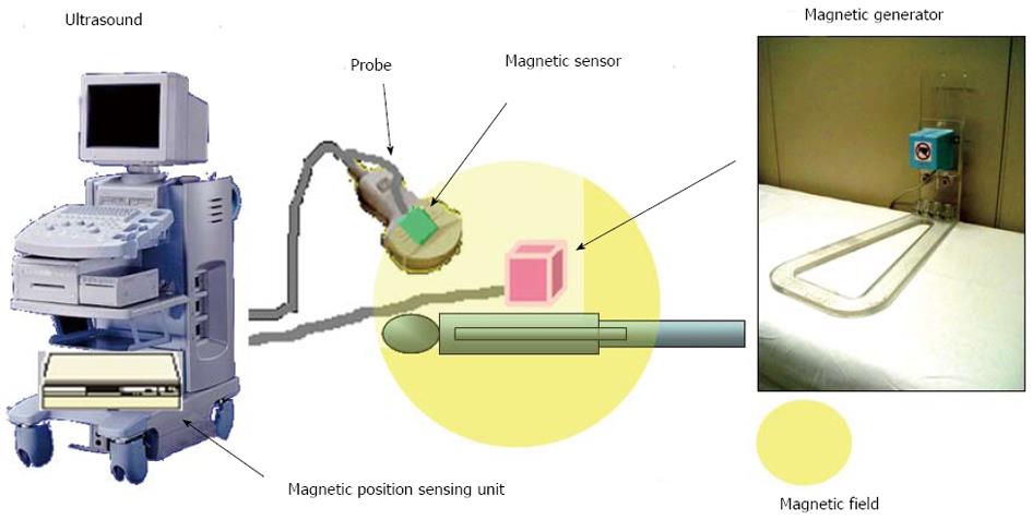
The RVS system (Figure 1) is made up of an ultrasound diagnostic device EUB-8500 (Hitachi-Aloka Medical Ltd., Tokyo, Japan), the EUP-C514 probe (Hitachi-Aloka Medical Ltd., Tokyo, Japan), which is a convex type, a personal computer (PC) and a magnetic position sensor unit to detect the position of the probe. Pre-scanned CT volume data is stored in a medium such as a compact disc read-only memory or PC using digital imaging and communication in medicine specifications via a local area network. The PC can display the images at a high speed of approximately 11 frames/s, with each frame being 256 × 256 pixels of virtual sonography. The PC is equipped with a magnetic position sensor unit to obtain information on the position of the probe. The magnetic position sensor unit consists of the main unit, a magnetism generator and a magnetic sensor. When the magnetism generator is placed close to the patient, the magnetic sensor attaches to the probe. On CT, by performing imaging of very thin slices (approximately 1 mm) by multidetector CT (MDCT) of at least 16 rows, the minimum volume data required to reconstruct MPR images can be collected. In our department, 32-row MDCT is used to collect volume data by 3-planar contrast imaging.
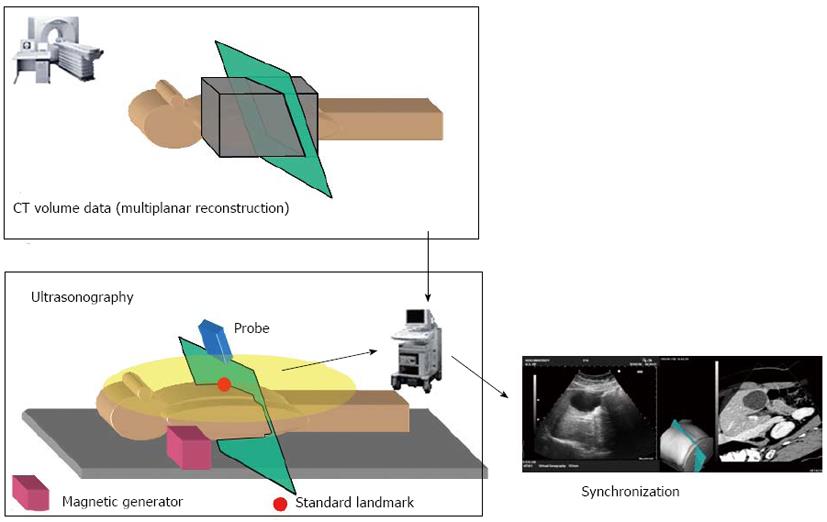
Information of the ultrasound probe position is obtained by the magnetic position sensor connected to the PC. The information obtained by this sensor is on its relative position to the magnetic sensor. Therefore, to prepare an MPR image that fits the ultrasound plane, it is necessary to decide on the pre-stored CT volume data and the position (reference point) of the ultrasound image. Ensiform cartilage that is palpable from the body surface is the ideal reference point. This is used to adjust the position (calibration). Apart from ensiform cartilage, calibration can also be performed using metal markers placed in the vasculature, kidneys, umbilical cord or body surface. In our department, calibration is mainly performed using ensiform cartilage. After the calibration, the ultrasound image obtained from the ultrasound scanning is displayed simultaneously. The virtual sonography-processing program in the PC detects the probe position and angle, reconstructs the corresponding planar image from the CT volume data, and displays it as an MPR image (Figure 2).
The respective RVS images are evaluated based on 3 levels, namely, Excellent: hardy any displacement in position, Good: midway between Excellent and Poor, and Poor: major displacement in position. The RVS images were stored in video form and were evaluated by 3 gastroenterologists (Sofuni A, Itoi T, Itokawa F) with at least 8 years of experience in abdominal sonography evaluation.
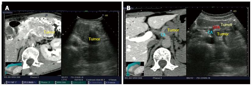
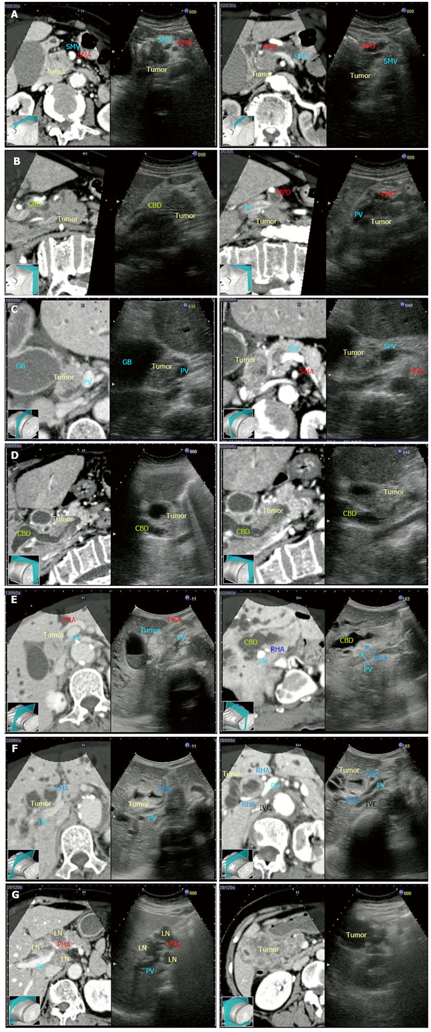
The results were based on the decisions of the majority, and the result of an actual case is shown in Table 1. The resulting evaluation levels of the RVS images were 12 excellent (pancreatic cancer, bile duct cancer, cholecystolithiasis and cholangiocellular carcinoma) and 3 good (pancreatic cancer and gallbladder cancer). The evaluation of the RVS image in the patient with pancreatic endocrine tumor was excellent (Figure 3). The diagnosis by endoscopic ultrasound-guided fine needle aspiration was poorly differentiated neuroendocrine carcinoma. The relationship between the tumor and the celiac artery posed a problem in terms of choice of treatment. With the RVS image, the ultrasound showed no vascular irregularity, and the boundary between the tumor, celiac artery (CA) and splenic artery (SPA) remained intact, thus surgery was performed. Even when surgery was performed, there was no invasion into the arteries. With RVS, the positional relationship between the CA and the tumor could be evaluated objectively and was useful in judging whether or not surgery should be performed. Similarly, the positional relationship between the tumor mass and the vasculature could be assessed objectively, even in the cases where images of pancreatic cancer (Figure 4A, B) and bile duct cancer (Figure 4C-F) were judged as excellent. It was also useful in understanding the stage of progression and anatomical relationship. A good case (gall bladder cancer) is shown in Figure 4G.
| No. | Case | Age (yr) | Sex | BMI (kg/m2) | Breath-holding | Evaluation |
| 1 | Pancreatic cancer | 76 | M | 18.1 | Possible | Excellent |
| 2 | Pancreatic endocrine tumor | 65 | F | 23.2 | Possible | Excellent |
| 3 | Pancreatic cancer | 73 | F | 21.3 | Possible | Excellent |
| 4 | Pancreatic cancer | 77 | M | 19.2 | Possible | Excellent |
| 5 | Pancreatic cancer | 65 | M | 22.2 | Possible | Good |
| 6 | Pancreatic cancer | 49 | M | 19.7 | Possible | Excellent |
| 7 | Pancreatic cancer | 58 | F | 21.4 | Possible | Excellent |
| 8 | Bile duct cancer | 81 | F | 12.7 | Impossible | Excellent |
| 9 | Bile duct cancer | 79 | F | 22.2 | Possible | Excellent |
| 10 | Bile duct cancer | 76 | F | 21.5 | Possible | Excellent |
| 11 | Bile duct cancer | 68 | M | 23.3 | Possible | Excellent |
| 12 | Gallbladder cancer | 80 | M | 25.1 | Possible | Good |
| 13 | Gallbladder cancer | 63 | F | 21.2 | Possible | Good |
| 14 | Cholecystolithiasis | 73 | M | 19.6 | Possible | Excellent |
| 15 | Cholangiocellular carcinoma | 68 | F | 21.7 | Possible | Excellent |
US is a generally subjective examination and is considered to lack objectivity, particularly in the staging of malignant tumors. By combining the objectivity of CT with the obscureness in the free scanning of US, it was possible to easily interpret the relationship between the lesions and the surrounding organs, as well as the position of vascular structures. Based on the results, incorporation of the objectivity of CT into voluntary scanning with an ultrasound probe facilitated understanding of the positional relationships among the lesion, the surrounding organs and vasculature, and the structure of the lesion itself. According to the study, this RVS system, combined with US and CT, also confirmed the comparatively precise diagnosis in US imaging. Moreover, this system compensates for each of the deficiencies of US and CT, and it becomes possible to provide more detailed information and make a more precise diagnosis.
US is usually more useful than CT in judging detailed imaging and the relationships with surrounding structures, and moreover, it is possible to consider the RVS system as providing images of detailed processes conveniently and in real time. For example, in Figure 3, it seems that the tumor invaded the blood vessel on CT imaging, but the RVS system showed that the tumor had not invaded the blood vessel. Therefore, RVS can aid in making a precise diagnosis when CT is unable to make judgments on the staging.
Compared with conventional B-mode US and CT, RVS images achieved a rate of 80% superior visualization and 20% better visualization, according to the statistics. The RVS visualization of lesions and the surrounding organs of all patients provided the objectivity and detailed imaging information for diagnosis lacking in conventional B-mode US and CT. It is also suggested to be useful when applied to making a diagnosis on the existence and stage of progression of malignant tumors, and the evaluation of infiltration into major blood vessels. Further studies in more patients are required to examine the applicability of this method to the diagnosis of malignant diseases and the extent of disease progression, including assessment of infiltration into major blood vessels. This needs to be investigated further in a larger number of patients.
Considering the investigations we have performed using RVS on patients in our department to date, the advantages and disadvantages of RVS are as outlined below. Five advantages are recognized at present: (1) CT objectivity in real-time sonographic images makes it easy to understand the structure and positional relationship between the lesion and the surrounding organs and vasculature; (2) it provides supportive education for surgeons and physicians that lack experience; (3) MPR images obtained by multi-slicing using the familiar operations of the ultrasound probe can be visualized instantly; (4) the display screen can be switched between the arterial phase, portal vein phase, and equilibrium phase (applies to the hemodynamics of the lesion and evaluation of the vasculature) by a simple operation; and (5) the common bile duct and pancreas are not affected by variations in breathing as much as the liver. There are 5 disadvantages: (1) the positions of the images during CT scanning and those of the US images are easily displaced due to breathing variations (liver > biliopancreatic area); (2) with only 1 reference point, synchronization of the position tends to be inadequate; (3) progression, infiltration and vascular evaluation are time-consuming and may put a strain on patients; (4) the focus is mainly on CT, thus US evaluation tends to be poor; and (5) the calibration is time-consuming. It is preferable to perform RVS after considering these advantages and disadvantages.
The main problem in the present RVS system is that when the position is displaced, calibration becomes time-consuming. At Hitachi-Aloka Medical Ltd., an ultrasound phantom was prepared to examine the precision, and RVS was performed using simulated CT. As a result, displacement of the position of the slicing plane tended to depend on the distance between the magnetism generator and the magnetic sensor. However, the distance was reported to be approximately ± 5 mm within a range of 70 cm with the magnetism generator as the center. In addition, with only 1-point calibration, there is displacement of the position. Therefore, multiple-point calibration is important. Precision for calibration needs to be improved by taking these factors into consideration. Moreover, in actual clinical settings, the position of organs may also vary according to the posture and depth of respiration of the subject. Errors can also arise due to these factors; therefore, a corrective function for positional displacement as in the synchronization of breathing needs to be installed in the device. In addition, recent advances in computer and laser technologies have enabled the construction of MPR images of MDCT to be performed easily even with a notebook PC. Taking these points into consideration, one can say that the superiority of RVS is its potential in the areas of therapy and education.
Apart from diagnosis, RVS is now commonly used to assist treatment under ultrasound guidance in liver disease. Even in the biliopancreatic area, particularly with pancreatic cancer, focused ultrasound treatment and high intensity focused ultrasound (HIFU) treatment are now being performed[14]. In the current HIFU, treatment progresses while looking at the 2D ultrasound image. The application of this method allows easy understanding of the range of treatment, and safe treatment can be performed objectively.
This study suggests that RVS is useful when applied to making a diagnosis on the existence and stage of progression of malignant tumors, and making evaluations of infiltration into major blood vessels, in spite of some disadvantages. Further improvement and progress of RVS is expected in the field of therapy.
Ultrasonography (US) is non-invasive, and images can be obtained in real time. However, visualization is sometimes difficult with the presence of bone, gas and air, and thus has the problem of diminished objectivity. Real-time virtual sonography (RVS) is a technological system that was developed in an attempt to resolve such problems. This is one method of compensating for the decreased objectivity in ultrasound diagnosis. This method is used during navigation in ultrasound-guided treatment and its usefulness has been reported in the field of liver disease; however, no data has been presented in the field of biliopancreatic disease.
The frontiers of research are in the fields of diagnosis and therapy for biliopancreatic disease.
The usefulness of the RVS system has been reported in the field of liver disease; however, no data has been presented in the field of biliopancreatic disease. RVS is useful when applied to making a precise diagnosis on the existence and stage of progression of malignant tumors, and making evaluations of infiltration into major blood vessels. Recently, therapies using ultrasound for biliopancreatic disease have been developed. Further improvement and progress of RVS is expected in the field of therapy.
The RVS system combined with US and computed tomography (CT) compensates for each of the deficiencies of US and CT, and it becomes possible to provide more detailed information and make a more precise diagnosis. Moreover, it is possible to consider the RVS system as providing images of detailed processes conveniently and in real time. Therefore, this system is useful in the case in which the detailed information and positional relationships among the lesions are needed for a precise diagnosis.
RVS can be used to observe an ultrasound image in real time by merging the ultrasound image with a multiplanar reconstruction CT image, using pre-scanned CT volume data. The RVS system combined with US and CT compensates for each of the deficiencies of US and CT.
This is a good study of the usefulness of RVS in biliary and pancreatic diseases. Twelve patients with biliary and pancreatic diseases in whom the combination of the objectivity of CT with free scanning using RVS was observed. This study showed that the visualization in biliary and pancreatic diseases using RVS had the objectivity and the detailed imaging for diagnosis lacking in conventional B-mode US and CT. This new technique has potential usefulness in visualizing and diagnosing biliary and pancreatic diseases in the future. Even though the patient numbers were small, the paper has the potential data to be used at the clinic for appropriate diagnosis.
P- Reviewers: Chowdhury P, Pezzilli R S- Editor: Gou SX L- Editor: Cant MR E- Editor: Wu HL
| 1. | Oshio K, Shinmoto H. Simulation of US imaging by using a CT data set. Radiology. 1996;201 Suppl:517 Available from: http://serials.unibo.it/cgi-ser/start/en/spogli/df-s.tcl?prog_art=2201452&language=ENGLISH&view=articoli. |
| 2. | Minami Y, Kudo M, Chung H, Inoue T, Takahashi S, Hatanaka K, Ueda T, Hagiwara H, Kitai S, Ueshima K. Percutaneous radiofrequency ablation of sonographically unidentifiable liver tumors. Feasibility and usefulness of a novel guiding technique with an integrated system of computed tomography and sonographic images. Oncology. 2007;72 Suppl 1:111-116. [RCA] [PubMed] [DOI] [Full Text] [Cited by in Crossref: 34] [Cited by in RCA: 37] [Article Influence: 2.1] [Reference Citation Analysis (0)] |
| 3. | Kawasoe H, Eguchi Y, Mizuta T, Yasutake T, Ozaki I, Shimonishi T, Miyazaki K, Tamai T, Kato A, Kudo S. Radiofrequency ablation with the real-time virtual sonography system for treating hepatocellular carcinoma difficult to detect by ultrasonography. J Clin Biochem Nutr. 2007;40:66-72. [RCA] [PubMed] [DOI] [Full Text] [Full Text (PDF)] [Cited by in Crossref: 34] [Cited by in RCA: 36] [Article Influence: 2.6] [Reference Citation Analysis (0)] |
| 4. | Kitada T, Murakami T, Kuzushita N, Minamitani K, Nakajo K, Osuga K, Miyoshi E, Nakamura H, Kishino B, Tamura S. Effectiveness of real-time virtual sonography-guided radiofrequency ablation treatment for patients with hepatocellular carcinomas. Hepatol Res. 2008;38:565-571. [RCA] [PubMed] [DOI] [Full Text] [Cited by in Crossref: 44] [Cited by in RCA: 42] [Article Influence: 2.5] [Reference Citation Analysis (0)] |
| 5. | Crocetti L, Lencioni R, Debeni S, See TC, Pina CD, Bartolozzi C. Targeting liver lesions for radiofrequency ablation: an experimental feasibility study using a CT-US fusion imaging system. Invest Radiol. 2008;43:33-39. [RCA] [PubMed] [DOI] [Full Text] [Cited by in Crossref: 101] [Cited by in RCA: 91] [Article Influence: 5.4] [Reference Citation Analysis (0)] |
| 6. | Sandulescu L, Saftoiu A, Dumitrescu D, Ciurea T. Real-time contrast-enhanced and real-time virtual sonography in the assessment of benign liver lesions. J Gastrointestin Liver Dis. 2008;17:475-478. [PubMed] |
| 7. | Sandulescu L, Saftoiu A, Dumitrescu D, Ciurea T. The role of real-time contrast-enhanced and real-time virtual sonography in the assessment of malignant liver lesions. J Gastrointestin Liver Dis. 2009;18:103-108. [PubMed] |
| 8. | Nakai M, Sato M, Sahara S, Takasaka I, Kawai N, Minamiguchi H, Tanihata H, Kimura M, Takeuchi N. Radiofrequency ablation assisted by real-time virtual sonography and CT for hepatocellular carcinoma undetectable by conventional sonography. Cardiovasc Intervent Radiol. 2009;32:62-69. [RCA] [PubMed] [DOI] [Full Text] [Cited by in Crossref: 60] [Cited by in RCA: 65] [Article Influence: 3.8] [Reference Citation Analysis (0)] |
| 9. | Okamoto E, Sato S, Sanchez-Siles AA, Ishine J, Miyake T, Amano Y, Kinoshita Y. Evaluation of virtual CT sonography for enhanced detection of small hepatic nodules: a prospective pilot study. AJR Am J Roentgenol. 2010;194:1272-1278. [RCA] [PubMed] [DOI] [Full Text] [Cited by in Crossref: 20] [Cited by in RCA: 22] [Article Influence: 1.5] [Reference Citation Analysis (0)] |
| 10. | Kasuya K, Sugimoto K, Kyo B, Nagakawa Y, Ikeda T, Mori Y, Wada T, Suzuki M, Nagai T, Itoi T. Ultrasonography-guided hepatic tumor resection using a real-time virtual sonography with indocyanine green navigation (with videos). J Hepatobiliary Pancreat Sci. 2011;18:380-385. [PubMed] |
| 11. | Liu FY, Yu XL, Liang P, Cheng ZG, Han ZY, Dong BW, Zhang XH. Microwave ablation assisted by a real-time virtual navigation system for hepatocellular carcinoma undetectable by conventional ultrasonography. Eur J Radiol. 2012;81:1455-1459. [RCA] [PubMed] [DOI] [Full Text] [Cited by in Crossref: 60] [Cited by in RCA: 64] [Article Influence: 4.6] [Reference Citation Analysis (0)] |
| 12. | Minami Y, Kitai S, Kudo M. Treatment response assessment of radiofrequency ablation for hepatocellular carcinoma: usefulness of virtual CT sonography with magnetic navigation. Eur J Radiol. 2012;81:e277-e280. [RCA] [PubMed] [DOI] [Full Text] [Cited by in Crossref: 17] [Cited by in RCA: 21] [Article Influence: 1.5] [Reference Citation Analysis (0)] |
| 13. | Sandulescu DL, Dumitrescu D, Rogoveanu I, Saftoiu A. Hybrid ultrasound imaging techniques (fusion imaging). World J Gastroenterol. 2011;17:49-52. [RCA] [PubMed] [DOI] [Full Text] [Full Text (PDF)] [Cited by in CrossRef: 28] [Cited by in RCA: 27] [Article Influence: 1.9] [Reference Citation Analysis (3)] |
| 14. | Sofuni A, Moriyasu F, Sano T, Yamada K, Itokawa F, Tsuchiya T, Tsuji S, Kurihara T, Ishii K, Itoi T. The current potential of high-intensity focused ultrasound for pancreatic carcinoma. J Hepatobiliary Pancreat Sci. 2011;18:295-303. [RCA] [PubMed] [DOI] [Full Text] [Cited by in Crossref: 24] [Cited by in RCA: 26] [Article Influence: 1.9] [Reference Citation Analysis (0)] |


