Published online Jan 28, 2013. doi: 10.3748/wjg.v19.i4.586
Revised: November 19, 2012
Accepted: November 24, 2012
Published online: January 28, 2013
Processing time: 152 Days and 19 Hours
Tubular duplication of the colon is very rare especially in adulthood, because it is frequently symptomatic earlier in newborn life, so only few cases are reported in literature. Several theories are proposed to explain the onset and the evolution of gut malformations as the aberrant lumen recanalization or the diverticular theory, the alteration of the lateral closure of the embryonal disk or finally the dorsal protrusion of the yolk-sac for herniation or adhesion to the ectoderm for an abnormality of the longitudinal line, but none clarifies the exact genesis of duplication. We present a case of “Y-shaped” tubular duplication of the transverse colon in a 21-year-old adult, with a history of chronic pain and constipation, referred to our department for abdominal pain with retrosternal irradiation, treated with the resection of the aberrant bowel.
- Citation: Banchini F, Delfanti R, Begnini E, Tripodi MC, Capelli P. Duplication of the transverse colon in an adult: Case report and review. World J Gastroenterol 2013; 19(4): 586-589
- URL: https://www.wjgnet.com/1007-9327/full/v19/i4/586.htm
- DOI: https://dx.doi.org/10.3748/wjg.v19.i4.586
Duplications of gastrointestinal tract are infrequent in general population with an incidence of two or three cases per year in referred pediatric center[1]. The most frequent localization is the ileum 30% and the ileocecal valve 30%, followed by the jejunum with 8%, the colon 6%-7% and the rectum 5%[2,3]. Since 1950, less than 50 colonic duplication have been reported in literature[4] and they were more often discovered in the first two years of life (80%)[5]. If no associated malformation is present, they remain frequently hidden for several years until a complication occurs[6]. The symptoms are unspecific and depend on the type of duplication and on the associated abnormality. More often they are abdominal mass, chronic pain, constipation, occlusion, and less frequently volvulus[7] intussusception, bleeding[8] or perforation (especially in sigmoid colon mimicking diverticulitis). Also the arising of carcinoma is observed: the most frequent is adenocarcinoma[9-13] followed by squamous carcinoma and carcinoid tumors[14-16]. The carcinoembryonic antigen expression can be observed in association with the arising of adenocarcinoma[17], but the presence of the carbohydrate antigen 19-9 is not well defined yet.
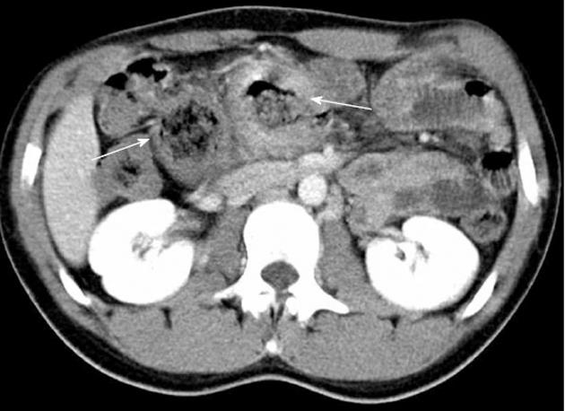
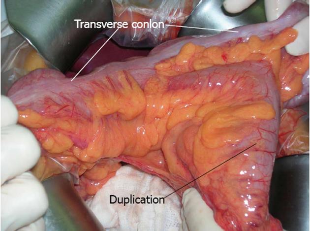
A 21-year-old man with a history of chronic constipation was referred to our department for abdominal pain with retrosternal irradiation. During childhood he was hospitalized for the same symptoms without a clear diagnosis: alimentary intolerance or celiac disease were excluded, and all the abdominal ultrasounds were always negative. The mother had no particular problems during pregnancy and labour, she had no exposition to pollution, but the patient was an eight months premature birth. At the hospitalization the blood samples showed mild leukocytosis and the abdominal Rx plane was normal. Abdominal sonography was performed but it was not diagnostic, because of air hiding; an abdominal computed tomography (CT) scan (Figure 1) revealed an oval mass with wall enhancement and similar stool material inside, suggestive for intestinal duplication. A small-bowel contrast study was performed but no alteration could be demonstrated. The patient was treated with antibiotics and was discharged asymptomatic with a planned intervention for the following week. At the explorative laparotomy a transverse colon Y-type duplication was shown (Figure 2); the duplication was bent down at the origin of the transverse mesocolon and an own mesocolon was seen. After dissection from the mesocolic stamp, the duplication was unbent revealing that the blood supply origin from a distal division of the middle colic artery with a trifurcation for right and left transverse colon and one branch for the duplication. The upper part of the common mesocolon presented a crabbed fusion of small vessels. To preserve the left marginal arcade, we dissected the artery for the duplication with conservation of the two branches for the native transverse colon and with resection of the duplicated bowel and 5 cm of the native transverse colon. A colo-colic anastomosis was performed. The post operative period was uneventful and the patient was discharged; at the moment he lies symptoms free. Histopathology confirmed the tubular duplication with multiple mucosal ulceration.
Digestive duplication is defined by the three Rowling’s criteria[18]: (1) the wall of the duplication is in continuity with one of the duplicated organ; (2) the cyst is surrounded by a smooth muscular layer; and (3) a layer of digestive mucosa is present, more often typical or heterotopic as gastric mucosa, colonic mucosa, bronchial or pancreatic structure[19,20]. Smith[21] well defined the term of duplication as “these formations, either tubular or spherical, which lie in contiguity with normal bowel and which share with it a common blood supply and usually a common muscle coat. There may or may not be a communication between the normal and anomalous lumina and the dividing septum may be muscular or merely a double layer of epithelium”. Gross et al[22] described four variations in the shape of duplications: (1) a tubular structure that branches out from the intestine and extends for some distance between the mesenteric leaves; (2) a double-barreled structure communicating with the intestinal lumen at one or both ends; (3) a cystic structure lying in the peritoneal cavity, attached by a mesenteric stalk; and (4) a spherical lesion contiguous with some part of the bowel, particularly along the ileum. Cystic types are the most frequent with 90%-95% of the cases and the tubular type 5%-10%; the latter can be distinguished in double barreled (80%) or Y-shaped form (20%)[23-26]. For colonic duplication McPherson makes another classification as type I (simple cysts), type II (diverticula) or the most common type III (tubular colonic duplication)[27]. Several theories have been proposed to describe this abnormality in which environmental factors, such as trauma or hypoxia, can play a role[28]. The aberrant lumen recanalization theory exposed by Bremer[29] seems to be the most comprehensive. As described by Johnson[30] in 1910 the gut grows and obliterates the lumen at the sixth week with subsequent internal vacuolization and coalescence of vacuoles with the formation of the single lumen. The aberrant vacuolization well explains that vacuoles can remain separated and create one or more duplication that can be separated or communicating with the original gut. This theory can be easily associated to the double barrel duplication. Another theory is the diverticular one, described by Lewis et al[31], criticized by some authors[1] because it doesn’t explain the forms of entire duplication of the colon (for example) with the presence of circular and longitudinal muscle coats. Blinder describes more simply that the tubular forms can be caused by an alteration of the lateral closure of the embryonal disk at the stage 9 of Steeter for an abnormality of the longitudinal line, instead of cystic forms that originate from diverticulation later in the evolution[32,33]. Another explanation, well described by Smith[21], is the dorsal protrusion of the yolk-sac caused by its herniation or adhesion to the ectoderm in same phase of presomite development taking to a “dorsal remnants” structure, that can be divided into: congenital dorsal enteric fistula, congenital dorsal enteric sinus, congenital dorsal enteric cyst, congenital dorsal enteric divericulum[34]. This last theory can also explain the association with other abnormalities, especially the vertebral alteration. The association of other malformations, as vertebral or genitourinary alterations, can be classified in the notochordodysraphies: duplication or alteration of the hindgut is associated with genitourinary problem in 60% of cases[35,36] and high malformation with vertebral problems[37-40]. In particular the hindgut duplication can be explained by an insult to the caudal cell mass at the days 23-25 resulting in the “split notochord syndrome[41,42]. Until 2011 only 18 cases of transverse colon duplication have been reported. The particular localisation sometimes can give problems in diagnosis, like mismatching with pancreatic cyst[5,43]. In tubular type, CT scan, contrast enema and colonoscopy, could be the best diagnostic tools: in fact the presence of air and stool can facilitate the diagnosis; in ultrasonography the presence of air doesn’t actually allow a good vision. Probably sonography, instead of CT scan, can be more performant in the cystic type, where the diagnosis is more difficult as described above. In fact in the cystic type of the transverse colon, the lesion have fluid inside and it could be adherent to the mesocolon or the pancreas, mimicking other alterations. The small-bowel contrast study, as in our case, is not so performant also in tubular type. The management of colonic duplication consists in the resection of the duplicated bowel with an extension of 2 cm in the normal bowel, due to the common blood supply at the two organs, the presence of fibrosis in the conjunction area[6,44] and the probability of arising tumor in the duplication. Our report shows that the symptomatology could be hidden for a long time, especially in absence of other anatomical alterations; probably for the presence of a big communication with the native bowel. The presence of stool inside the duplication helped us in the diagnosis, but it remains not easy to do, as reported by some authors[43]. In conclusion the colon duplication is not frequent especially in adulthood; the differential diagnosis is not easy; it must be resected even if asymptomatic, because of the risk of arising tumor.
P- Reviewer Augustin G S- Editor Zhai HH L- Editor A E- Editor Xiong L
| 1. | Blickman JG, Rieu PH, Buonomo C, Hoogeveen YL, Boetes C. Colonic duplications: clinical presentation and radiologic features of five cases. Eur J Radiol. 2006;59:14-19. [PubMed] |
| 2. | Kekez T, Augustin G, Hrstic I, Smud D, Majerovic M, Jelincic Z, Kinda E. Colonic duplication in an adult who presented with chronic constipation attributed to hypothyroidism. World J Gastroenterol. 2008;14:644-646. [RCA] [PubMed] [DOI] [Full Text] [Cited by in CrossRef: 25] [Cited by in RCA: 12] [Article Influence: 0.7] [Reference Citation Analysis (0)] |
| 3. | Puligandla PS, Nguyen LT, St-Vil D, Flageole H, Bensoussan AL, Nguyen VH, Laberge JM. Gastrointestinal duplications. J Pediatr Surg. 2003;38:740-744. [RCA] [PubMed] [DOI] [Full Text] [Cited by in Crossref: 158] [Cited by in RCA: 150] [Article Influence: 6.8] [Reference Citation Analysis (0)] |
| 4. | Vargas-de la Llata RG, García-Huerta G, Gutiérrez-Mata F, Noriega-Maldonado O, Dorantes-González HJ, López-Giacoman C. [Sigmoid colon duplication: an adult case report.]. Rev Gastroenterol Mex. 2009;74:259-262. [PubMed] |
| 5. | Mourra N, Chafai N, Bessoud B, Reveri V, Werbrouck A, Tiret E. Colorectal duplication in adults: report of seven cases and review of the literature. J Clin Pathol. 2010;63:1080-1083. [RCA] [PubMed] [DOI] [Full Text] [Cited by in Crossref: 31] [Cited by in RCA: 42] [Article Influence: 2.8] [Reference Citation Analysis (0)] |
| 6. | Correia-Pinto J, Romero R, Carvalho JL, Silva G, Guimarães H, Estevão-Costa J. Neonatal perforation of a Y-shaped sigmoid duplication. J Pediatr Surg. 2001;36:1422-1424. [RCA] [PubMed] [DOI] [Full Text] [Cited by in Crossref: 13] [Cited by in RCA: 17] [Article Influence: 0.7] [Reference Citation Analysis (0)] |
| 7. | Salvador II, Modelli ME, Pereira CR. [Tubular duplication of the colon: a case report and review of the literature]. J Pediatr (Rio J). 1996;72:254-257. [PubMed] |
| 8. | Fotiadis C, Genetzakis M, Papandreou I, Misiakos EP, Agapitos E, Zografos GC. Colonic duplication in adults: report of two cases presenting with rectal bleeding. World J Gastroenterol. 2005;11:5072-5074. [PubMed] |
| 9. | Hattori H. Adenocarcinoma occurring just at the attached site of colonic duplication in an adult man. Dig Dis Sci. 2005;50:1754. [PubMed] |
| 10. | Delladetsima J, Papachristodoulou A, Zografos G. Carcinoma arising in a duplicated colon. Am Surg. 1992;58:782-783. [PubMed] |
| 11. | Arkema KK, Calenoff L. Adenocarcinoma in tubular duplication of the sigmoid colon. Gastrointest Radiol. 1977;2:137-139. [PubMed] |
| 12. | Hsu H, Gueng MK, Tseng YH, Wu CC, Liu PH, Chen CC. Adenocarcinoma arising from colonic duplication cyst with metastasis to omentum: A case report. J Clin Ultrasound. 2011;39:41-43. [PubMed] |
| 13. | Letarte F, Sideris L, Leblanc G, Leclerc YE, Dubé P. Pseudomyxoma peritonei arising from intestinal duplication. Am Surg. 2011;77:233-234. [PubMed] |
| 14. | Hickey WF, Corson JM. Squamous cell carcinoma arising in a duplication of the colon: case report and literature review of squamous cell carcinoma of the colon and of malignancy complicating colonic duplication. Cancer. 1981;47:602-609. [RCA] [PubMed] [DOI] [Full Text] [Cited by in RCA: 1] [Reference Citation Analysis (0)] |
| 15. | Horie H, Iwasaki I, Takahashi H. Carcinoid in a gastrointestinal duplication. J Pediatr Surg. 1986;21:902-904. [RCA] [PubMed] [DOI] [Full Text] [Cited by in Crossref: 27] [Cited by in RCA: 20] [Article Influence: 0.5] [Reference Citation Analysis (0)] |
| 16. | Lee J, Jeon YH, Lee S. Papillary adenocarcinoma arising in a duplication of the cecum. Abdom Imaging. 2008;33:601-603. [RCA] [PubMed] [DOI] [Full Text] [Cited by in Crossref: 16] [Cited by in RCA: 16] [Article Influence: 0.9] [Reference Citation Analysis (0)] |
| 17. | Inoue Y, Nakamura H. Adenocarcinoma arising in colonic duplication cysts with calcification: CT findings of two cases. Abdom Imaging. 1998;23:135-137. [RCA] [PubMed] [DOI] [Full Text] [Cited by in Crossref: 43] [Cited by in RCA: 45] [Article Influence: 1.7] [Reference Citation Analysis (0)] |
| 19. | Glaser C, Kuzinkovas V, Maurer C, Glättli A, Mouton WG, Baer HU. A large duplication cyst of the stomach in an adult presenting as pancreatic pseudocyst. Dig Surg. 1998;15:703-706. [RCA] [PubMed] [DOI] [Full Text] [Cited by in Crossref: 12] [Cited by in RCA: 20] [Article Influence: 0.8] [Reference Citation Analysis (0)] |
| 20. | Godlewski G, Bossy J, Baumel H. [Anatomical and embryological aspects of gastric duplications]. Bull Assoc Anat (Nancy). 1980;64:229-241. [PubMed] |
| 21. | Smith JR. Accessory enteric formations: a classification and nomenclature. Arch Dis Child. 1960;35:87-89. [RCA] [PubMed] [DOI] [Full Text] [Cited by in Crossref: 45] [Cited by in RCA: 40] [Article Influence: 0.6] [Reference Citation Analysis (0)] |
| 22. | Gross RE, Holcomb GW, Farber S. Duplications of the alimentary tract. Pediatrics. 1952;9:448-468. [PubMed] |
| 23. | Bona R, Costamagna D, Gentilli S, Mioli PR, Pinna Pintor M, Spampinato L. Diagnosis and surgical treatment of colonic duplication in a young woman. Tech Coloproctol. 2005;9:169. [RCA] [PubMed] [DOI] [Full Text] [Cited by in Crossref: 4] [Cited by in RCA: 5] [Article Influence: 0.3] [Reference Citation Analysis (0)] |
| 24. | Haouimi A, Ouslimane D, Berair R. Duplications coliques. Paris, France: Encycl Med Chir Radiodiagnostic-Appareil digestif 1999; 4. |
| 25. | Iyer CP, Mahour GH. Duplications of the alimentary tract in infants and children. J Pediatr Surg. 1995;30:1267-1270. [RCA] [PubMed] [DOI] [Full Text] [Cited by in Crossref: 123] [Cited by in RCA: 100] [Article Influence: 3.3] [Reference Citation Analysis (0)] |
| 26. | Pintér AB, Schubert W, Szemlédy F, Göbel P, Schäfer J, Kustos G. Alimentary tract duplications in infants and children. Eur J Pediatr Surg. 1992;2:8-12. [RCA] [PubMed] [DOI] [Full Text] [Cited by in Crossref: 23] [Cited by in RCA: 20] [Article Influence: 0.6] [Reference Citation Analysis (0)] |
| 27. | McPherson AG, Trapnell JE, Airth GR. Duplication of the colon. Br J Surg. 1969;56:138-142. [RCA] [PubMed] [DOI] [Full Text] [Cited by in Crossref: 32] [Cited by in RCA: 33] [Article Influence: 0.6] [Reference Citation Analysis (0)] |
| 28. | Bishop HC, Koop CE. Surgical management of duplications of the alimentary tract. Am J Surg. 1964;107:434-442. [RCA] [PubMed] [DOI] [Full Text] [Cited by in Crossref: 51] [Cited by in RCA: 42] [Article Influence: 1.4] [Reference Citation Analysis (0)] |
| 29. | Bremer JL. Diverticula and duplications of the intestinal tract. Arch Pathol. 1944;38:132-140. |
| 30. | Johnson FP. The development of the mucous membrane of the œsophagus, stomach and small intestine in the human embryo. Am J Anat. 1910;10:521–575. [RCA] [DOI] [Full Text] [Cited by in Crossref: 133] [Cited by in RCA: 108] [Article Influence: 0.9] [Reference Citation Analysis (0)] |
| 31. | Lewis FT, Thyng FW. Regular occurrence of intestinal diverticula in embryos of pig, rabbit, and man. Am J Anat. 1908;7:505-519. [RCA] [DOI] [Full Text] [Cited by in Crossref: 184] [Cited by in RCA: 154] [Article Influence: 1.3] [Reference Citation Analysis (0)] |
| 32. | Blinder G, Hiller N, Adler SN. A double stomach in an adult. Am J Gastroenterol. 1999;94:1100-1102. [PubMed] |
| 33. | Baumel H, Godlewski G, Pignodel C. [Gastric duplications. A case report (author’s transl)]. J Chir (Paris). 1980;117:629-633. [PubMed] |
| 34. | Bentley JF, Smith JR. Developmental posterior enteric remnants and spinal malformations: the split notochord syndrome. Arch Dis Child. 1960;35:76-86. [RCA] [PubMed] [DOI] [Full Text] [Cited by in Crossref: 320] [Cited by in RCA: 288] [Article Influence: 4.4] [Reference Citation Analysis (0)] |
| 35. | Craigie RJ, Abbaraju JS, Ba’ath ME, Turnock RR, Baillie CT. Anorectal malformation with tubular hindgut duplication. J Pediatr Surg. 2006;41:e31-e34. [PubMed] |
| 36. | Yousefzadeh DK, Bickers GH, Jackson JH, Benton C. Tubular colonic duplication--review of 1876-1981 literature. Pediatr Radiol. 1983;13:65-71. [PubMed] |
| 37. | Scotte M, Testart J. [Cystic duplications of the esophagus in adults. Report of 2 cases]. J Chir (Paris). 1989;126:583-585. [PubMed] |
| 38. | Anderson MC, Silberman WW, Shields TW. Duplications of the alimentary tract in the adult. Arch Surg. 1962;85:94-108. [RCA] [PubMed] [DOI] [Full Text] [Cited by in Crossref: 50] [Cited by in RCA: 56] [Article Influence: 2.1] [Reference Citation Analysis (0)] |
| 39. | Guivarc’h M, Granveau C. [Esophageal duplication in the adult. A case detected by x-ray computed tomography and review of the literature]. Chirurgie. 1987;113:154-165. [PubMed] |
| 40. | Ildstad ST, Tollerud DJ, Weiss RG, Ryan DP, McGowan MA, Martin LW. Duplications of the alimentary tract. Clinical characteristics, preferred treatment, and associated malformations. Ann Surg. 1988;208:184-189. [PubMed] |
| 41. | Dominguez R, Rott J, Castillo M, Pittaluga RR, Corriere JN. Caudal duplication syndrome. Am J Dis Child. 1993;147:1048-1052. [PubMed] |
| 42. | Stern LE, Warner BW. Gastrointestinal duplications. Semin Pediatr Surg. 2000;9:135-140. [RCA] [PubMed] [DOI] [Full Text] [Cited by in Crossref: 87] [Cited by in RCA: 81] [Article Influence: 3.2] [Reference Citation Analysis (0)] |
| 43. | Carneiro FP, de Nazareth Sobreira M, de Azevedo AE, Alves AP, Campos KM. Colonic duplication in an adult mimicking a tumor of pancreas. World J Gastroenterol. 2008;14:966-968. [PubMed] |


