Published online Sep 21, 2013. doi: 10.3748/wjg.v19.i35.5943
Revised: July 28, 2013
Accepted: August 17, 2013
Published online: September 21, 2013
Processing time: 82 Days and 4.6 Hours
In order to improve lesion localisation in small-bowel capsule endoscopy, a modified capsule design has been proposed incorporating localisation and - in theory - stabilization capabilities. The proposed design consists of a capsule fitted with protruding wheels attached to a spring-mechanism. This would act as a miniature odometer, leading to more accurate lesion localization information in relation to the onset of the investigation (spring expansion e.g., pyloric opening). Furthermore, this capsule could allow stabilization of the recorded video as any erratic, non-forward movement through the gut is minimised. Three-dimensional (3-D) printing technology was used to build a capsule prototype. Thereafter, miniature wheels were also 3-D printed and mounted on a spring which was attached to conventional capsule endoscopes for the purpose of this proof-of-concept experiment. In vitro and ex vivo experiments with porcine small-bowel are presented herein. Further experiments have been scheduled.
Core tip: In order to improve localisation, capsule endoscopy developers proposed the use of an applied external magnetic field. However, this affords only a rough estimate of capsule - hence lesion - localization. Therefore, a modified capsule design was proposed in 2010. It consists of a capsule fitted with protruding wheels attached to a spring-mechanism. This allows the wheels to retract or expand to fit the lumen, whilst the capsule passes through the intestine, acting as a miniature odometer. Using three-dimensional printing technology, we built a conceptual capsule prototype and miniature wheels; with the latter, we perform in vitro and ex vivo proof-of-concept experiments.
- Citation: Karargyris A, Koulaouzidis A. Capsule-odometer: A concept to improve accurate lesion localisation. World J Gastroenterol 2013; 19(35): 5943-5946
- URL: https://www.wjgnet.com/1007-9327/full/v19/i35/5943.htm
- DOI: https://dx.doi.org/10.3748/wjg.v19.i35.5943
Since its commercialization in the early 2000s, wireless capsule endoscopy (CE) has become a mainstream investigation for the small-bowel[1]. However, as with any technological advancement, there are performance limitations. In CE, the two main issues are low video resolution and reduced image capture rate[2]. Additionally, current systems offer somewhat crude localisation software - of questionable help - in mapping the imaged small-bowel. To address these issues, “smarter’’ CE designs have been proposed using enhanced digital circuits[2] with more efficient antennae[2-7], and on-board video-compression[8] to conserve battery energy and increase video resolution and image capture frame rate.
In order to improve capsule localisation, capsule endoscope developers have proposed the use of an applied external magnetic field powerful enough to manoeuvre a capsule[9]. In addition, by using geometric algorithms and applying magnetic forces, measurement of the location of the capsule as a vector in the coordinate’s space is achievable[9]. However, this method affords only a rough estimate of capsule position due to: (1) constant changes in the anatomy of the human intestine; and (2) all measurements are taken relative to the externally applied magnetic field.
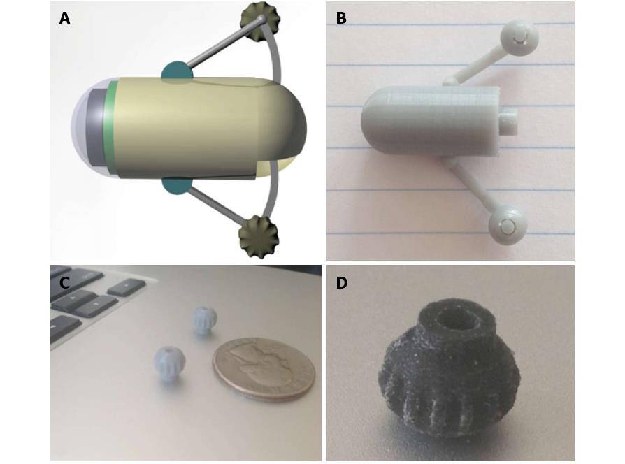
In 2010[10], we proposed a modified capsule design incorporating localisation and stabilization capabilities. The proposed design consists of a capsule fitted with protruding wheels attached to a spring-mechanism (Figure 1A). These springs allow the wheels to retract or expand to fit the lumen whilst the capsule passes through the intestine. This would then lead to more accurate location information, hence accurate lesion localisation, in relation to the onset (pyloric opening) of the investigation. Furthermore, this capsule could theoretically allow stabilization of the recorded video as any erratic, non-forward movement through the gut is minimised.
Therefore, we aim to test the feasibility of the proposed design in vitro and ex vivo. Three-dimensional (3-D) printing technology[11,12] was used to build a conceptual capsule prototype (herein called ODOcaps) (Figure 1B). Furthermore, miniature wheels were initially produced with UV Curable Acrylic Plastic (Figure 1C). They were later re-designed and made of a synthetic resilient, textile-like material, chosen for its known strength, flexibility and durability (TPU 92A-1) (Figure 1D). This material is produced via laser sintering, a process similar to stereo-lithography that uses temperature-sensitive powder (instead of UV-sensitive liquid), causing the powder to become a coherent mass without melting[13]. For the tread area of the wheels, two designs were considered: smooth or tractor-tread and tested on various types of surfaces. The tractor-tread design of the wheels was selected because it could - theoretically - achieve better traction and full rotation of the wheels when in contact with the intestinal mucosa.
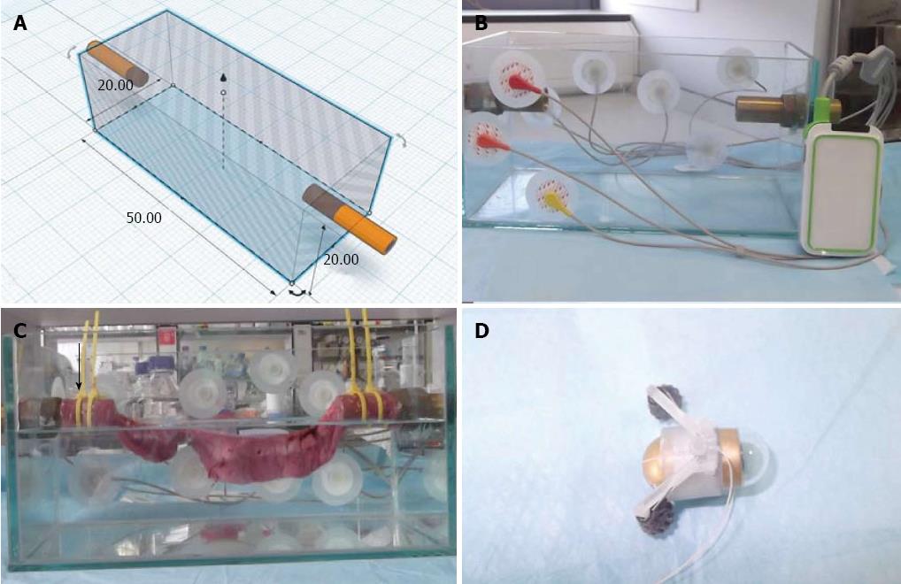
Thereafter, the wheels were inserted in 3-D printed, L-shaped miniature tubes (UV Curable Acrylic Plastic; Figure 2A) allowing almost frictionless rotation. The tubes along with the wheels where attached to a spring (stainless steel torsion spring[14] with 90o deflection and 0.093 inch pounds torque from Associated Spring Raymond) to allow extension/retraction of the wheels, Figure 2B. Thereafter, spring with wheels was clipped onto a 3-D printed ring (11.5 mm in diameter made of UV Curable Acrylic Plastic; Figure 2B and C). The ring has two (1 mm) holes, diametrically opposite to each other. The ring was designed to tightly fit on conventional CE systems (PillCam® SB2, Given® Imaging Ltd., Israel; MiroCam®, IntroMedic Ltd., South Korea) and the holes were used for the insertion of a 0.5 mm silk string.
For the in vitro experiment, a translucent polycarbonate tube (internal diameter 22 mm and 50 cm long; Figure 2D) was used. The tube characteristics mimic the diameter and slippery surface of an adult small-bowel. The assembled ring with wheels on spring was fitted on a demo PillCam® SB2 and was inserted into one end of the tube; thereafter, it was pulled through by the string (Figure 2D). While the capsule was pulled along the tube, the wheels were observed rotating constantly without any skidding effect. A video that demonstrates this experiment is available at: http://dl.dropbox.com/u/7591304/Capsule.mov.
For the ex vivo experiment; a glass tank (50 cm × 20 cm × 20 cm) with fix points for the intestine (metal tubes) and entry points for the assembled device was constructed (Figure 3A and B)[15]. A standard simulated intestinal environment (Figure 3C) was created by mounting a 32 cm long, freshly harvested porcine (Large White x Landrace 15-mo-old female sow) small-intestine to both ends of a fluid-filled tank[15]. The tank was filled with Normal Tyrode’s Solution - Base1 (NTS-1) and Base2 (NTS-2), (Dr. Lohmann Diaclean GmbH, Germany), diluted with 9l of sterile water. Thereafter, the assembled ring with wheels on spring fitted on a MiroCam®) was inserted into the suspended bowel via one of the metal tubes (Figure 3D). Consistency of wheels’ rotation was validated by utilising an all-purpose endoscope, Findoo MircoCam (dnt® GmbH, Germany). The latter was introduced from the same end as the capsule and recorded the wheels movement while following the moving capsule. A video is available at: http://dl.dropboxusercontent.com/u/7591304/1035301R.AVI
In conclusion, in vitro and ex vivo, ‘‘proof of concept’’ experimentation based on a conceptual CE design (Figure 1A and B) - that at least in theory offers enhanced localisation capabilities - showed promising preliminary results. Further elaborate experiments (i.e., at first stage, force measurements and construction of a functional prototype)[16] and in vivo experiments with this prototype are essential and currently under way.
We feel truly indebted to Dr Adrian Thompson, as without his invaluable help this work would not have been possible. We also like to help Dr Dimitrios Sigounas for his help with the ex vivo experiment.
P- Reviewers Figueiredo P, Kopacova M S- Editor Zhai HH L- Editor A E- Editor Ma S
| 1. | Koulaouzidis A, Rondonotti E, Karargyris A. Small-bowel capsule endoscopy: a ten-point contemporary review. World J Gastroenterol. 2013;19:3726-3746. [RCA] [PubMed] [DOI] [Full Text] [Full Text (PDF)] [Cited by in CrossRef: 125] [Cited by in RCA: 125] [Article Influence: 10.4] [Reference Citation Analysis (1)] |
| 2. | Khan TH, Wahid KA. Low power and low complexity compressor for video capsule endoscopy. IEEE T CIRC SYST VID. 2011;21:1534-1546. [DOI] [Full Text] |
| 3. | Than TD, Alici G, Zhou H, Li W. A review of localization systems for robotic endoscopic capsules. IEEE Trans Biomed Eng. 2012;59:2387-2399. [RCA] [PubMed] [DOI] [Full Text] [Cited by in Crossref: 178] [Cited by in RCA: 91] [Article Influence: 7.0] [Reference Citation Analysis (0)] |
| 4. | Thone J, Radiom S, Turgis D, Carta R, Gielen G, Puers R. Design of a 2 Mbps FSK near-field transmitter for wireless capsule endoscopy. Sensors and Actuators A. 2009;156:43-48. [DOI] [Full Text] |
| 5. | Gao Y, Zheng Y, Diao S, Toh WD, Ang CW, Je M, Heng CH. Low-power ultrawideband wireless telemetry transceiver for medical sensor applications. IEEE Trans Biomed Eng. 2011;58:768-772. [RCA] [PubMed] [DOI] [Full Text] [Cited by in Crossref: 109] [Cited by in RCA: 112] [Article Influence: 7.5] [Reference Citation Analysis (0)] |
| 6. | Diao S, Gao Y, Toh W, Alper C, Zheng Y, Je M, Heng CM. A low-power, high data-rate CMOS ASK transmitter for wireless capsule endoscopy. IEEE. 2011;1-4. [DOI] [Full Text] |
| 7. | Lee SH, Lee J, Yoon YJ, Park S, Cheon C, Kim K, Nam S. A wideband spiral antenna for ingestible capsule endoscope systems: experimental results in a human phantom and a pig. IEEE Trans Biomed Eng. 2011;58:1734-1741. [RCA] [PubMed] [DOI] [Full Text] [Cited by in Crossref: 107] [Cited by in RCA: 27] [Article Influence: 1.9] [Reference Citation Analysis (0)] |
| 8. | Kim K, Yun S, Lee S, Nam S, Yoon YJ, Cheon C. A design of a high-speed and high-efficiency capsule endoscopy system. IEEE Trans Biomed Eng. 2012;59:1005-1011. [RCA] [PubMed] [DOI] [Full Text] [Cited by in Crossref: 34] [Cited by in RCA: 35] [Article Influence: 2.5] [Reference Citation Analysis (0)] |
| 9. | Basar MR, Malek F, Juni KM, Shaharom Idris M, Iskandar M Saleh M. Ingestible Wireless Capsule Technology: A Review of Development and Future Indication. Int J Antenn Propag. 2012;2012:1-14. [DOI] [Full Text] |
| 10. | Bourbakis N, Giakos G, Karargyris A. Design of new-generation robotic capsules for therapeutic and diagnostic endoscopy. IEEE. 2010;1-6. [DOI] [Full Text] |
| 11. | 3D Printing service i. materialise. Available from: http: //i.materialise.com. |
| 12. | Available from: http: //www.shapeways.com/. |
| 13. | Titsch M. Materialise Launches Flexible Media TPU 92A-1. Available from: http: //www.3dprinter-world.com/article/materialise-launches-flexible-media-tpu-92a-1. |
| 14. | Titsch M; SPEC. 316 stainless steel torsion springs. Available from: http: //www.asraymond.com/documents/316 Stainless Steel Torsion Springs.pdf. |
| 15. | Glass P, Cheung E, Sitti M. A legged anchoring mechanism for capsule endoscopes using micropatterned adhesives. IEEE Trans Biomed Eng. 2008;55:2759-2767. [RCA] [PubMed] [DOI] [Full Text] [Cited by in Crossref: 126] [Cited by in RCA: 82] [Article Influence: 5.1] [Reference Citation Analysis (0)] |
| 16. | Tachecí I, Kvetina J, Bures J, Osterreicher J, Kunes M, Pejchal J, Rejchrt S, Spelda S, Kopácová M. Wireless capsule endoscopy in enteropathy induced by nonsteroidal anti-inflammatory drugs in pigs. Dig Dis Sci. 2010;55:2471-2477. [RCA] [PubMed] [DOI] [Full Text] [Cited by in Crossref: 7] [Cited by in RCA: 12] [Article Influence: 0.8] [Reference Citation Analysis (0)] |


