Published online Jan 21, 2013. doi: 10.3748/wjg.v19.i3.422
Revised: November 30, 2012
Accepted: December 15, 2012
Published online: January 21, 2013
Processing time: 132 Days and 9.3 Hours
Histiocytic sarcoma (HS) is a rare malignant neoplasm that originates from a histiocytic hematopoietic lineage characterized by histiocytic differentiation and its corresponding immunophenotypic features. We herein reported a case of primary HS of the stomach which was confirmed through histopathologic examination and immunohistochemical staining. A 52-year-old woman presented with progressive difficulty in feeding and dull pain in the epigastric region. Gastroscopy, endoscopic ultrasonography, double contrast examination, and computed tomography revealed a mass located on the posterior wall of fundus and lesser curvature of the stomach. Microscopically, the cytoplasm of the tumor cells was abundant and eosinophilic. Immunohistochemical staining revealed that the tumor cells were positive for CD45RO and CD68. It is difficult to differentiate HS of stomach from other gastric malignancies by radiological evaluation alone. However, HS may be considered when a protruding and ulcerated mass in stomach shows heterogeneous hypervascular features. To the best of our knowledge, this is the first report in English language literature that emphasizes the imaging findings of human gastric HS.
- Citation: Shen XZ, Liu F, Ni RJ, Wang BY. Primary histiocytic sarcoma of the stomach: A case report with imaging findings. World J Gastroenterol 2013; 19(3): 422-425
- URL: https://www.wjgnet.com/1007-9327/full/v19/i3/422.htm
- DOI: https://dx.doi.org/10.3748/wjg.v19.i3.422
Histiocytic sarcoma (HS) is an exceedingly rare lymphohematopoietic malignant neoplasm composed of tumor cells showing morphologic and immunophenotypic features of mature histiocytes[1,2]. Most patients are adults (median age 46 years). Male predilection is found in some studies. About one-third of cases present in lymph nodes, about one-third in skin, and about one-third in a variety of other extranodal sites, most commonly the intestinal tract[3].
HS of the stomach is extremely rare and it has rarely been described in the English literature[3,4]. The imaging findings of gastric HS are not well known. Thus, it is still indistinguishable from other gastric neoplasms both clinically and radiographically[5]. To the best of our knowledge, only ten cases of gastric HS have been reported in the international medical literature[3-8].
We report herein a case of primary HS of the stomach with an emphasis on the imaging findings of endoscopic ultrasonography (EUS), gastroscope, double contrast examination, and computed tomography (CT).
A 52-year-old woman presented with a one-month history of progressive difficulty in feeding and dull pain in the epigastric region for 3 d, without haematemesis or black dejection. At physical examination, she was slightly tender in the middle of the upper abdomen, without abdominal distension. There was no history of previous known stomach disease.
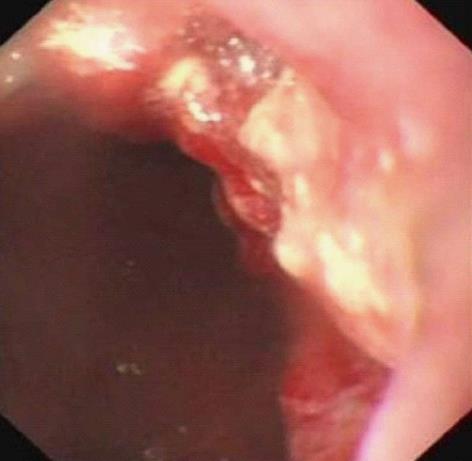
Gastroscopic examination revealed a large irregular ulcer with apophysis and erosive hemorrhage located on the posterior wall of fundus and lesser curvature of the stomach (Figure 1).
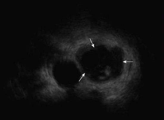
EUS detected thickening about the semi-perimeter of the gastric fundal mucosa, and a heterogenous hypoechoic mass, and the whole layer of the local gastric wall was infiltrated (Figure 2).
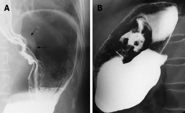
The upper digestive pneumobarium double imaging demonstrated a filling defect and a shadow of soft tissue mass around the cardia and lesser curvature of the stomach, and the abdominal part of esophagus was involved. There was destruction of the mucosa, rigidity of the gastric wall, and intracavitary niches in the lesion area (Figure 3).
Plain and contrast-enhanced CT scans of the abdomen showed a focal irregular thickened gastric wall and a poorly marginated, ulcerated soft tissue mass formation with heterogeneously obvious enhancement in the lesser gastric curvature and fundus area (Figure 4). Swelling of the adjacent regional lymph nodes was also found.
Radical total gastrectomy was performed, and a 7 cm × 6 cm irregularly protruded tumor in the gastric curvature and fundus with the distal esophagus involved was found during the operation, associated with enlargement of the 9th and 11th groups of perigastric lymph nodes.
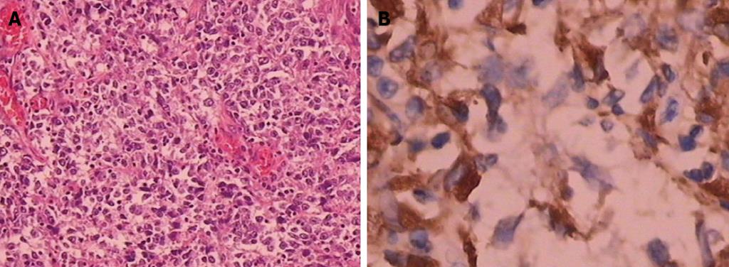
The surgical specimens were sent to histopathologic examination. The greater omentum did not show any tumor invasion, and tumor metastasis was not found in any of the 22 examined perigastric lymph nodes. The tumors showed infiltrative borders and areas of ulceration, with invasion into the serosa. The tumor cells were monomorphic to pleomorphic in shape. The cytoplasm was abundant and eosinophilic. The nuclei were large, and round to oval. Bizarre multinuclear giant cells were scattered. Immunohistochemical analysis indicated that the tumor cells expressed CD68, CD45RO, CD31 and Cyclin D1, and approximately 90% of the cells stained for Ki-67, but Bcl-2, Bcl-6, CD10, CD23, CD35, CD56, EMA, CK, Mum-1 and Pax-5 were negative (Figure 5). The histological and immunohistochemical diagnosis for the malignant tumor of the stomach was histiocytic sarcoma.
The patient received three cycles of chemotherapy after radical surgery. She has been free of recurrence or metastasis for 4 mo.
Histiocytic sarcoma, also known as true histiocytic lymphoma, is an exceedingly rare lymphohematopoietic malignant neoplasm composed of tumor cells showing morphologic and immunophenotypic features of mature histiocytes[2]. These malignant tumors most commonly present in lymph nodes or at extranodal sites, such as the gastrointestinal (GI) system, bone marrow and skin. The vast majority of GI HS are located in the intestinal tract; only few cases are found in the stomach. Hornick et al[3] and Copie-Bergman et al[4] once described 14 cases of extranodal HS and 13 cases of true histiocytic lymphomas, respectively; among these cases, a total of 12 lesions in 11 patients were located in the GI tract, but only one lesion arose in the stomach. Gastric HS can be located anywhere in the stomach: the cardia, the fundus, the antrum, the lesser curvature, or the greater curvature, but most arise in the lesser curvature and involve the adjacent area. There were ulcers in almost all lesions, and perigastric regional lymphadenopathies were also the common associated findings in the patients with gastric HS[5-8].
Sundersingh et al[7] reported a multifocal HS of the gastrointestinal tract, and a CT scan revealed multifocal, circumferential gastrointestinal wall thickening involving the stomach and jejunal loops. Gastric HS can also exist together with HS of the colon[2]. In our case, the distal esophagus had also been infiltrated. Akiba et al[9] reported a case of HS arising in the parotid gland region; the patient had recurrent lesions in the pelvis and stomach 5 mo after parotidectomy.
On CT or magnetic resonance imaging enhanced scans, extranodal HS lesions often present with notably heterogeneous contrast enhancement, because generally the neoplasm has a plentiful blood supply[10,11]. Fant et al[12] reported one case of primary gastric HS in a dog, a large, protruding and ulcerated mass was observed on tomographic and endoscopic examinations, moreover, the lesion was also apparently strengthened on enhanced CT scan. The CT manifestations in our case were exceedingly similar with those of the above case.
The imaging features of gastric HS are nonspecific. Abdominal radiographs may show a soft tissue mass with an irregular ulcer and heterogeneous hypervascular features, mostly arising in the lesser curvature and involving the adjacent area.
In conclusion, HS of the stomach is extremely rare, and, to our knowledge, its imaging features have not been previously detailed and summarized in the English-language literature in humans[5-8]. The gastric HS can be primary or secondary, but the primary tumor seems to be more prevalent, and it can also coexist with other sites of HS. It is difficult to differentiate HS of the stomach from other gastric malignancies by radiological evaluation alone. However, HS may be considered when a protruding and ulcerated mass in the stomach shows heterogeneous hypervascular features, particularly when it arises in the lesser curvature.
P- Reviewers Greenawalt DM, Okita NT, Polymeros D S- Editor Song XX L- Editor A E- Editor Li JY
| 1. | World Health Organization Classification of Tumors: Pathology and Genetics of Tumours of Haematopoietic and Lymphoid Tissue. Lyon: IARC Press 2001; 274-289. |
| 2. | Grogan TM, Pileri SA, Chan JK, Weiss LM, Fletcher CD. Histiocytic sarcoma. WHO Classification of Tumours of Haematopoeitic and Lymphoid Tissues. Lyon: IARC Press 2008; 356-357. |
| 3. | Hornick JL, Jaffe ES, Fletcher CD. Extranodal histiocytic sarcoma: clinicopathologic analysis of 14 cases of a rare epithelioid malignancy. Am J Surg Pathol. 2004;28:1133-1144. [RCA] [PubMed] [DOI] [Full Text] [Cited by in Crossref: 218] [Cited by in RCA: 213] [Article Influence: 10.1] [Reference Citation Analysis (0)] |
| 4. | Copie-Bergman C, Wotherspoon AC, Norton AJ, Diss TC, Isaacson PG. True histiocytic lymphoma: a morphologic, immunohistochemical, and molecular genetic study of 13 cases. Am J Surg Pathol. 1998;22:1386-1392. [RCA] [PubMed] [DOI] [Full Text] [Cited by in Crossref: 81] [Cited by in RCA: 61] [Article Influence: 2.3] [Reference Citation Analysis (0)] |
| 5. | Yoo KD, Han DS, Chung SM, Kim SM, Bae JH, Eun CS, Paik SS, Oh YH. A case of extranodal histiocytic sarcoma of stomach mimicking gastric adenocarcinoma. Korean J Gastroenterol. 2010;55:127-132. [RCA] [PubMed] [DOI] [Full Text] [Cited by in Crossref: 2] [Cited by in RCA: 3] [Article Influence: 0.2] [Reference Citation Analysis (0)] |
| 6. | Congyang L, Xinggui W, Hao L, Weihua H. Synchronous histiocytic sarcoma and diffuse large B cell lymphoma involving the stomach: a case report and review of the literature. Int J Hematol. 2011;93:247-252. [RCA] [PubMed] [DOI] [Full Text] [Cited by in Crossref: 13] [Cited by in RCA: 16] [Article Influence: 1.1] [Reference Citation Analysis (0)] |
| 7. | Sundersingh S, Majhi U, Seshadhri RA, Tenali GS. Multifocal histiocytic sarcoma of the gastrointestinal tract. Indian J Pathol Microbiol. 2012;55:233-235. [RCA] [PubMed] [DOI] [Full Text] [Cited by in Crossref: 9] [Cited by in RCA: 11] [Article Influence: 0.8] [Reference Citation Analysis (0)] |
| 8. | Feng T, He MX, Gu WY, Bai CG, Ma DL, Zheng JM, Zhu MH. Histiocytic sarcoma of stomach: report of a case. Zhonghua Binglixue Zazhi. 2012;41:130-131. [RCA] [PubMed] [DOI] [Full Text] [Cited by in RCA: 1] [Reference Citation Analysis (0)] |
| 9. | Akiba J, Harada H, Kawahara A, Arakawa F, Mihashi H, Mihashi R, Ohshima K, Yano H. Histiocytic sarcoma of the parotid gland region. Pathol Int. 2011;61:373-376. [RCA] [PubMed] [DOI] [Full Text] [Cited by in Crossref: 9] [Cited by in RCA: 14] [Article Influence: 1.0] [Reference Citation Analysis (0)] |
| 10. | Bell SL, Hanzely Z, Alakandy LM, Jackson R, Stewart W. Primary meningeal histiocytic sarcoma: a report of two unusual cases. Neuropathol Appl Neurobiol. 2012;38:111-114. [RCA] [PubMed] [DOI] [Full Text] [Cited by in Crossref: 15] [Cited by in RCA: 17] [Article Influence: 1.3] [Reference Citation Analysis (0)] |
| 11. | Makis W, Ciarallo A, Derbekyan V, Lisbona R. Histiocytic sarcoma involving lymph nodes: imaging appearance on gallium-67 and F-18 FDG PET/CT. Clin Nucl Med. 2011;36:e37-e38. [RCA] [PubMed] [DOI] [Full Text] [Cited by in Crossref: 8] [Cited by in RCA: 13] [Article Influence: 0.9] [Reference Citation Analysis (0)] |
| 12. | Fant P, Caldin M, Furlanello T, De Lorenzi D, Bertolini G, Bettini G, Morini M, Masserdotti C. Primary gastric histiocytic sarcoma in a dog--a case report. J Vet Med A Physiol Pathol Clin Med. 2004;51:358-362. [RCA] [PubMed] [DOI] [Full Text] [Cited by in Crossref: 18] [Cited by in RCA: 18] [Article Influence: 0.9] [Reference Citation Analysis (0)] |


