CASE REPORT
A 56-year-old Korean man was seen in the gastroenterology clinic at this hospital for management of signet ring cell carcinoma of the colon.
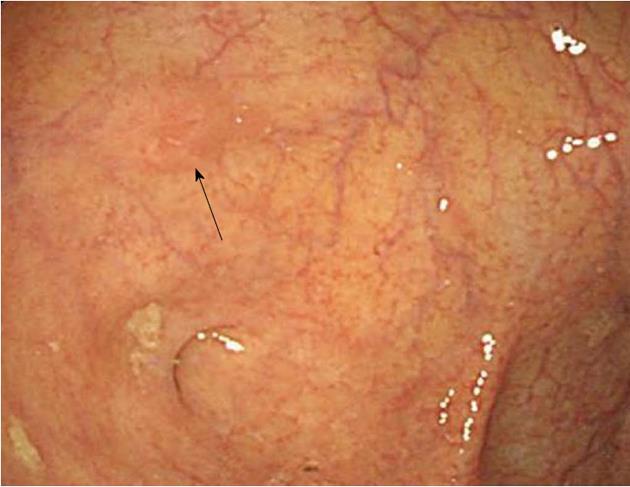
The patient had been healthy up to the time of his presentation. Ten days before his evaluation at this hospital, the patient saw a gastroenterologist at another hospital and underwent colonoscopy for a health checkup. Colonoscopic examination revealed a IIa-like, ill-defined and flatly elevated 5-mm tumor in the cecum (Figure 1). In addition, several polyps were observed in the ascending colon extending to the transverse colon. Snare polypectomy of the tumor in the cecum was performed incompletely, and a biopsy of several polyps was also conducted. Pathological examination of a biopsy specimen of the cecal tumor showed signet ring cell carcinoma, while biopsy specimens of several polyps showed tubular adenoma with low grade dysplasia. The patient was referred to the gastroenterology clinic at this hospital on October 6, 2009.
Figure 1 Initial colonoscopic findings (from another hospital).
Colonoscopic examination revealed a IIa-like, ill-defined and flatly elevated 5-mm tumor in the cecum (arrow).
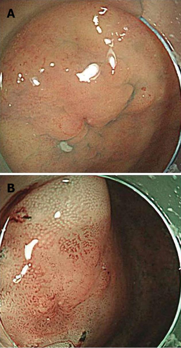
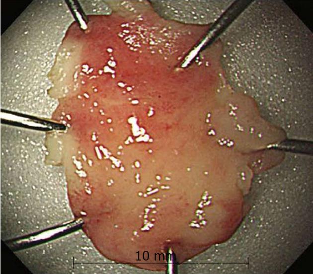
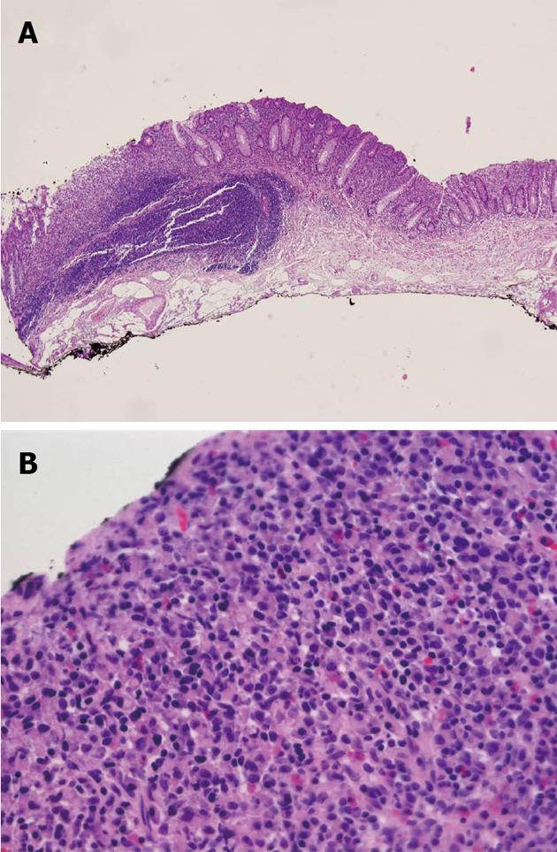
At presentation, the patient was active and was experiencing no symptoms. He took no medications and had no known allergies to medications. He drank alcohol, had a history of smoking (20 packs per-year), and did not use illicit drugs. His past history was unremarkable, and there was no family history of cancer. On examination, his body weight was 59.0 kg and height was 154 cm; the vital signs and remainder of the physical examination were normal. The level of carcinoembryonic antigen was 2.26 ng/mL (reference value, < 3.4). Results of a complete blood count, plasma levels of electrolytes, tests of coagulation and kidney and liver function, and a urinalysis were normal. One day later, upper endoscopic examination revealed no primary lesions, and colonoscopic examination revealed a IIa-like, ill-defined and flatly elevated 9-mm residual tumor in the cecum (Figure 2). In addition, several lateral spreading tumors were observed in the ascending colon extending to the transverse colon. Endoscopic mucosal resection (EMR) of the residual tumor in the cecum was performed. The tumor was successfully removed en bloc by EMR without complications. The resected specimen was 10 mm in diameter (Figure 3). Histologically, the tumor was composed of carcinoma with lymphoid hyperplasia and, favored signet ring cell carcinoma that had invaded the lamina propria without venous or perineural invasion (Figure 4). The cut end of the resected specimen was examined for safety. The other tumors were removed by polypectomy; histologically, the specimens were composed of tubular adenoma with low grade dysplasia. Abdominal computed tomography (CT) and CT scanning with positron-emission tomography (PET-CT) showed no evidence of primary lesions or distant metastasis. Two weeks later, the patient underwent an additional laparoscopic right-hemicolectomy, because of the high incidence of peritoneal metastasis associated with signet ring cell carcinoma and the possibility of recurrence of several lateral spreading tumors. The resected specimen revealed no residual carcinoma at the EMR site and showed no lymph node metastasis (Figure 5).
Figure 2 Colonoscopic findings at this hospital.
A: Colonoscopic examination revealed a IIa-like, ill-defined and flatly elevated 9-mm residual tumor in the cecum; B: Narrow band imaging shows the lesion more clearly.
Figure 3 Resected specimen by endoscopic mucosal resection.
The resected specimen was 10 mm in diameter.
Figure 4 Scanning view of the endoscopic mucosal resection site.
Histologically, the resected specimen showed diffusely infiltrated signet ring cells in the lamina propria without venous or perineural invasion. A: hematoxylin/eosin staining, × 40; B: hematoxylin/eosin staining, × 400.
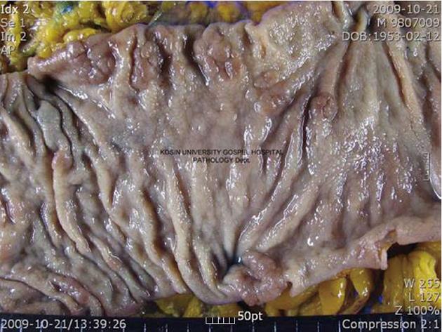
Figure 5 Gross findings.
The mucosal surface of the cecum showing an ill-defined irregularly-shaped scar (probably the endoscopic mucosal resection site). The remaining mucosa showing multiple polyps and previous polypectomy sites.
The patient has been in good health for two years and has shown no recurrence after the operation.
DISCUSSION
Primary signet ring cell carcinoma of the colon and rectum was first described by Laufman and Saphir in 1951[5]. Its characteristics include more advanced stages at presentation[1,4,6], younger age at presentation[3,4], chiefly peritoneal dissemination[1,4,7], lymphatic spread[5,6], few liver metastases[1,4,8], and poor prognosis[1,4,6-8]. Because its clinical symptoms develop late, most cases are usually detected at an advanced stage. Bonello et al[9] described three factors for this delay in diagnosis: (1) the rarity of the tumor; (2) intramucosal tumor spread with relative sparing of the mucosa, accounting for minimal symptoms and heme-negative stools; and (3) radiographic tumor resemblance to inflammatory processes. Because most cases are detected at an advanced stage, the prognosis of such tumors is dismal. Median and mean survival times are reported as 20 and 45 mo, respectively[6-8], and 5-year survival rates are between 9% and 36%[1,6,8]. Makino et al[10] found that all 17 patients with stage 0/I disease were alive at the latest follow-up evaluation, and the 5-year survival rate of patients with T2 disease was 75.0%. Therefore, to improve outcome, early diagnosis is very important. The patient in this case report was diagnosed with primary signet ring cell carcinoma of the colon at an early stage, and a thorough workup did not reveal any other sites of involvement and the tumor invaded only lamina propria without venous or perineural invasion. A review of the literature revealed that there are only 27 cases of primary signet ring cell carcinoma of the colon and rectum detected at an early stage, including the patient in this case report[11-17]. Of these 27 patients, 22 were males and 5 were females, with a mean age of 57.1 years (range: 6-79 years). The mean size of the tumor was 15.7 mm (range: 2-45 mm). Regarding the location of the tumors, 14 cases were in the right-side of the colon, 7 cases were in the left-side of the colon, and 6 cases were in the rectum. Macroscopically, 10 cases were polypoid type, 4 cases were flat type, and 13 cases were depressed type. Microscopically, 20 cases had submucosal invasion and 7 cases had intramucosal invasion. Makino et al[10] reported that all 69 patients with scirrhous and polypoid tumors had stage T3 or T4 disease, while 83.3% of patients with superficial tumors had stage T0 or T1 disease. In this case report, the patient’s tumor was located in the mucosa, and the gross shape of the tumor was IIa-like, ill-defined and flatly elevated. Thus, for early detection of signet ring cell carcinoma of the colorectum, we believe that careful observation of easily overlooked lesions such as superficial tumors, flatly elevated, or flatly depressed lesions during colonoscopic examinations is important.
Some authors have suggested an association between ulcerative colitis and signet ring cell carcinoma of the colorectum[5,18]. Ojeda et al[19] reported that 7 of 60 patients (12%) had both ulcerative colitis and primary colorectal signet ring cell carcinoma. Anthony et al[1] found that 4 of 29 patients (14%) had a previous history of inflammatory bowel disease. The diagnosis in 2 of these patients was ulcerative colitis; the other 2 patients were diagnosed with Crohn’s disease. The patient in this case report had no inflammatory bowel disease. Although the role of chronic inflammatory bowel disease in the development of this tumor is still undetermined, regular colonic examination of patients with history of chronic inflammatory bowel disease could be important.
A positive family history is a risk factor for ordinary colorectal cancer. However, in accordance with some studies, a positive family history may not be a predictive factor for signet ring cell carcinoma[2,4]. This finding could be attributed to the relatively small number of cases or could represent variability of this type of tumor. The patient in this case report had no family history of colorectal cancer.
Little is known about the early stages of signet ring cell carcinoma. It is unclear whether signet ring cell carcinoma develops from a pre-existing adenomatous polyp or as a so-called de novo carcinoma. To our knowledge, only 3 cases of an association between signet ring cell carcinoma and adenomatous polyps have been reported. Hamazaki et al[12] reported a case of a 6-year-old boy with a signet ring cell carcinoma in a polyp of the colon, Nakamura et al[14] described a case of a 4.5 cm rectal adenoma with multiple foci of signet ring cell carcinoma, and Tandon et al[20] reported a case of a sigmoid colon adenoma with a focus of signet ring cell carcinoma. The patient in this case report had several lateral spreading tumors in the ascending colon extending to the transverse colon; however, histologically, none of the specimens were composed of foci of signet ring cell carcinoma. Although controlled studies about the association of adenomas and signet ring cell carcinomas are lacking, it could be possible that progression of an adenoma to signet ring cell carcinoma is occuring.
In the present case report, we have described a rare case of primary signet ring cell carcinoma of the colon detected and treated at an early stage, and provided a review of the literature.


