Editorial Open Access
Copyright ©2012 Baishideng Publishing Group Co., Limited. All rights reserved.
World J Gastroenterol. Oct 7, 2012; 18(37): 5145-5150
Published online Oct 7, 2012. doi: 10.3748/wjg.v18.i37.5145
Cell sheet technology for regeneration of esophageal mucosa
Ryo Takagi, Masayuki Yamato, Nobuo Kanai, Daisuke Murakami, Makoto Kondo, Takaaki Ishii, Takeshi Ohki, Teruo Okano, Institute of Advanced Biomedical Engineering and Science, Tokyo Women’s Medical University, Tokyo 162-8666, Japan
Nobuo Kanai, Takeshi Ohki, Masakazu Yamamoto, Department of Surgery, Institute of Gastroenterology, Tokyo Women’s Medical University, Tokyo 162-8666, Japan
Daisuke Murakami, Makoto Kondo, Takaaki Ishii, Hideo Namiki, Graduate School of Science and Engineering, Waseda University, Tokyo 162-8480, Japan
Author contributions: Yamato M, Ohki T, Namiki H, Yamamoto M and Okano T designed this research; Takagi R, Kanai N, Murakami D, Kondo M and Ishii T fabricated cell sheets; Kanai N and Ohki T performed endoscopic surgery; and Takagi R and Yamato M wrote this manuscript.
Supported by The Formation of Innovation Center for Fusion of Advanced Technologies in the Special Coordination Funds for Promoting Science and Technology from the Ministry of Education, Culture, Sports, Science, and Technology, Japan
Correspondence to: Teruo Okano, PhD, Institute of Advanced Biomedical Engineering and Science, Tokyo Women’s Medical University, TWIns, 8-1 Kawada-cho, Shinjuku-ku, Tokyo 162-8666, Japan. tokano@abmes.twmu.ac.jp
Telephone: +81-3-53679945 Fax: +81-3-33596046
Received: December 24, 2011
Revised: April 18, 2012
Accepted: April 27, 2012
Published online: October 7, 2012

Abstract

The progress of tissue-engineering technology has realized development of new therapies to treat various disorders by using cultured cells. Cell- and tissue-based therapies have been successfully applied to human patients, and several tissue-engineered products have been approved by the regulatory agencies and are commercially available. In the review article, we describe our experience of development and clinical application of cell sheet-based regenerative medicine. Endoscopic mucosal resection (EMR) and endoscopic submucosal dissection (ESD) have been shown to be useful for removal of gastrointestinal neoplasms with less invasiveness compared with open surgery, especially in esophageal surgery. However, postoperative inflammation and stenosis are major complications observed after intensive mucosal resection. Therefore, we have developed novel regenerative medicine to prevent such complications and promote wound healing of esophageal mucosa after EMR or ESD. Transplantable oral mucosal epithelial cell sheets were fabricated from patients’ own oral mucosa. Immediately after EMR or ESD, fabricated autologous cell sheets were endoscopically transplanted to the ulcer sites. We performed a preclinical study with a canine model. In human clinical settings, cell culture and cell sheet fabrication were performed in clean rooms according to good manufacturing practice guidelines, and pharmaceutical drugs were used as supplements to culture medium in place of research regents used in animal study. We believe that cell-based regenerative medicine would be useful to improve quality of life of patients after EMR or ESD.

Key Words: Cell sheet; Endoscopic resection; Esophageal stenosis; Oral mucosa; Good manufacturing practice


INTRODUCTION

Endoscopic mucosal resection (EMR) is one of the standard techniques for removing superficial early-stage cancers in the gastrointestinal tract with low invasiveness compared with open surgery[1,2]. In the case of the esophagus, EMR is a useful operation for removing m1 (epithelial cancers restricted to the intraepithelial layer) and m2 (mucosal cancers with invasion of intermediate depth) carcinomas without esophageal reconstruction. To permit large en bloc resection of cancerous lesions without technical restriction of EMR, endoscopic submucosal dissection (ESD) was developed as a new mucosal resection technique with development of endoscopic devices[3-5]. Early-stage esophageal cancers are removed using a hook-knife, which is an endoscopic device used to perform ESD[6]. ESD is a more impressive operative procedure than EMR for reducing the recurrence of esophageal squamous cell carcinoma[7]. Although these developments of endoscopic surgery contribute low invasive cancer resection for patients suffering from esophageal cancer, there are some postoperative complications after ESD. Esophageal stenosis is a major complication caused by endoscopic resection, and the stenosis is significantly associated with the mucosal defect involving over three-fourths circumference of the esophagus lumen[8]. The esophageal stenosis caused by aggressive ESD considerably affects the patient’s quality of life, since the patient has to receive treatment with balloon dilation or temporary stents to expand the esophageal stricture with further inflammation and postoperative pain. These physical dilations carry a risk of perforation[9]. Treatment with anti-inflammatory drugs after endoscopic resection may be an effective therapy for preventing stricture after ESD[10,11].

With recent progression of tissue engineering and regenerative medicine, there are some reports proposing new technologies using biological scaffolds[12] or cell suspensions[13,14] for preventing the esophageal stenosis caused by mucosal defects. We have developed a novel method of endoscopic transplantation of autologous epithelial cell sheets immediately after ESD to prevent the postoperative complications[15]. Transplantable tissue-like epithelial cell grafts are fabricated by cell sheet technology. On the basis of results obtained with canine and porcine models, we have used this technology with human patients since 2008.

CELL SHEET TECHNOLOGY FOR REGENERATION OF ESOPHAGEAL MUCOSA
Tissue engineering by using cell sheet technology

The concept of tissue engineering was originally proposed by Langer et al[16]. Conventionally, biodegradable polymer scaffolds have been used to reconstruct tissue architecture, and cells are seeded on them. The technique should be useful to reconstruct bone and cartilage, having a large amount of extracellular matrices (ECM) and few cells. However, scaffold-based tissue engineering would not be optimal for the regeneration of parenchymal tissues filled with a huge amount of cells and faint ECM. Therefore, we have proposed an alternative method of tissue reconstruction by using transplantable cell sheets to eliminate biodegradable scaffolds.

In order to fabricate transplantable cell sheets without any scaffolds, we employ temperature-responsive culture surfaces, onto which poly (N-isopropylacrylamide) is covalently immobilized to control cell adhesion/detachment with a simple temperature change[17]. Cells adhere, spread, and proliferate on temperature-responsive surfaces at 37 °C, which is the normal temperature for mammalian cell culture. By reducing temperature below 32 °C, cells spontaneously detach from the surfaces without proteolytic enzyme such as trypsin, since the grafted polymer becomes hydrophilic. When the temperature is reduced after cells reach confluence, all the cells are harvested as a single contiguous cell sheet. Because this technique eliminates trypsin for cell harvest, all the cell membrane proteins including growth factor receptors, ion channels and cell-to-cell junction proteins are intact even after cell and cell sheet harvest. Furthermore, ECM deposited during cell culture is retained under cell sheets[18]. Therefore, cell sheets easily integrate to transplanted sites. With this technique, many types of epithelial cell sheets are fabricated and subjected to regenerative medicine of skin[19], cornea[20], urinary bladder[21], and trachea[22].

Cultured epithelial cells for clinical application

Cultured autologous epidermal keratinocytes have been used as cell grafts to treat burns as the first therapy using cultured cells[23]. A transplantable epidermal cell graft is fabricated using murine 3T3 feeder layer cells[24]. Human keratinocytes co-cultured with 3T3 feeder cells proliferate and show stratification as in vivo with characteristics of native epithelial tissues including tonofilaments and desmosomes. Interestingly, human epidermal keratinocytes can be serially cultured to more than 150th passages in vitro[25]. The cultivation of human keratinocytes achieved clinical treatments for severe burns[26] and giant congenital nevi[27] by transplantation of cultured autologous keratinocyte grafts. Moreover, allogeneic keratinocyte grafts are also used for skin ulcers[28]. In these cases, keratinocyte sheets are harvested with bacteria-derived dispase treatment.

Oral mucosal epithelium is also stratified squamous epithelium, and on the basis of the feeder layer method, the epithelial cells have been used as a cell source to fabricate transplantable epithelial cell grafts for treating oral mucosa[29-31]. Moreover, since extraction of oral mucosal tissue is easy, cultured human oral mucosal epithelial cells are selected as useful cell grafts for ectopic transplantation, such as skin[30,32]. We have successfully applied cultured autologous oral mucosal epithelial cell sheets fabricated on temperature-responsive culture inserts to treat human patient cornea suffering from limbal epithelial stem cell deficiency[33,34]. Furthermore, we utilized cultured autologous oral mucosal epithelial cell sheets for bladder reconstruction by the transplantation onto a demucosalized gastric flap in a canine model[35]. We believe that cultured oral mucosal epithelial cell sheets fabricated on temperature-responsive surfaces are useful for treatment of many types of epithelial diseases as well as reconstruction of defective tissues.

Fabrication of transplantable oral mucosal epithelial cell sheets in the clinical setting

To promote healing of esophageal ulcers after ESD for the prevention of postoperative complications, we performed endoscopic transplantation of cultured autologous oral mucosal epithelial cell sheets in the clinical setting. From the safety view point, animal-derived materials, such as bovine serum and 3T3 feeder cells should be eliminated as much as possible. For example, human embryonic stem cells cultured under a typical culture condition using animal-derived serum replacements and mouse feeder layer express an immunogenic nonhuman sialic acid[36]. Moreover, cells co-cultured with a mouse feeder layer are classified as xenogeneic products by United States Food and Drug Administration.

We reported that oral mucosal epithelial cells make transplantable stratified cell sheets on temperature-responsive cell culture inserts having micropores to supply culture medium from the basal cell surfaces even without a 3T3 feeder layer and allogeneic serum[37]. The method can, in principle, permit the cultured human oral mucosal epithelial cell sheets to be used as an autologous epithelial cell sheet graft for human patients[38]. Therefore, the autologous epithelial cell sheets are not, in principle, rejected after transplantation onto the esophageal ulcer and may be replaced by native epithelium in the esophagus. Moreover, the culture medium for the preparation of cultured oral mucosal epithelial cell sheets is modified to exclude laboratory reagents as much as possible for the clinical use[39].

Transplantable epithelial cell sheets are fabricated in a cell-processing center (CPC) with clean rooms according to the good manufacturing practice guideline in the clinical study. The cleanliness of the CPC is controlled by air conditioning systems using high efficiency particulate air filters, and standard operation procedures (SOP) are documented to keep a sterile environment of cell culture rooms in the CPC. The environment of the CPC is continuously monitored with various monitors including aerosol particles, temperature, and humidity, and various sterilization tests are performed to validate the cleanliness of the environment. In these tests culture rooms in the CPC were successfully kept sterile[40,41]. SOP of the culture method was also documented for the prevention of human error of operators to fabricate cell sheets highly reproducibly. Consequently, transplantable human epithelial cell sheets were successfully fabricated in a clinical study to treat ten human patients, and validation tests to demonstrate the quality assurance were performed one day before the ESD and transplantation.

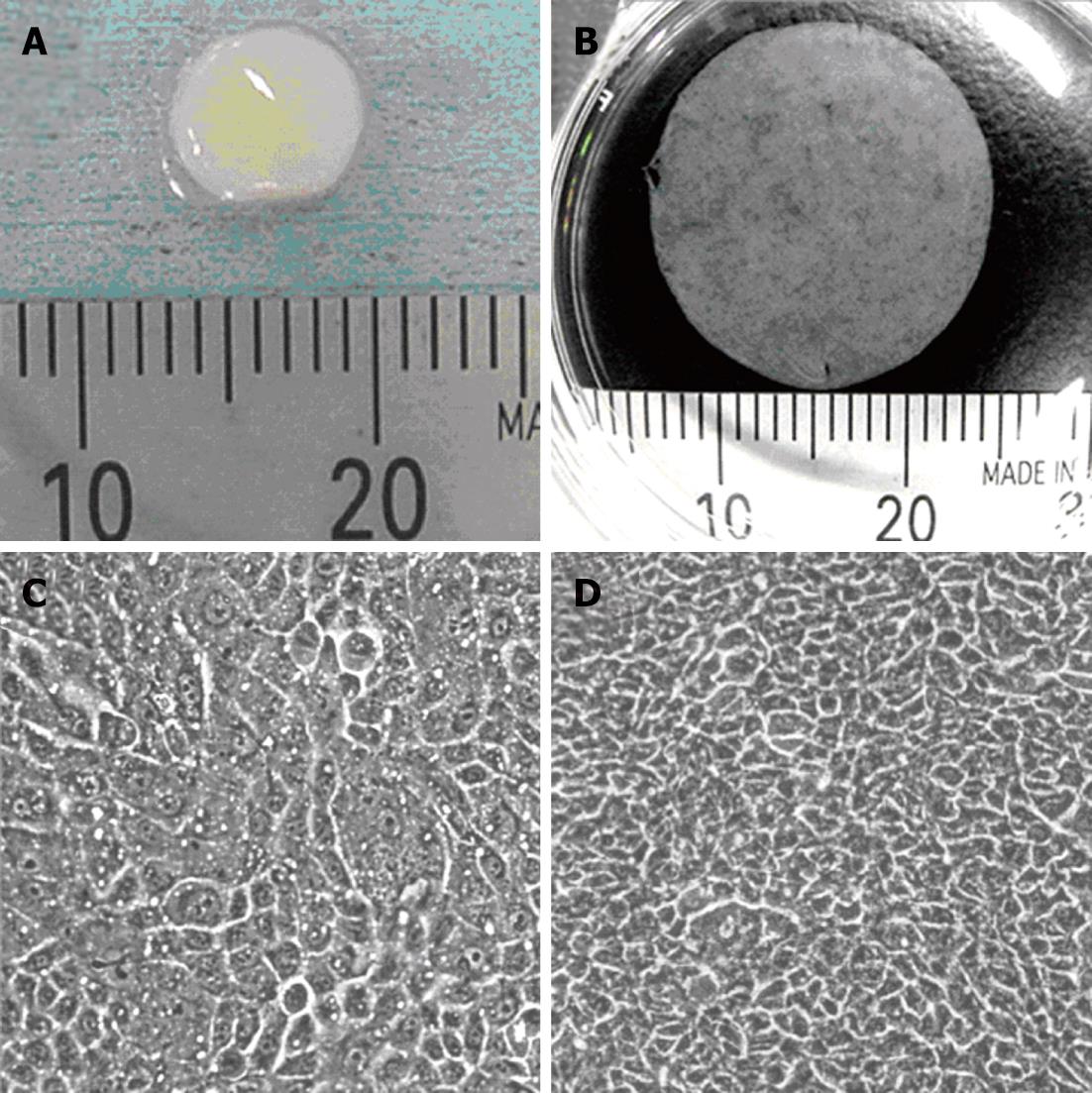
From the average yield of the cells isolated from excised human oral mucosal tissues of healthy volunteer donors, we concluded that approximately 0.3 cm2 of oral mucosal tissue is needed to fabricate one transplantable epithelial cell sheet having an area of 2.8 cm2 (Figure 1A and B). The degree of cell stratification is an important point to enable reproducible cell sheet harvesting from temperature-responsive culture inserts. Figure 1C and D show the growth of seeded human oral mucosal epithelial cells. These cells became confluent on the surface of temperature-responsive culture inserts, and elevation of cell density accompanied with cell stratification was observed after a further 5 d.

Figure 1
Figure 1 Transplantable oral mucosal epithelial cell sheet. A: Swine buccal oral mucosal tissue taken by punch biopsy; B: Human oral mucosal epithelial cell sheet harvested from temperature-responsive cell culture insert; C: Cultured human oral mucosal epithelial cells just become confluent on the surface of temperature responsive culture insert; D: The oral mucosal epithelial cells cultured for more than 5 d.
Endoscopic transplantation of epithelial cell sheet onto esophageal ulcer immediately after ESD

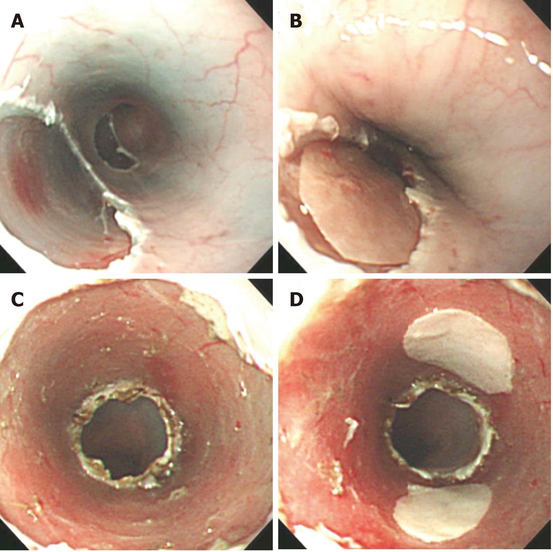
Since 2008, we have performed a clinical study using cultured autologous human oral mucosal epithelial cell sheets to treat esophageal ulcer after endoscopic resection of a mucosal neoplasm. Endoscopic transplantation of the cultured epithelial cell sheets is performed using a support membrane to grasp by endoscopic forceps[42]. Epithelial cell sheets transplanted onto esophageal ulcer with a support membrane in a swine model are shown in Figure 2. One epithelial cell sheet fabricated on temperature-responsive culture insert (23 mm in diameter) covers the ulcer surface involving approximately one-third circumference of the esophagus lumen (Figure 2A and B), and three or four epithelial cell sheets were needed to cover the full circumferential ulcer (Figure 2C and D).

Figure 2
Figure 2 Transplantation of oral mucosal epithelial cell sheets on esophageal ulcer surface in animal model. A: Swine esophageal ulcer after endoscopic mucosal resection (EMR); B: Swine oral mucosal epithelial cell sheet transplanted on esophageal ulcer after EMR; C: Swine esophageal ulcer after endoscopic submucosal dissection (ESD); D: Two swine oral mucosal epithelial cell sheets transplanted on esophageal ulcer after ESD.

Although more research is needed for understanding the mechanism and correlation between ulcer size and number of transplanted epithelial cell sheets for preventing inflammation and postoperative stenosis, serial cultivation is a useful method of preparation of epithelial cells from small tissue for fabricating cell grafts to transplant onto a large ulcer after aggressive ESD. Serial culture using low calcium concentration medium (LCM) is a useful method to amplify a cell number of stratified squamous epithelial cells without using 3T3 feeder cells[43,44]. Interestingly, although normal human epidermal keratinocytes cultured in LCM show some mesenchymal-like phenotype, the keratinocytes can express the differentiation marker and stratify in differentiation-inducing culture conditions[45]. Since the conventional culture medium for serial cultivation of epithelial cells typically includes animal-derived material, the medium is not useful for clinical use. The modification of the LCM would be important for fabricating epithelial cell sheets to transplant onto large esophageal ulcers after aggressive ESD without any complications.

CONCLUSION

This article introduces cell sheet technology to fabricate stratified epithelial cell sheet grafts for preventing postoperative inflammation and stenosis after endoscopic resection for removing early-stage cancers from the esophagus. The efficacy of the oral mucosal epithelial cell sheet has been demonstrated, and the method for fabricating the cell sheet has also developed to use the cell graft as a clinical application for treating esophageal ulcers. This new therapy has been developed by integration of endoscopic technology and cell sheet technology. We believe that cell sheet technology will meet new technology and then the integration will create new therapies for treating patients without any specific therapies.

ACKNOWLEDGMENTS

The author thanks Mr. Mizutani M, Mr. Ichikura S, Mr. Sugiyama H, Ms. Nohmi Y, Izawa Y and Oguri S (CellSeed Inc., Tokyo, Japan) for their useful comments and technical criticism.

Footnotes

Peer reviewers: Dr. Noriko Nakajima, School of Medicine, Nihon University, 1-8-13 Kandasurugadai, Tokyo 1018309, Japan; Dr. Marco Scarpa, Oncological Surgery Unit, Via Gattamelata 64, 35128 Padova, Italy

S- Editor Gou SX L- Editor O’Neill M E- Editor Li JY

References
1.  Soetikno RM, Gotoda T, Nakanishi Y, Soehendra N. Endoscopic mucosal resection. Gastrointest Endosc. 2003;57:567-579.  [PubMed]  [DOI]  [Cited in This Article: ]
2.  Soetikno R, Kaltenbach T, Yeh R, Gotoda T. Endoscopic mucosal resection for early cancers of the upper gastrointestinal tract. J Clin Oncol. 2005;23:4490-4498.  [PubMed]  [DOI]  [Cited in This Article: ]
3.  Hirao M, Masuda K, Asanuma T, Naka H, Noda K, Matsuura K, Yamaguchi O, Ueda N. Endoscopic resection of early gastric cancer and other tumors with local injection of hypertonic saline-epinephrine. Gastrointest Endosc. 1988;34:264-269.  [PubMed]  [DOI]  [Cited in This Article: ]
4.  Gotoda T, Kondo H, Ono H, Saito Y, Yamaguchi H, Saito D, Yokota T. A new endoscopic mucosal resection procedure using an insulation-tipped electrosurgical knife for rectal flat lesions: report of two cases. Gastrointest Endosc. 1999;50:560-563.  [PubMed]  [DOI]  [Cited in This Article: ]
5.  Gotoda T, Yamamoto H, Soetikno RM. Endoscopic submucosal dissection of early gastric cancer. J Gastroenterol. 2006;41:929-942.  [PubMed]  [DOI]  [Cited in This Article: ]
6.  Oyama T, Tomori A, Hotta K, Morita S, Kominato K, Tanaka M, Miyata Y. Endoscopic submucosal dissection of early esophageal cancer. Clin Gastroenterol Hepatol. 2005;3:S67-S70.  [PubMed]  [DOI]  [Cited in This Article: ]
7.  Ishihara R, Iishi H, Takeuchi Y, Kato M, Yamamoto S, Yamamoto S, Masuda E, Tatsumi K, Higashino K, Uedo N. Local recurrence of large squamous-cell carcinoma of the esophagus after endoscopic resection. Gastrointest Endosc. 2008;67:799-804.  [PubMed]  [DOI]  [Cited in This Article: ]
8.  Katada C, Muto M, Manabe T, Boku N, Ohtsu A, Yoshida S. Esophageal stenosis after endoscopic mucosal resection of superficial esophageal lesions. Gastrointest Endosc. 2003;57:165-169.  [PubMed]  [DOI]  [Cited in This Article: ]
9.  Takahashi H, Arimura Y, Okahara S, Uchida S, Ishigaki S, Tsukagoshi H, Shinomura Y, Hosokawa M. Risk of perforation during dilation for esophageal strictures after endoscopic resection in patients with early squamous cell carcinoma. Endoscopy. 2011;43:184-189.  [PubMed]  [DOI]  [Cited in This Article: ]
10.  Ono S, Fujishiro M, Kodashima S, Minatsuki C, Hirano K, Niimi K, Goto O, Yamamichi N, Fukuda T, Seto Y. High-dose dexamethasone may prevent esophageal stricture after endoscopic submucosal dissection. Clin J Gastroenterol. 2010;3:155-158.  [PubMed]  [DOI]  [Cited in This Article: ]
11.  Yamaguchi N, Isomoto H, Shikuwa S, Nakayama T, Hayashi T, Ohnita K, Takeshima F, Kohno S, Nakao K. Effect of oral prednisolone on esophageal stricture after complete circular endoscopic submucosal dissection for superficial esophageal squamous cell carcinoma: a case report. Digestion. 2011;83:291-295.  [PubMed]  [DOI]  [Cited in This Article: ]
12.  Badylak SF, Vorp DA, Spievack AR, Simmons-Byrd A, Hanke J, Freytes DO, Thapa A, Gilbert TW, Nieponice A. Esophageal reconstruction with ECM and muscle tissue in a dog model. J Surg Res. 2005;128:87-97.  [PubMed]  [DOI]  [Cited in This Article: ]
13.  Sakurai T, Miyazaki S, Miyata G, Satomi S, Hori Y. Autologous buccal keratinocyte implantation for the prevention of stenosis after EMR of the esophagus. Gastrointest Endosc. 2007;66:167-173.  [PubMed]  [DOI]  [Cited in This Article: ]
14.  Honda M, Hori Y, Nakada A, Uji M, Nishizawa Y, Yamamoto K, Kobayashi T, Shimada H, Kida N, Sato T. Use of adipose tissue-derived stromal cells for prevention of esophageal stricture after circumferential EMR in a canine model. Gastrointest Endosc. 2011;73:777-784.  [PubMed]  [DOI]  [Cited in This Article: ]
15.  Ohki T, Yamato M, Murakami D, Takagi R, Yang J, Namiki H, Okano T, Takasaki K. Treatment of oesophageal ulcerations using endoscopic transplantation of tissue-engineered autologous oral mucosal epithelial cell sheets in a canine model. Gut. 2006;55:1704-1710.  [PubMed]  [DOI]  [Cited in This Article: ]
16.  Langer R, Vacanti JP. Tissue engineering. Science. 1993;260:920-926.  [PubMed]  [DOI]  [Cited in This Article: ]
17.  Okano T, Yamada N, Sakai H, Sakurai Y. A novel recovery system for cultured cells using plasma-treated polystyrene dishes grafted with poly(N-isopropylacrylamide). J Biomed Mater Res. 1993;27:1243-1251.  [PubMed]  [DOI]  [Cited in This Article: ]
18.  Kushida A, Yamato M, Konno C, Kikuchi A, Sakurai Y, Okano T. Decrease in culture temperature releases monolayer endothelial cell sheets together with deposited fibronectin matrix from temperature-responsive culture surfaces. J Biomed Mater Res. 1999;45:355-362.  [PubMed]  [DOI]  [Cited in This Article: ]
19.  Yamato M, Utsumi M, Kushida A, Konno C, Kikuchi A, Okano T. Thermo-responsive culture dishes allow the intact harvest of multilayered keratinocyte sheets without dispase by reducing temperature. Tissue Eng. 2001;7:473-480.  [PubMed]  [DOI]  [Cited in This Article: ]
20.  Nishida K, Yamato M, Hayashida Y, Watanabe K, Maeda N, Watanabe H, Yamamoto K, Nagai S, Kikuchi A, Tano Y. Functional bioengineered corneal epithelial sheet grafts from corneal stem cells expanded ex vivo on a temperature-responsive cell culture surface. Transplantation. 2004;77:379-385.  [PubMed]  [DOI]  [Cited in This Article: ]
21.  Shiroyanagi Y, Yamato M, Yamazaki Y, Toma H, Okano T. Urothelium regeneration using viable cultured urothelial cell sheets grafted on demucosalized gastric flaps. BJU Int. 2004;93:1069-1075.  [PubMed]  [DOI]  [Cited in This Article: ]
22.  Kanzaki M, Yamato M, Hatakeyama H, Kohno C, Yang J, Umemoto T, Kikuchi A, Okano T, Onuki T. Tissue engineered epithelial cell sheets for the creation of a bioartificial trachea. Tissue Eng. 2006;12:1275-1283.  [PubMed]  [DOI]  [Cited in This Article: ]
23.  O’Connor NE, Mulliken JB, Banks-Schlegel S, Kehinde O, Green H. Grafting of burns with cultured epithelium prepared from autologous epidermal cells. Lancet. 1981;1:75-78.  [PubMed]  [DOI]  [Cited in This Article: ]
24.  Rheinwald JG, Green H. Serial cultivation of strains of human epidermal keratinocytes: the formation of keratinizing colonies from single cells. Cell. 1975;6:331-343.  [PubMed]  [DOI]  [Cited in This Article: ]
25.  Mathor MB, Ferrari G, Dellambra E, Cilli M, Mavilio F, Cancedda R, De Luca M. Clonal analysis of stably transduced human epidermal stem cells in culture. Proc Natl Acad Sci USA. 1996;93:10371-10376.  [PubMed]  [DOI]  [Cited in This Article: ]
26.  Gallico GG, O’Connor NE, Compton CC, Kehinde O, Green H. Permanent coverage of large burn wounds with autologous cultured human epithelium. N Engl J Med. 1984;311:448-451.  [PubMed]  [DOI]  [Cited in This Article: ]
27.  Gallico GG, O’Connor NE, Compton CC, Remensnyder JP, Kehinde O, Green H. Cultured epithelial autografts for giant congenital nevi. Plast Reconstr Surg. 1989;84:1-9.  [PubMed]  [DOI]  [Cited in This Article: ]
28.  Phillips TJ, Kehinde O, Green H, Gilchrest BA. Treatment of skin ulcers with cultured epidermal allografts. J Am Acad Dermatol. 1989;21:191-199.  [PubMed]  [DOI]  [Cited in This Article: ]
29.  de Luca M, Albanese E, Megna M, Cancedda R, Mangiante PE, Cadoni A, Franzi AT. Evidence that human oral epithelium reconstituted in vitro and transplanted onto patients with defects in the oral mucosa retains properties of the original donor site. Transplantation. 1990;50:454-459.  [PubMed]  [DOI]  [Cited in This Article: ]
30.  Hata K, Ueda M. Fabrication of cultured epithelium using oral mucosal cells and its clinical applications. Hum Cell. 1996;9:91-96.  [PubMed]  [DOI]  [Cited in This Article: ]
31.  Tsai CY, Ueda M, Hata K, Horie K, Hibino Y, Sugimura Y, Toriyama K, Torii S. Clinical results of cultured epithelial cell grafting in the oral and maxillofacial region. J Craniomaxillofac Surg. 1997;25:4-8.  [PubMed]  [DOI]  [Cited in This Article: ]
32.  Ueda M, Hata K, Horie K, Torii S. The potential of oral mucosal cells for cultured epithelium: a preliminary report. Ann Plast Surg. 1995;35:498-504.  [PubMed]  [DOI]  [Cited in This Article: ]
33.  Pellegrini G. Changing the cell source in cell therapy? N Engl J Med. 2004;351:1170-1172.  [PubMed]  [DOI]  [Cited in This Article: ]
34.  Nishida K, Yamato M, Hayashida Y, Watanabe K, Yamamoto K, Adachi E, Nagai S, Kikuchi A, Maeda N, Watanabe H. Corneal reconstruction with tissue-engineered cell sheets composed of autologous oral mucosal epithelium. N Engl J Med. 2004;351:1187-1196.  [PubMed]  [DOI]  [Cited in This Article: ]
35.  Watanabe E, Yamato M, Shiroyanagi Y, Tanabe K, Okano T. Bladder augmentation using tissue-engineered autologous oral mucosal epithelial cell sheets grafted on demucosalized gastric flaps. Transplantation. 2011;91:700-706.  [PubMed]  [DOI]  [Cited in This Article: ]
36.  Martin MJ, Muotri A, Gage F, Varki A. Human embryonic stem cells express an immunogenic nonhuman sialic acid. Nat Med. 2005;11:228-232.  [PubMed]  [DOI]  [Cited in This Article: ]
37.  Murakami D, Yamato M, Nishida K, Ohki T, Takagi R, Yang J, Namiki H, Okano T. The effect of micropores in the surface of temperature-responsive culture inserts on the fabrication of transplantable canine oral mucosal epithelial cell sheets. Biomaterials. 2006;27:5518-5523.  [PubMed]  [DOI]  [Cited in This Article: ]
38.  Murakami D, Yamato M, Nishida K, Ohki T, Takagi R, Yang J, Namiki H, Okano T. Fabrication of transplantable human oral mucosal epithelial cell sheets using temperature-responsive culture inserts without feeder layer cells. J Artif Organs. 2006;9:185-191.  [PubMed]  [DOI]  [Cited in This Article: ]
39.  Takagi R, Yamato M, Murakami D, Kondo M, Yang J, Ohki T, Nishida K, Kohno C, Okano T. Preparation of keratinocyte culture medium for the clinical applications of regenerative medicine. J Tissue Eng Regen Med. 2011;5:e63-e73.  [PubMed]  [DOI]  [Cited in This Article: ]
40.  Takagi R, Murakami D, Kondo M, Ohki T, Sasaki R, Mizutani M, Yamato M, Nishida K, Namiki H, Yamamoto M. Fabrication of human oral mucosal epithelial cell sheets for treatment of esophageal ulceration by endoscopic submucosal dissection. Gastrointest Endosc. 2010;72:1253-1259.  [PubMed]  [DOI]  [Cited in This Article: ]
41.  Takagi R, Yamato M, Murakami D, Kondo M, Ohki T, Sasaki R, Nishida K, Namiki H, Yamamoto M, Okano T. Fabrication and validation of autologous human oral mucosal epithelial cell sheets to prevent stenosis after esophageal endoscopic submucosal dissection. Pathobiology. 2011;78:311-319.  [PubMed]  [DOI]  [Cited in This Article: ]
42.  Ohki T, Yamato M, Ota M, Okano T, Yamamoto M. Application of cell sheet technology for esophageal endoscopic submucosal dissection. Techniques in Gastrointestinal Endoscopy. 2011;13:105-109.  [PubMed]  [DOI]  [Cited in This Article: ]
43.  Hennings H, Michael D, Cheng C, Steinert P, Holbrook K, Yuspa SH. Calcium regulation of growth and differentiation of mouse epidermal cells in culture. Cell. 1980;19:245-254.  [PubMed]  [DOI]  [Cited in This Article: ]
44.  Boyce ST, Ham RG. Calcium-regulated differentiation of normal human epidermal keratinocytes in chemically defined clonal culture and serum-free serial culture. J Invest Dermatol. 1983;81:33s-40s.  [PubMed]  [DOI]  [Cited in This Article: ]
45.  Takagi R, Yamato M, Murakami D, Sugiyama H, Okano T. Low calcium culture condition induces mesenchymal cell-like phenotype in normal human epidermal keratinocytes. Biochem Biophys Res Commun. 2011;412:226-231.  [PubMed]  [DOI]  [Cited in This Article: ]