Copyright
©2011 Baishideng Publishing Group Co.
World J Gastroenterol. May 21, 2011; 17(19): 2372-2378
Published online May 21, 2011. doi: 10.3748/wjg.v17.i19.2372
Published online May 21, 2011. doi: 10.3748/wjg.v17.i19.2372
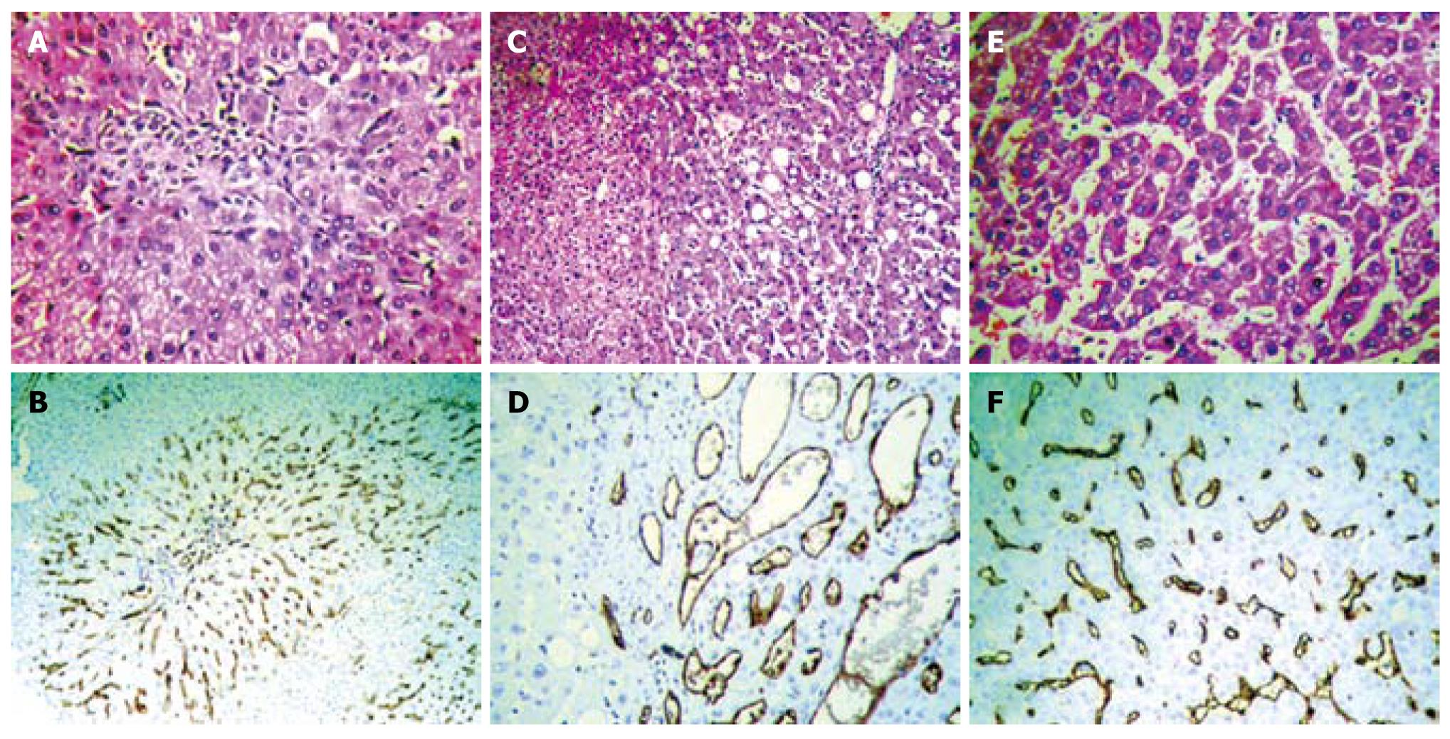
Figure 1 Atypical focal nodular hyperplasia with minimal fibrous septa (A, HE stain, × 200) shows focal microvessels around the periphery of the fibrous septa (B, CD34 immunostaining, × 200).
Hepatocellular adenoma is composed of benign-looking hepatocytes with mild steatosis, without a capsule around the periphery (C, HE stain, × 200), and shows a chaotic microvessel distribution pattern with thin-walled vascular staining (D, CD34 immunostaining, × 200). Highly differentiated hepatocellular carcinoma is arranged in a thin trabecular pattern (E, HE stain, × 400) and shows a sinusoidal capillarization pattern (F, CD34 immunostaining, × 200).
- Citation: Cong WM, Dong H, Tan L, Sun XX, Wu MC. Surgicopathological classification of hepatic space-occupying lesions: A single-center experience with literature review. World J Gastroenterol 2011; 17(19): 2372-2378
- URL: https://www.wjgnet.com/1007-9327/full/v17/i19/2372.htm
- DOI: https://dx.doi.org/10.3748/wjg.v17.i19.2372


