Editorial Open Access
Copyright ©2010 Baishideng Publishing Group Co., Limited. All rights reserved.
World J Gastroenterol. Nov 7, 2010; 16(41): 5148-5161
Published online Nov 7, 2010. doi: 10.3748/wjg.v16.i41.5148
Growth factor- and cytokine-driven pathways governing liver stemness and differentiation
Aránzazu Sánchez, School of Pharmacy, Complutense University, 28040 Madrid, Spain
Isabel Fabregat, Bellvitge Biomedical Research Institute (IDIBELL) and University of Barcelona, Molecular Oncology Laboratory, Hospital Duran i Reynals. Gran Via de Hospitalet, 199, L’Hospitalet, 08907 Barcelona, Spain
Author contributions: Both authors have equally contributed to the elaboration of this manuscript.
Supported by Grants from the Ministerio de Ciencia e Innovación, MICINN, Spain (SAF2009-12477 to Sánchez A; BFU2009-07219 and ISCIII-RTICC RD06/0020 to Fabregat I), AGAUR-Generalitat de Catalunya (2009SGR-312 to Fabregat I) and UCM-BSCH (920359 to Sánchez A)
Correspondence to: Dr. Isabel Fabregat, PhD, Bellvitge Biomedical Research Institute (IDIBELL) and University of Barcelona, Molecular Oncology Laboratory, Hospital Duran i Reynals. Gran Via de Hospitalet, 199, L’Hospitalet, 08907 Barcelona, Spain. ifabregat@idibell.cat
Telephone: +34-93-2607828 Fax: +34-93-2607426
Received: June 9, 2010
Revised: July 21, 2010
Accepted: July 28, 2010
Published online: November 7, 2010

Abstract

Liver is unique in its capacity to regenerate in response to injury or tissue loss. Hepatocytes and other liver cells are able to proliferate and repopulate the liver. However, when this response is impaired, the contribution of hepatic progenitors becomes very relevant. Here, we present an update of recent studies on growth factors and cytokine-driven intracellular pathways that govern liver stem/progenitor cell expansion and differentiation, and the relevance of these signals in liver development, regeneration and carcinogenesis. Tyrosine kinase receptor signaling, in particular, c-Met, epidermal growth factor receptors or fibroblast growth factor receptors, contribute to proliferation, survival and differentiation of liver stem/progenitor cells. Different evidence suggests a dual role for the transforming growth factor (TGF)-β signaling pathway in liver stemness and differentiation. On the one hand, TGF-β mediates progression of differentiation from a progenitor stage, but on the other hand, it contributes to the expansion of liver stem cells. Hedgehog family ligands are necessary to promote hepatoblast proliferation but need to be shut off to permit subsequent hepatoblast differentiation. In the same line, the Wnt family and β-catenin/T-cell factor pathway is clearly involved in the maintenance of liver stemness phenotype, and its repression is necessary for liver differentiation during development. Collectively, data indicate that liver stem/progenitor cells follow their own rules and regulations. The same signals that are essential for their activation, expansion and differentiation are good candidates to contribute, under adequate conditions, to the paradigm of transformation from a pro-regenerative to a pro-tumorigenic role. From a clinical perspective, this is a fundamental issue for liver stem/progenitor cell-based therapies.

Key Words: Hepatocyte growth factor; Epidermal growth factor; Fibroblast growth factor; Transforming growth factor-β; Hedgehog and β-catenin; Liver; Stem cell


INTRODUCTION

Liver is unique in its capacity to regenerate in response to injury or tissue loss. Following two-thirds partial hepatectomy, hepatocytes exit the G0 phase of the cell cycle and synchronously re-enter the cell cycle to regenerate the liver mass completely in 6-7 d in rodents, or 3-4 mo in humans[1-4]. Liver stem/progenitor cells do not seem to be required for this process. However, when this response is impaired, as in the case of a hepatocyte-selective proliferative defect or acute liver failure, the contribution of liver progenitors becomes much more relevant[5,6]. A population of small cells with a low cytoplasmic/nuclear ratio that arose from a small number of portal cells, not from hepatocytes, were first observed in rodents and became known as “oval cells”[7,8]. Although several lines of evidence suggest that oval cells derive from the biliary compartment, other origins have been also suggested[4].

Significant work in the past few years has focused on the signaling pathways, as well as cell-cell interactions, that control the initial proliferation/expansion and the terminal differentiation of liver stem/progenitor cells. Oval cells express MET, the receptor of the hepatocyte growth factor (HGF). The superabundance of HGF-producing cells in the immediate vicinity of oval cell proliferation and differentiation suggests that this growth factor is involved in all aspects of stem cell behavior, proliferation, migration, and differentiation, through a paracrine mechanism[9]. Oval cells also express receptors for epidermal growth factor (EGF)-like ligands, and in vivo infusion of a combination of HGF and EGF enhance the mitogenic response of oval cells after administration of 2-acetylaminofluorene[10], which reveals the relevance of both growth factors in liver stem/progenitor cell biology. Additionally, different studies have revealed that oval cells also respond to other growth factors in an autocrine/paracrine manner[11]. The transforming growth factor (TGF)-β family of cytokines play a relevant role in the maintenance of embryonic stem cell identity, and it has been shown that the specification of pancreas and liver progenitors is restricted by the TGF-β pathway[12]. All these growth factors and cytokines might modulate not only proliferation of liver stem/progenitor cells, but also cell death, as well as contributing to their terminal differentiation.

This review gives an update on recent relevant studies of the growth factors and cytokine-driven intracellular pathways that govern liver stem/progenitor cell expansion and differentiation, and the relevance of these signals in liver development, regeneration and carcinogenesis.

TYROSINE KINASE RECEPTOR-MEDIATED SIGNALING PATHWAYS
HGF

HGF was firstly identified in the 1980s as a potent mitogen for hepatocytes[13-15]. A factor secreted by fibroblasts and smooth muscle cells was discover separately, which promoted epithelial cell scattering[16]. Later studies unraveled that HGF and scatter factor were indistinguishable[17,18]. HGF is a growth factor that induces a wide range of biological activities, including stimulation of proliferation, migration, morphogenesis, and survival of a variety of cell types[19-24], which plays a major role in tissue formation and homeostasis. HGF acts through binding to its tyrosine kinase receptor, Met. Ligand-receptor binding results in autophosphorylation of the receptor in specific tyrosine residues located in the C-terminal domain, and subsequent phosphorylation/activation of multiple adapter and signal transducing proteins, such as growth factor receptor-bound protein 2 (Grb2)/Sos, Ras-mitogen-activated protein kinase, Grb2-associated binding protein 1 (Gab1), phosphoinositide 3-kinase (PI3K), phospholipase C-γ, p38, and signal transducer and activator of transcription (STAT)-3, among others, which mediate the biological activities of HGF/c-Met[25,26]. For decades, HGF has been recognized as a growth factor involved in the hepatocyte proliferative response during liver regeneration (a recent review on the role of HGF in liver regeneration can be found in[3]), but an unequivocal demonstration of an essential role of the HGF/c-Met signaling in liver regeneration has only been provided recently. Thus, liver specific c-Met and HGF conditional knock-out mice show an impairment of the regenerative response[27-29]. Hepatocytes that lack a functional c-Met display reduced basal survival and a higher sensitivity to Fas-induced liver damage, both in vivo and in vitro. Moreover, after toxic liver injury induced by exposure to CCl4, c-met-/- livers showed delayed healing from necrotic injury[28]. Complete abolition of the cell cycle has also been demonstrated when c-Met is deleted after partial hepatectomy, by using RNA interference techniques[30]. In addition to the effects on proliferation, these authors have described an alteration in expression of apoptosis-related genes, particularly, increased expression of pro-apoptotic genes, and decreased expression of anti-apoptotic genes, and enhanced activation of caspase 3. Altogether, these studies provide clear evidences of a role for this ligand/receptor system in promoting hepatocyte proliferation, survival, and tissue remodeling during liver regeneration.

HGF/c-Met signaling is also essential during fetal liver development. Both HGF- and c-Met-deficient embryos show abnormally small livers and liver-to-body weight ratios and massive hepatocyte apoptosis[31-33]. Recent studies from Dr. Maina’s laboratory have demonstrated HGF/c-Met survival properties in primary embryonic hepatocytes, as shown by the ability of HGF to impair Fas-induced apoptosis by acting through PI3K and AKT to prevent FLICE inhibitory protein degradation[34]. Additionally, in the context of the adult liver, numerous works have reported an important contribution of the pro-survival activity of HGF in protecting liver during fibrosis and other hepatic dysfunction[35-40], thus expanding the scenarios in which the anti-apoptotic activity of HGF plays an active role.

Although hepatocytes have long been considered the prime target of the actions of HGF, there is accumulating evidence of an important role for HGF/c-Met signaling on liver stem/progenitor cell function and behavior. Liver stem/progenitor cells express c-met[41]. Furthermore, during oval cell activation induced by N-acetyl-2-aminofluorene/partial hepatectomy (AAF/PH) in rats, HGF expression increases coincidentally with oval cell proliferation, mainly on the periportal regions where oval cells are located[9,41]. These data suggest that the HGF/c-Met system regulates some aspects of liver stem/progenitor cell biology. In support of this, in vivo infusion of HGF during AAF/PH-induced liver regeneration stimulates oval cell expansion into the liver lobules[10]. Similar results have been obtained by in vivo transfer of HGF cDNA into liver subjected to the Solt-Farber regime[42]. HGF-dependent mitogenic activity has also been shown in rat and mouse oval cell lines in vitro by either adenovirus-mediated transfer of the HGF gene or addition of exogenous HGF[43-46]. The molecular mechanisms that mediate the mitogenic effects of HGF in liver progenitors appear to be cell-type specific, because PI3K/AKT activation[43] and nuclear factor-κB activation, downstream of p38 and extracellular signal-regulated kinase (ERK) MAPKs[44], are involved. Additionally, bi-potential hepatoblast cell lines (precursors of both hepatocytes and cholangiocytes) have been established from transgenic animals expressing a constitutively active human Met[47]. However, HGF is much more than a mitogen for liver stem/progenitor cells. HGF effectively protects WB-F344 cell from apoptosis induced by tumor necrosis factor-α in a dose-dependent manner[44]. More recently, using a novel in vitro model of genetically modified oval cells that harbor an inactivated Met tyrosine kinase, we have demonstrated that loss of Met increases sensitivity to apoptosis caused by serum deprivation or treatment with TGF-β[46]. By virtue of these results, we hypothesize that Met-driven anti-apoptotic activity plays an important role supporting the expansion of liver progenitors following liver injury, by helping them to overcome the local tissue injuries and inhibitory signals. Therefore, the HGF/c-Met signaling pathway might be a major survival pathway in liver that operates during liver development, homeostasis, and regeneration (both hepatocyte and oval cell-mediated).

The role of HGF as a morphogen is already established[48]. Morphogenesis is a complex invasive growth program that plays a fundamental role in normal development[49,50]. Not surprisingly, morphogenic properties of HGF are exhibited in embryonic and postnatal mouse non-parenchymal epithelial cell-derived cell lines. In these cells, HGF induces a morphogenic response that includes cell scattering and ductal branching in collagen gels. These changes are not observed by treatment with other liver growth factors including EGF, TGF-β, acidic fibroblast growth factor (aFGF) and insulin, and are not inhibited by TGF-β[51]. A similar response has also been described in pancreatic oval cells in vitro; a cell population closely related to its hepatic counterpart[52]. Recently, HGF has been directly involved in promoting motility and invasiveness of human liver progenitor cells through Matrigel; an effect that is partially mediated by matrix-metalloproteinase-mediated extracellular matrix (ECM) proteolytic degradation and activation of the MAPK/ERK pathway[53].

Another important biological activity of HGF in liver progenitor cells that cannot be overlooked is its modulatory effect on the differentiation capacity of these cells. HGF has been reported to be required not only for an efficient proliferation and survival, but for hepatocytic differentiation of embryonic hepatic stem cells in vitro[54-57]. Furthermore, virtually all the strategies for in vitro differentiation of embryonic and adult stem/progenitor cells of different origin into hepatocytes include HGF as a hepatic-inducing factor (a thorough compilation of in vitro differentiation methods is found in[58]). Successful differentiation is generally achieved by step by step addition of growth factors, cytokines, and hormones, trying to emulate the sequence of events taking place during in vivo hepatogenesis. The requirement of HGF in this process relies on the specific role played by this growth factor at different liver developmental stages. During the commitment phase, HGF might antagonize differentiation of bi-potential hepatoblasts along the cholangiocytic lineage, which results in support of growth and differentiation of fetal hepatocytes[59]. In fact, results from Suzuki et al[57] have demonstrated that HGF can initiate differentiation of albumin-negative liver stem cells into albumin-positive hepatic precursors at the same time that allows their expansion and decreases expression of cholangiocyte-lineage markers, such as CK19 or γ-glutamyl transferase, but it cannot induce latter markers of hepatocyte differentiation, such as glucose-6-phosphatase or tryptophan-2,3-dioxygenase.

Later, after birth, the expression of both HGF and c-Met significantly increases in the liver, and activation of this pathway crucially assists during complete functional hepatic maturation[41,60]. We have also described that HGF combined with TGF-β helps to maintain the expression of hepatocyte differentiation markers in rat fetal hepatocytes in culture[61,62]. In rat primary neonatal hepatocytes, HGF promotes scattering, but this effect is not associated with a dedifferentiation process, as shown by an increase in the expression of hepatic differentiation markers using HGF treatment[63,64]. Although a role for HGF in hepatocyte differentiation seems to be beyond reasonable doubt, it should be pointed out that HGF has also been directly implicated in transdifferentiation of hepatocytes to biliary epithelial cells[65]. These data highlight the enormous complexity in terms of signaling networks and molecular mechanisms associated with regulation of phenotypic transitions in liver epithelial cells.

All these results provide sufficient evidence to support a crucial role for an HGF/c-Met-induced signaling pathway in liver stem/progenitor cell biology. What it is not totally clear, however, is its mode of action. HGF is mainly produced by mesenchymal cells, and c-Met is expressed in epithelial cells, therefore, this ligand-receptor system is generally considered to act in a paracrine fashion[66]. Consistent with this, the main producers of HGF in the liver are the stellate cells[67,68], although sinusoidal endothelial cells also express HGF[69]. c-Met is expressed in hepatocytes, biliary epithelial cells, and liver progenitor cells[41,54,70,71]. As mentioned above, in rat models of oval cell activation in vivo, HGF mRNA has been identified in the desmin-positive stellate cells that surround the ductal structures of oval cells, or interspersed with the oval cells, whereas c-Met expression is detected in oval cells[9,41]. These results have prompted the conclusion that the HGF/c-Met system operates in oval cells via a paracrine mechanism. However, in vitro studies carried out in our laboratory in oval cell lines have shown an autocrine regulatory mechanism for HGF/c-Met involved in protection against apoptosis[46]. Whether the autocrine regulation plays a role in vivo has not been explored, but these data suggest that oval cells might respond to both autocrine and paracrine Met signaling in a context-dependent fashion.

EGF receptor ligands

Another tyrosine kinase receptor family that regulates liver pathophysiology is the EGF receptor (EGFR) family. This receptor is part of a complex signaling system that includes multiple ligands, namely TGF-α, EGF, heparin-binding EGF, amphiregulin, betacellulin, epiregulin, epigen, and crypto; and four transmembrane receptors: EGFR (Her1/ErbB-1), ErbB-2 (Her2/neu), ErbB-3 (Her3), and ErbB-4 (Her4). Complexity of this pathway relies on differential ligand binding affinity to the receptors, as well as formation of receptor homo- and hetero-dimers, all of which leads to activation of distinct intracellular signaling cascade and diverse biological activities. Among all the ligands, TGF-α and EGF are the most widely studied. Both TGF-α and EGF bind and activate the same receptor, EGFR. Ligand binding results in dimerization and autophosphorylation of EGFR in tyrosine residues of the cytoplasmic domain. This leads to recruitment of adapter proteins, such as Grb2 and Shc, and subsequent activation of multiple downstream pathways, including PI3K, Ras-MAPK, c-Jun N-terminal kinase, p38, protein kinase C and STAT-3, which mediate cell proliferation, migration, differentiation and evasion from apoptosis[72-76]. TGF-α and EGF are well-known regulators of hepatocyte proliferation. Infusion of either one of the two factors initiates DNA synthesis in liver of adult rats[77,78], and when added exogenously, they stimulate growth of primary hepatocytes at all developmental stages: fetal, neonatal, and adult[79-82]. Together with HGF, EGFR ligands represent the only complete mitogens in adult hepatocytes in serum-free medium, and the most important growth factors involved in the proliferative response during liver regeneration[3]. Demonstration of a critical role for EGFR in hepatocyte proliferation during the initial phases of liver regeneration has recently been provided by generating mice with a liver-specific EGFR deficiency[83]. In addition to the regulatory role in proliferation, and similarly to Met, EGFR is a major survival pathway in the liver[84]. EGF-mediated EGFR activation is able to abolish completely the apoptotic response induced by TGF-β in fetal rat hepatocytes[62] or by Fas receptor stimulation in mouse hepatocytes[85]. Furthermore, EGFR ligands appear to be important modulators of hepatocyte differentiation as well. Morphological and gene expression studies from our laboratory and collaborators have shown that EGF, acting in cooperation with other cytokines and hormones, maintains primary fetal and neonatal hepatocyte differentiation[64,86,87].

Expression studies have suggested that ErbB1-triggered signaling also plays a role in stem/progenitor-cell-mediated liver regeneration. Indeed, both TGF-α and EGFR are transcriptionally upregulated during the period of active proliferation and differentiation of progenitor cells in the rat liver subjected to the 2-AAF/PH protocol[88], and they appear to drive the early proliferation of the progenitor cell compartment[89,10]. Although both HGF and EGF promote the expansion of oval cells in vivo, some differences are observed. HGF increased number of both ductal and Ito cells at a similar rate, whereas infusion of EGF mostly increases ductal cells. Our laboratory and others have also shown that EGF and TGF-α are mitogens in mouse and rat oval cells in vitro[11,46,90].

The effects of this signaling pathway on liver stem/progenitor cells are not restricted to mitogenesis. In vivo infusion with either EGF or HGF not only amplifies liver progenitor expansion following liver injury but also decreases apoptosis[10]. We also have evidence of an important role for EGFR-mediated signaling in regulating oval cell survival in vitro. Thus, inhibition of EGFR via treatment with a synthetic inhibitor increases basal apoptosis (in the absence of serum and exogenous stimuli) and strongly amplifies TGF-β-induced apoptosis (our unpublished results). EGFR-mediated motogenic, morphogenic, and differentiation activities have also been reported. EGF, combined with TGF-β, triggers a scattering response in mouse oval cell lines in vitro[11]. In addition to this, EGF is often included as a hepatogenic factor in strategies to induce in vitro differentiation of stem/progenitor cells into hepatocyte-like cells[58]. Consistently, TβT-FH cell lines - a cell population of fetal hepatocytes that has suffered an epithelial mesenchymal transition (EMT) and a dedifferentiation process after TGF-β treatment - recover the original epithelial phenotype and gain cytokeratin-19 expression by treatment with EGF and DMSO, which suggests conversion to hepatoblast-like cells[91].

It should be noted that we have seen autocrine regulation for EGFR-dependent signaling in oval cell lines (our unpublished results). Different to the HGF/c-Met system, an EGFR-ligand-mediated autocrine mechanism has been suggested, based on the detection of EGFR ligand transcripts in oval cells during liver regeneration[88]. In vivo and in vitro studies have shown that choline-deficient ethionine-supplemented diet-treated mouse livers express cytokines such as lymphotoxin-β, interferon-γ, and interleukin (IL)-6[92,93]. Collectively, these data strongly support that autocrine regulatory mechanisms are important in liver progenitor cells. The significance of the c-Met and EGFR autocrine loops in oval cells is not yet totally understood. Certainly, autocrine signaling has been mostly associated with malignancy. This seems to apply to stem/progenitor cells as well, because tumorigenic conversion of mouse oval cell has been associated with growth factor production and alteration in growth factor responsiveness[11]. More specifically, an HGF/c-Met autocrine loop has been identified in spontaneously transformed WB-F344 rat liver stem-like cells, which contributes to drive autonomous cell proliferation[94]. Spontaneously transformed oval cells express TGF-α. These cells form tumors when injected into nude mice, a capacity that is significantly reduced by transfection with TGF-α antisense gene[95]. A cross-talk between Met and EGFR has also been proposed. Thus, rat liver-epithelial-cell-derived tumor cell lines that constitutively express TGF-α display increased levels of both c-met gene and protein, as well as an amplified response to HGF[96]. However, 3,5-diethoxycarbonyl-1,4-dihydro-collidine (DDC)-treated mouse liver-derived oval cell lines, which show autocrine activation of Met and EGFR-dependent pathways, do not show any sign of transformation[46] (and our unpublished results). Whether this is due to a matter of time, dose, or lack of critical partners for the neoplastic transformation, it is not yet known. For now, awaiting further conclusive experiments, a direct link between the establishment of functional c-Met and EGFR-dependent autocrine loops and neoplastic transformation cannot be firmly established in liver progenitor cells.

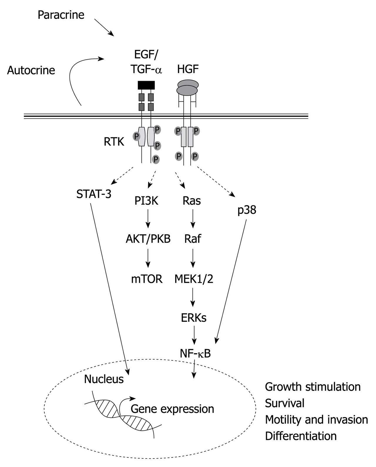
All the data summarized above highlight not only a relevant role of Met and EGFR-triggered signaling pathways in regulating the liver progenitor cell compartment, but a striking parallelism between the two pathways, both of which mediate growth, survival, migration and hepatocytic differentiation (Figure 1). Further studies will clarify which of these biological activities are totally overlapping and which are not. In this sense, Met and EGFR mutant mice and cell models should provide very useful and invaluable tools in this regard.

Figure 1
Figure 1 Schematic illustration of the major signaling pathways and biological activities induced by hepatocyte growth factor and epidermal growth factor receptor ligands in liver progenitor cells. HGF: Hepatocyte growth factor; EGF: Epidermal growth factor; TGF: Transforming growth factor; STAT: Signal transducer and activator of transcription; PI3K: Phosphoinositide 3-kinase; NF-κB: Nuclear factor κB; mTOR: Mammalian target of rapamycin; MEK: Mitogen-activated protein kinase kinase; ERKs: Extracellular-signal-regulated kinases.
FGFs

The FGFs are a family of growth factors with high affinity for heparan sulfate proteoglycans, which bind to transmembrane tyrosine kinase FGF receptors (FGFRs). The role of these growth factors in hepatic fate specification of pre-hepatic endoderm cells during liver development is well known. There are a number of excellent reviews on this subject, some of which are cited here[97-99]. Activation of adult stem/progenitor cells is believed to use similar, if not identical, genetic programs as the embryonic progenitors, therefore, FGF is among the main targets to be analyzed in rodent models of oval cell expansion. Indeed, aFGF (also known as FGF-1) is upregulated at the stage of oval cell progression[89,100], being expressed by both oval cells and Ito cells. FGFR1 and FGFR2 are also expressed at high levels during the period of active proliferation and differentiation of oval cells, but exhibit a different pattern. Although FGFR1 is mainly expressed in oval cells, FGFR2 is expressed in both oval and Ito cells[101]. These results suggest a differential role for these two receptors during liver stem/progenitor-cell-mediated regeneration, as well as the establishment of both autocrine and paracrine signaling. In addition to these in vivo observations, in vitro studies have shown that aFGF is able to push Met murine hepatocytes-bi-potential precursors isolated from transgenic livers, which express a constitutively active human Met - to progress from a very early state of differentiation to a more mature state, associated with the expression of liver functions[47]. Moreover, FGF1-pretreated cells are resistant to the TGF-β dedifferentiation effect[102]. Essentially, FGFs have proved to be effective in mediating early hepatic differentiation, and therefore are included in most in vitro differentiation protocols[58].

TGF-β FAMILY

The TGF-β family of cytokines regulates hepatocyte proliferation and death, and plays relevant roles during liver regeneration[103]. However, TGF-β and other pro-inflammatory cytokines are important inducers of fibro-carcinogenesis, due to their ability to induce myofibroblast differentiation and ECM deposition[104,105]. Recent evidence has shown that TGF-β might also regulate liver stemness and phenotype[106].

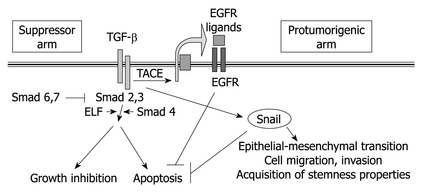
TGF-β active form acts as a dimer, and signals by bringing together two receptors with serine-threonine kinase activity, which are known as type I and type II receptors (Figure 2). After TGF-β binding, the type II receptor phosphorylates and activates the type I receptor, which is responsible for phosphorylation of the receptor-regulated Smad family of transcription factors (R-Smads), i.e. Smad 2 and 3 in the case of TGF-β1, β2 and β3[107]. After phosphorylation, Smad 2 or 3 forms a complex with Smad 4 and shuttles to the nucleus, where it possesses DNA-binding activity, although it must associate with other DNA-binding co-factors to achieve high-affinity binding. Indeed, through combination with different transcription factors, the same TGF-β stimulus can induce or repress many different target genes. Activation of the Smad transcriptional activity can be counteracted by the presence of other members of the Smad family that play inhibitory effects (inhibitory Smads), such as Smad 6 and Smad 7, which compete with Smad 2 and 3 for binding either to the receptor complex or to Smad 4.

Figure 2
Figure 2 Overview of the major signaling pathways, and their cellular effects, induced by transforming growth factor-β in liver cells. TGF: Transforming growth factor; EGFR: Epidermal growth factor receptor; ELF: Embryonic liver fodrin; TACE: Tumor necrosis factor-converting enzyme.

The most relevant Smad-mediated cell responses to TGF-β are growth inhibition and apoptosis. TGF-β inhibits proliferation in adult[108,109], as well as fetal or regenerating hepatocytes[82,109], and it is a well-known inducer of hepatocyte cell death[110,111]. The inhibitory effects of TGF-β on liver stem/progenitor cell growth and survival are somewhat unclear. TGF-β overexpression results in impairment of oval cell expansion induced by DDC[112]. Consistently, we have shown that treatment with TGF-β inhibits growth and induces apoptosis in DDC-derived oval cell lines in vitro[46]. However, when compared to adult hepatocytes, other authors have reported that the majority of the adult liver progenitor cells are more resistant to endogenously produced TGF-β anti-proliferative and pro-apoptotic effects in vitro[113], which indicates a differential sensitivity to TGF-β between liver progenitors and mature hepatocytes. Indeed, several mechanisms have been proposed that would explain this differential response. On the one hand, the ratio between R-Smads and inhibitory Smads might be different in oval cells. In this sense, Nguyen et al[114] have recently reported that oval cell lines show higher levels of Smad 6 than do adult hepatocytes. On the other hand, oval cells might show over-activation of survival signals, such as the MAPK/ERK pathway[115] that might be related to the autocrine production of growth/survival factors, such as HGF[46]. It is worth noting that, when combined with growth factors such as EGF or HGF, TGF-β might contribute to cellular scattering and morphological differentiation of hepatoblasts and liver stem cells[87,116,117]. In this same line of evidence, hepatocytes are undifferentiated and liver architecture is lost in Smad 2+/- and Smad 3+/- mice, as well as in ELF (a β-spectrin that is crucial for the propagation of the TGF-β signal) mutants[118-121]. Defects in Smad 2+/- and Smad 3+/- mutants are restored by HGF treatment. Although mechanisms by which TGF-β signals can be supplanted by HGF are unknown, data seem to support cooperation between two independent pathways, that converge on β1-integrin expression, rather than direct pathway crosstalk[119]. Emerging new data have reinforced the idea that TGF-β is important for stem cell transitioning to a progenitor cell phenotype, and ultimately, its conversion to a fully differentiated phenotype. Indeed, TGF-β signaling, and particularly ELF, appears to play a crucial role in hepatocyte proliferation and transitional phenotype during human liver regeneration, and its loss is associated with activation of liver progenitor cells[106]. It has also been shown recently that TGF-β might be a determinant in the appearance of progenitor/stem cells in hepatocarcinogenesis. The examination of human hepatocellular carcinoma (HCC) has revealed that cells that are labeled with stem-cell markers have unexpectedly lost the TGF-β receptor II and ELF, and show marked activation of the IL-6 pathway, a major stem-cell signaling pathway[121,122]. These data support that absence of TGF-β-driven epithelial differentiation favors carcinogenesis.

However, TGF-β is a pleiotropic cytokine inducing several, and sometimes contradictory, signals in epithelial cells. Indeed, in addition to the well-known Smad-mediated transcriptional responses that predominantly address tumor suppressor actions, TGF-β induces other Smad-dependent or independent effects that contribute to tumor progression[107]. Among these, related to the topic of this review, one of the more relevant is the capacity to induce EMT processes. EMT is a physiological process during embryogenesis, in which an epithelial cell loses expression of adhesion molecules, such as E-cadherin, and other components responsible for cell polarity. Instead, they express mesenchymal components of the cytoskeleton and acquire motility and scattering properties[123]. A closely related phenotypic conversion is also detected in fibrosis and neoplasia and is associated with disease progression[124]. Members of the TGF-β family can initiate and maintain EMT in a variety of biological systems and pathophysiological situations, through activation of major signaling pathways and transcriptional regulators integrated in extensive signaling networks[125,126]. In culture, hepatocytes and hepatoma cells undergo EMT in response to TGF-β[127-130], which support a potentially crucial role for TGF-β in the development and progression of hepatic fibrogenesis and cancer. The mechanisms that allow cells to escape from the apoptotic effects of TGF-β and undergo EMT are not completely understood. However, recent results have indicated that, in some epithelial cells, including fetal rat hepatocytes and hepatoma cells, TGF-β might induce both pro- and anti-apoptotic signals and their balance defines the cell fate[129,131-133]. In fact, certain evidence indicates a crosstalk between the genetic programs that control TGF-β-induced growth arrest/apoptosis and those that regulate EMT, because once the cell has adopted a mesenchymal phenotype, it does not respond to TGF-β suppressor effects[128,134,135]. Thus, TGF-β might regulate its own signaling to switch from tumor suppression to tumor progression. Indeed, it is interesting to point out that transcription factors of the Snail family, repressors of the E-cadherin gene, are required for cell survival during EMT[136,137].

In recent works from our group, we have demonstrated that transdifferentiation of liver cells from an epithelial to a mesenchymal phenotype, such as that mediated by TGF-β, induces dedifferentiation and allows enrichment in a population of cells with putative liver progenitor properties[91,128,138]. The phenotypic characteristics observed (elongated phenotype, strong expression of Thy-1, vimentin or α-smooth muscle actin) are also highly reminiscent of myofibroblasts[139]. These finding are in agreement with the recently proven fact that EMT not only endows cells with migratory and invasive properties, but also induces stem cell properties[123]. Furthermore, additional works have proved the capacity of TGF-β to induce/maintain a stemness phenotype in other tissues[140]. Preliminary results have indicated that human fetal hepatocytes respond to TGF-β downregulation of E-cadherin and upregulation of Snail, thus acquiring a fibroblastic-like morphology and a liver progenitor cell phenotype (unpublished observations from our group in collaboration with Dr. N. Fausto, University of Washington, Seattle, USA).

These findings might have implications for regenerative biology of the liver and open new perspectives for the in vitro isolation of putative liver stem/progenitor cells to be used in basic and translational research in the liver. Moreover, a recent study has also suggested that TGF-β treatment of HCC cells might induce the selection of cells expressing high levels of CD133, a putative stem-cell marker in diverse hematopoietic and non-hematopoietic tissues and cancers[141], through a mechanism partially dependent on the Smads pathway, and that involves epigenetic regulation of the CD133 promoter. These results indicate that TGF-β might also play a role in transdifferentiating liver tumor cells to liver cancer stem cells.

In the embryo, progenitors to pancreatic β cells and hepatocyte lineages arise from neighboring domains of ventral foregut endoderm. It has been recently found that the specification of pancreas and liver progenitors is restricted by the TGF-β pathway[12]. It has been speculated that TGF-β signaling restrains lateral endoderm specification until the cells move sufficiently far from the heart and into a bone morphogenetic protein signaling domain, with the latter then becoming the dominant Smad 4-dependent pathway, which leads to pancreatic induction. TGF-β signaling appears to be strongly inhibitory to the pancreatic lineage and modestly inhibitory to the liver lineage, which suggests that TGF-β also plays a relevant role in maintaining the stemness phenotype of endodermal precursors during liver development.

All these results suggest a dual role for the TGF-β signaling pathway in liver stemness and differentiation. On the one hand, TGF-β mediates progression of differentiation from a stem or progenitor stage, but on the other hand, it contributes to the expansion of liver stem cells. Further work is necessary to understand better the relevance of both effects in liver development, regeneration and carcinogenesis.

HEDGEHOG FAMILY LIGANDS

Hedgehog (Hh) family ligands are widely acknowledged morphogens that regulate tissue remodeling during embryogenesis[142,143]. In particular, Indian hedgehog and Sonic hedgehog ligands, and their receptor, Patched, are expressed at different stages of liver organogenesis, and available data point to dynamic signal activation and temporarily restricted effects on hepatoblast regulation. Thus, Hh signaling is necessary to promote hepatoblast proliferation but it needs to be shut off to permit subsequent hepatoblast differentiation[143-148]. Hh signaling is also involved in the maintenance of a hepatic progenitor reservoir throughout life, both in humans and mice. Blockade of Hh activity in hepatic progenitors in vitro decreases survival, whereas stimulation of Hh activity inhibits endogenous apoptosis. Both autocrine and paracrine modes of action are suggested, based on simultaneous expression of Hh ligands and receptors in liver progenitor cells[149]. Additional observations suggest that Hh pathway activation is a common feature of various types of chronic liver injury associated with mobilization of hepatic progenitor populations. Participation of the Hh pathway has been demonstrated in the ductular reaction that is elicited by chronic alcohol-induced liver injury in mice and humans[150]; non-alcoholic steatohepatitis in humans[151]; methionine choline-deficient ethionine-supplemented diet[152] and fatty liver damage[153] in mice. Hh ligands are released by hepatocytes and myofibroblasts, and lead to enhanced viability and proliferation of bile ductular cells and hepatic progenitors, thus promoting the ductular response and fibrogenesis. Myofibroblasts, but not hepatocytes, also respond to Hh ligands with an increase in viability[152,154-157]. Either Hh paracrine signaling between myofibroblast and hepatic progenitors or autocrine signaling, or both, promote EMT in hepatic progenitors, which ultimately contributes to liver fibrosis[157-159].

The way in which Hh signaling interacts with other pathways to control the fate of hepatic progenitor populations during liver repair after damage or neoplasia is still not well understood. Nonetheless, some interesting pieces of information are emerging. Similar to that which has been described in embryogenesis[160], a functional crosstalk between the TGF-β and Hh signaling pathways has been proposed in the context of adult liver progenitor expansion during liver injury. In ethanol-fed mice, TGF-β induces production of Hh ligands in hepatocytes, which subsequently promotes survival and expansion of ductular/oval cell populations. This supports the concept that enhanced exposure to TGF-β contributes to the accumulation of cells with a ductular phenotype, which are protected from TGF-β-mediated apoptosis[155,161]. Reciprocally, Hh-induced EMT might depend, at least partially, on induction of TGF-β[159]. Further research is required to delineate the Hh-TGF-β interaction.

These findings suggest a role for the Hh pathway as a major mediator in the communication network established between myofibroblast and liver progenitors, which promotes progenitor accumulation and contributes to hepatic fibrosis pathogenesis through EMT induction.

WINGLESS/β-CATENIN PATHWAY

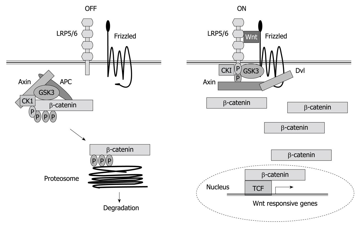
Proteins of the wingless (Wnt) family are secreted signaling molecules that regulate multiple processes in animal development and control tissue homeostasis. The Wnt signaling pathway is highly conserved throughout evolution and plays essential roles in controlling cell proliferation, cell to cell adhesion, and motility[162]. In the liver, it plays many crucial roles during hepatic development and regeneration, and its dysregulation is evident in aberrant growth during hepatocarcinogenesis[163]. Wnt signaling is itself inherently complex[164]. On the one hand, both the ligands and receptors involved in Wnt signal transduction belong to large multi-gene families, which allows for a large number of possible ligand-receptor interactions. On the other hand, Wnt-receptor interactions can elicit a variety of intracellular responses, the best-known of which results in the activation of β-catenin/T-cell factor (TCF) transcriptional complexes. In the absence of signals, β-catenin accumulates in adherens junctions and free β-catenin levels are very low because the tumor suppressor adenomatous polyposis coli forms a trimer complex with glycogen synthase kinase-3-β and axin/conductin, which interacts with and phosphorylates β-catenin, thus targeting it for degradation to the proteasome (Figure 3). Wnts bind a two-part receptor: a seven transmembrane Frizzled and low-density lipoprotein-related protein (LRP)5/6, both being required for canonical signaling. Ligand binding mediates phosphorylation of the cytoplasmic tail of LRP5/6, which creates an axin-binding site. Axin recruitment inactivates the destruction complex. This stabilizes β-catenin, which enters into the nuclei where it displaces Groucho from TCF, nucleating formation of a multiprotein activator complex, and activating Wnt target genes[165] (Figure 3). Furthermore, β-catenin is at the crossroads of growth factor and cytokine signaling. Indeed, certain evidence indicates that TGF-β might induce nuclear β-catenin accumulation, through induction of platelet-derived growth factor signaling[166]. β-catenin expression leads to elevated EGFR levels in hepatocytes and immunohistological analysis shows high correlation between the expression of nuclear/cytoplasmic β-catenin and EGFR in most hepatoblastomas[167]. β-catenin also participates in homotypic cell-cell interactions through its association with E-cadherin. Thus, nuclear β-catenin accumulation in HCC cells might contribute to impaired E-cadherin expression, which mediates the EMT process, migration and survival. It is also known that β-catenin and Met form a complex on hepatocyte membranes. Upon HGF stimulation, the complex is dissociated, in a β-catenin tyrosine phosphorylation-dependent but Wnt-independent manner, which results in β-catenin nuclear translocation[168]. In addition, a role for β-catenin as a downstream effector of HGF in HGF-induced hepatomegaly has been demonstrated[169].

Figure 3
Figure 3 Wnt/β-catenin signaling. Left figure shows how β-catenin is pushed to degradation in the absence of Wnt ligand. Right figure shows how in the presence of Wnt, β-catenin regulates gene expression. TCF: T-cell factor; LRP: Low density lipoprotein receptor-related protein; APC: Adenomatous polyposis coli; CK1: Casein kinase 1; GSK: Glycogen synthase kinase.

It has been recently suggested that oval cells respond to Wnt ligands in vitro with an increase in amino-terminus dephosphorylated β-catenin and cell cycle entry[170], and that canonical Wnt/β-catenin/TCF signaling plays a key role in the normal activation and proliferation of adult hepatic stem cells[171,172]. In this same line of evidence, repression of Wnt/β-catenin signaling in the anterior endoderm is essential for liver and pancreas specification[173], which indicates that turning Wnt signaling off is essential for liver differentiation. An opposite situation might be found during hepatocarcinogenesis, where reactivation of the Wnt/β-catenin pathway, or accumulation of nuclear β-catenin due to other alterations might contribute to the expansion of liver tumor stem cells. Indeed, recent reports have highlighted the relevance of Wnt/β-catenin signaling in the activation of tumorigenic liver progenitor cells[174] and the acquisition of hepatic stem-cell markers[175] in HCC. Accumulation of nuclear β-catenin has been shown to induce an early liver progenitor phenotype in HCC, which correlates with tumor recurrence[176].

In summary, Wnt family and β-catenin/TCF pathways are clearly involved in the maintenance of the liver stemness phenotype, and its repression is necessary for liver differentiation during development. Its overactivation during liver tumorigenesis might contribute to the acquisition of a liver cancer stem cell phenotype.

CONCLUSION

For the past 10-15 years, significant progress has been made in elucidating the cellular and molecular mechanisms that contribute to control of liver stem/progenitor cell behavior. Its complexity and multifaceted nature is evident. Among the diverse signaling pathways at play, the major growth factors, cytokines, and other ligand/receptor systems known to play an important role in liver development, homeostasis and regeneration, certainly occupy a central position. Many of the classical responses elicited by these factors are conserved. However, differences in the downstream mechanisms following ligand-receptor engagement, or signal interaction and crosstalk, as well as specific alterations in intracellular mediators, are being identified as contributing factors to the differential response of liver stem/progenitor cells compared with their mature counterparts. In addition, besides delivery of signals by the surrounding microenvironment, accumulating observations are pointing to co-existence of paracrine and autocrine modes of response as general regulatory mechanisms on this cell population. Collectively, these data indicate that liver stem/progenitor cells follow their own rules and regulations. It is also clear that the same signals that are essential for their activation, expansion, and differentiation, are good candidates to contribute, under adequate conditions, to the paradigm of transformation from a pro-regenerative to a pro-tumorigenic role. Unquestionably, from a clinical perspective, this is a fundamental issue for liver/stem progenitor cell-based therapies. Despite the fact that additional efforts are required to delineate perfectly how the functional switch takes place, the studies summarized here are steps in the right direction.

Footnotes

Peer reviewers: Dr. Thomas Kietzmann, Professor, Department of Biochemistry, University of Oulu, FI-90014 Oulu, Finland; Toshihiro Mitaka, MD, PhD, Professor, Department of Pathophysiology, Cancer Research Institute, Sapporo Medical University School of Medicine, South-1, West-17, Chuo-ku, Sapporo 060-8556, Japan

S- Editor Wang JL L- Editor Kerr C E- Editor Zheng XM

References
1.  Taub R. Liver regeneration: from myth to mechanism. Nat Rev Mol Cell Biol. 2004;5:836-847.  [PubMed]  [DOI]  [Cited in This Article: ]
2.  Fausto N, Campbell JS, Riehle KJ. Liver regeneration. Hepatology. 2006;43:S45-S53.  [PubMed]  [DOI]  [Cited in This Article: ]
3.  Michalopoulos GK. Liver regeneration. J Cell Physiol. 2007;213:286-300.  [PubMed]  [DOI]  [Cited in This Article: ]
4.  Michalopoulos GK. Liver regeneration: Alternative epithelial pathways. Int J Biochem Cell Biol. 2009;Epub ahead of print.  [PubMed]  [DOI]  [Cited in This Article: ]
5.  Fausto N. Liver regeneration and repair: hepatocytes, progenitor cells, and stem cells. Hepatology. 2004;39:1477-1487.  [PubMed]  [DOI]  [Cited in This Article: ]
6.  Duncan AW, Dorrell C, Grompe M. Stem cells and liver regeneration. Gastroenterology. 2009;137:466-481.  [PubMed]  [DOI]  [Cited in This Article: ]
7.  Grisham JW, Hartroft WS. Morphologic identification by electron microscopy of "oval" cells in experimental hepatic degeneration. Lab Invest. 1961;10:317-332.  [PubMed]  [DOI]  [Cited in This Article: ]
8.  Sell S, Osborn K, Leffert HL. Autoradiography of "oval cells" appearing rapidly in the livers of rats fed N-2-fluorenylacetamide in a choline devoid diet. Carcinogenesis. 1981;2:7-14.  [PubMed]  [DOI]  [Cited in This Article: ]
9.  Alison MR, Poulsom R, Jeffery R, Anilkumar TV, Jagoe R, Sarraf CE. Expression of hepatocyte growth factor mRNA during oval cell activation in the rat liver. J Pathol. 1993;171:291-299.  [PubMed]  [DOI]  [Cited in This Article: ]
10.  Nagy P, Bisgaard HC, Santoni-Rugiu E, Thorgeirsson SS. In vivo infusion of growth factors enhances the mitogenic response of rat hepatic ductal (oval) cells after administration of 2-acetylaminofluorene. Hepatology. 1996;23:71-79.  [PubMed]  [DOI]  [Cited in This Article: ]
11.  Isfort RJ, Cody DB, Richards WG, Yoder BK, Wilkinson JE, Woychik RP. Characterization of growth factor responsiveness and alterations in growth factor homeostasis involved in the tumorigenic conversion of mouse oval cells. Growth Factors. 1998;15:81-94.  [PubMed]  [DOI]  [Cited in This Article: ]
12.  Wandzioch E, Zaret KS. Dynamic signaling network for the specification of embryonic pancreas and liver progenitors. Science. 2009;324:1707-1710.  [PubMed]  [DOI]  [Cited in This Article: ]
13.  Michalopoulos G, Houck KA, Dolan ML, Leutteke NC. Control of hepatocyte replication by two serum factors. Cancer Res. 1984;44:4414-4419.  [PubMed]  [DOI]  [Cited in This Article: ]
14.  Nakamura T, Nawa K, Ichihara A. Partial purification and characterization of hepatocyte growth factor from serum of hepatectomized rats. Biochem Biophys Res Commun. 1984;122:1450-1459.  [PubMed]  [DOI]  [Cited in This Article: ]
15.  Russell WE, McGowan JA, Bucher NL. Partial characterization of a hepatocyte growth factor from rat platelets. J Cell Physiol. 1984;119:183-192.  [PubMed]  [DOI]  [Cited in This Article: ]
16.  Stoker M, Gherardi E, Perryman M, Gray J. Scatter factor is a fibroblast-derived modulator of epithelial cell mobility. Nature. 1987;327:239-242.  [PubMed]  [DOI]  [Cited in This Article: ]
17.  Naldini L, Weidner KM, Vigna E, Gaudino G, Bardelli A, Ponzetto C, Narsimhan RP, Hartmann G, Zarnegar R, Michalopoulos GK. Scatter factor and hepatocyte growth factor are indistinguishable ligands for the MET receptor. EMBO J. 1991;10:2867-2878.  [PubMed]  [DOI]  [Cited in This Article: ]
18.  Weidner KM, Arakaki N, Hartmann G, Vandekerckhove J, Weingart S, Rieder H, Fonatsch C, Tsubouchi H, Hishida T, Daikuhara Y. Evidence for the identity of human scatter factor and human hepatocyte growth factor. Proc Natl Acad Sci USA. 1991;88:7001-7005.  [PubMed]  [DOI]  [Cited in This Article: ]
19.  Zarnegar R, Michalopoulos GK. The many faces of hepatocyte growth factor: from hepatopoiesis to hematopoiesis. J Cell Biol. 1995;129:1177-1180.  [PubMed]  [DOI]  [Cited in This Article: ]
20.  Matsumoto K, Nakamura T. Emerging multipotent aspects of hepatocyte growth factor. J Biochem. 1996;119:591-600.  [PubMed]  [DOI]  [Cited in This Article: ]
21.  Birchmeier C, Gherardi E. Developmental roles of HGF/SF and its receptor, the c-Met tyrosine kinase. Trends Cell Biol. 1998;8:404-410.  [PubMed]  [DOI]  [Cited in This Article: ]
22.  Birchmeier C, Birchmeier W, Gherardi E, Vande Woude GF. Met, metastasis, motility and more. Nat Rev Mol Cell Biol. 2003;4:915-925.  [PubMed]  [DOI]  [Cited in This Article: ]
23.  Zhang YW, Vande Woude GF. HGF/SF-met signaling in the control of branching morphogenesis and invasion. J Cell Biochem. 2003;88:408-417.  [PubMed]  [DOI]  [Cited in This Article: ]
24.  Tulasne D, Foveau B. The shadow of death on the MET tyrosine kinase receptor. Cell Death Differ. 2008;15:427-434.  [PubMed]  [DOI]  [Cited in This Article: ]
25.  Furge KA, Zhang YW, Vande Woude GF. Met receptor tyrosine kinase: enhanced signaling through adapter proteins. Oncogene. 2000;19:5582-5589.  [PubMed]  [DOI]  [Cited in This Article: ]
26.  Stuart KA, Riordan SM, Lidder S, Crostella L, Williams R, Skouteris GG. Hepatocyte growth factor/scatter factor-induced intracellular signalling. Int J Exp Pathol. 2000;81:17-30.  [PubMed]  [DOI]  [Cited in This Article: ]
27.  Borowiak M, Garratt AN, Wüstefeld T, Strehle M, Trautwein C, Birchmeier C. Met provides essential signals for liver regeneration. Proc Natl Acad Sci USA. 2004;101:10608-10613.  [PubMed]  [DOI]  [Cited in This Article: ]
28.  Huh CG, Factor VM, Sánchez A, Uchida K, Conner EA, Thorgeirsson SS. Hepatocyte growth factor/c-met signaling pathway is required for efficient liver regeneration and repair. Proc Natl Acad Sci USA. 2004;101:4477-4482.  [PubMed]  [DOI]  [Cited in This Article: ]
29.  Phaneuf D, Moscioni AD, LeClair C, Raper SE, Wilson JM. Generation of a mouse expressing a conditional knockout of the hepatocyte growth factor gene: demonstration of impaired liver regeneration. DNA Cell Biol. 2004;23:592-603.  [PubMed]  [DOI]  [Cited in This Article: ]
30.  Paranjpe S, Bowen WC, Bell AW, Nejak-Bowen K, Luo JH, Michalopoulos GK. Cell cycle effects resulting from inhibition of hepatocyte growth factor and its receptor c-Met in regenerating rat livers by RNA interference. Hepatology. 2007;45:1471-1477.  [PubMed]  [DOI]  [Cited in This Article: ]
31.  Bladt F, Riethmacher D, Isenmann S, Aguzzi A, Birchmeier C. Essential role for the c-met receptor in the migration of myogenic precursor cells into the limb bud. Nature. 1995;376:768-771.  [PubMed]  [DOI]  [Cited in This Article: ]
32.  Schmidt C, Bladt F, Goedecke S, Brinkmann V, Zschiesche W, Sharpe M, Gherardi E, Birchmeier C. Scatter factor/hepatocyte growth factor is essential for liver development. Nature. 1995;373:699-702.  [PubMed]  [DOI]  [Cited in This Article: ]
33.  Uehara Y, Minowa O, Mori C, Shiota K, Kuno J, Noda T, Kitamura N. Placental defect and embryonic lethality in mice lacking hepatocyte growth factor/scatter factor. Nature. 1995;373:702-705.  [PubMed]  [DOI]  [Cited in This Article: ]
34.  Moumen A, Ieraci A, Patané S, Solé C, Comella JX, Dono R, Maina F. Met signals hepatocyte survival by preventing Fas-triggered FLIP degradation in a PI3k-Akt-dependent manner. Hepatology. 2007;45:1210-1217.  [PubMed]  [DOI]  [Cited in This Article: ]
35.  Ogasawara J, Watanabe-Fukunaga R, Adachi M, Matsuzawa A, Kasugai T, Kitamura Y, Itoh N, Suda T, Nagata S. Lethal effect of the anti-Fas antibody in mice. Nature. 1993;364:806-809.  [PubMed]  [DOI]  [Cited in This Article: ]
36.  Kosai KI, Finegold MJ, Thi-Huynh BT, Tewson M, Ou CN, Bowles N, Woo SL, Schwall RH, Darlington GJ. Retrovirus-mediated in vivo gene transfer in the replicating liver using recombinant hepatocyte growth factor without liver injury or partial hepatectomy. Hum Gene Ther. 1998;9:1293-1301.  [PubMed]  [DOI]  [Cited in This Article: ]
37.  Kosai K, Matsumoto K, Funakoshi H, Nakamura T. Hepatocyte growth factor prevents endotoxin-induced lethal hepatic failure in mice. Hepatology. 1999;30:151-159.  [PubMed]  [DOI]  [Cited in This Article: ]
38.  Ueki T, Kaneda Y, Tsutsui H, Nakanishi K, Sawa Y, Morishita R, Matsumoto K, Nakamura T, Takahashi H, Okamoto E. Hepatocyte growth factor gene therapy of liver cirrhosis in rats. Nat Med. 1999;5:226-230.  [PubMed]  [DOI]  [Cited in This Article: ]
39.  Phaneuf D, Chen SJ, Wilson JM. Intravenous injection of an adenovirus encoding hepatocyte growth factor results in liver growth and has a protective effect against apoptosis. Mol Med. 2000;6:96-103.  [PubMed]  [DOI]  [Cited in This Article: ]
40.  Xue F, Takahara T, Yata Y, Kuwabara Y, Shinno E, Nonome K, Minemura M, Takahara S, Li X, Yamato E. Hepatocyte growth factor gene therapy accelerates regeneration in cirrhotic mouse livers after hepatectomy. Gut. 2003;52:694-700.  [PubMed]  [DOI]  [Cited in This Article: ]
41.  Hu Z, Evarts RP, Fujio K, Marsden ER, Thorgeirsson SS. Expression of hepatocyte growth factor and c-met genes during hepatic differentiation and liver development in the rat. Am J Pathol. 1993;142:1823-1830.  [PubMed]  [DOI]  [Cited in This Article: ]
42.  Shiota G, Kunisada T, Oyama K, Udagawa A, Nomi T, Tanaka K, Tsutsumi A, Isono M, Nakamura T, Hamada H. In vivo transfer of hepatocyte growth factor gene accelerates proliferation of hepatic oval cells in a 2-acetylaminofluorene/partial hepatectomy model in rats. FEBS Lett. 2000;470:325-330.  [PubMed]  [DOI]  [Cited in This Article: ]
43.  Okano J, Shiota G, Matsumoto K, Yasui S, Kurimasa A, Hisatome I, Steinberg P, Murawaki Y. Hepatocyte growth factor exerts a proliferative effect on oval cells through the PI3K/AKT signaling pathway. Biochem Biophys Res Commun. 2003;309:298-304.  [PubMed]  [DOI]  [Cited in This Article: ]
44.  Yao P, Zhan Y, Xu W, Li C, Yue P, Xu C, Hu D, Qu CK, Yang X. Hepatocyte growth factor-induced proliferation of hepatic stem-like cells depends on activation of NF-kappaB. J Hepatol. 2004;40:391-398.  [PubMed]  [DOI]  [Cited in This Article: ]
45.  Kato H, Shimomura T, Murai R, Gonda K, Ishii K, Yoshida Y, Kanbe T, Hashiguchi K, Sakabe T, Takubo K. Regulation of hepatic oval cell proliferation by adenoviral mediated hepatocyte growth factor gene transfer and signal transduction inhibitors. Hepatogastroenterology. 2007;54:821-825.  [PubMed]  [DOI]  [Cited in This Article: ]
46.  del Castillo G, Factor VM, Fernández M, Alvarez-Barrientos A, Fabregat I, Thorgeirsson SS, Sánchez A. Deletion of the Met tyrosine kinase in liver progenitor oval cells increases sensitivity to apoptosis in vitro. Am J Pathol. 2008;172:1238-1247.  [PubMed]  [DOI]  [Cited in This Article: ]
47.  Spagnoli FM, Amicone L, Tripodi M, Weiss MC. Identification of a bipotential precursor cell in hepatic cell lines derived from transgenic mice expressing cyto-Met in the liver. J Cell Biol. 1998;143:1101-1112.  [PubMed]  [DOI]  [Cited in This Article: ]
48.  Montesano R, Matsumoto K, Nakamura T, Orci L. Identification of a fibroblast-derived epithelial morphogen as hepatocyte growth factor. Cell. 1991;67:901-908.  [PubMed]  [DOI]  [Cited in This Article: ]
49.  Comoglio PM, Trusolino L. Invasive growth: from development to metastasis. J Clin Invest. 2002;109:857-862.  [PubMed]  [DOI]  [Cited in This Article: ]
50.  Rosário M, Birchmeier W. How to make tubes: signaling by the Met receptor tyrosine kinase. Trends Cell Biol. 2003;13:328-335.  [PubMed]  [DOI]  [Cited in This Article: ]
51.  Johnson M, Koukoulis G, Matsumoto K, Nakamura T, Iyer A. Hepatocyte growth factor induces proliferation and morphogenesis in nonparenchymal epithelial liver cells. Hepatology. 1993;17:1052-1061.  [PubMed]  [DOI]  [Cited in This Article: ]
52.  Jeffers M, Rao MS, Rulong S, Reddy JK, Subbarao V, Hudson E, Vande Woude GF, Resau JH. Hepatocyte growth factor/scatter factor-Met signaling induces proliferation, migration, and morphogenesis of pancreatic oval cells. Cell Growth Differ. 1996;7:1805-1813.  [PubMed]  [DOI]  [Cited in This Article: ]
53.  Delgado JP, Vanneaux V, Branger J, Touboul T, Sentilhes L, Mainot S, Lainas P, Leclerc P, Uzan G, Mahieu-Caputo D. The role of HGF on invasive properties and repopulation potential of human fetal hepatic progenitor cells. Exp Cell Res. 2009;315:3396-3405.  [PubMed]  [DOI]  [Cited in This Article: ]
54.  Suzuki A, Zheng Y, Kondo R, Kusakabe M, Takada Y, Fukao K, Nakauchi H, Taniguchi H. Flow-cytometric separation and enrichment of hepatic progenitor cells in the developing mouse liver. Hepatology. 2000;32:1230-1239.  [PubMed]  [DOI]  [Cited in This Article: ]
55.  Minguet S, Cortegano I, Gonzalo P, Martínez-Marin JA, de Andrés B, Salas C, Melero D, Gaspar ML, Marcos MA. A population of c-Kit(low)(CD45/TER119)- hepatic cell progenitors of 11-day postcoitus mouse embryo liver reconstitutes cell-depleted liver organoids. J Clin Invest. 2003;112:1152-1163.  [PubMed]  [DOI]  [Cited in This Article: ]
56.  Suzuki A, Zheng YW, Kaneko S, Onodera M, Fukao K, Nakauchi H, Taniguchi H. Clonal identification and characterization of self-renewing pluripotent stem cells in the developing liver. J Cell Biol. 2002;156:173-184.  [PubMed]  [DOI]  [Cited in This Article: ]
57.  Suzuki A, Iwama A, Miyashita H, Nakauchi H, Taniguchi H. Role for growth factors and extracellular matrix in controlling differentiation of prospectively isolated hepatic stem cells. Development. 2003;130:2513-2524.  [PubMed]  [DOI]  [Cited in This Article: ]
58.  Snykers S, De Kock J, Rogiers V, Vanhaecke T. In vitro differentiation of embryonic and adult stem cells into hepatocytes: state of the art. Stem Cells. 2009;27:577-605.  [PubMed]  [DOI]  [Cited in This Article: ]
59.  Kinoshita T, Miyajima A. Cytokine regulation of liver development. Biochim Biophys Acta. 2002;1592:303-312.  [PubMed]  [DOI]  [Cited in This Article: ]
60.  Kamiya A, Kinoshita T, Miyajima A. Oncostatin M and hepatocyte growth factor induce hepatic maturation via distinct signaling pathways. FEBS Lett. 2001;492:90-94.  [PubMed]  [DOI]  [Cited in This Article: ]
61.  Sanchez A, Alvarez AM, Benito M, Fabregat I. Transforming growth factor beta modulates growth and differentiation of fetal hepatocytes in primary culture. J Cell Physiol. 1995;165:398-405.  [PubMed]  [DOI]  [Cited in This Article: ]
62.  Fabregat I, Sánchez A, Alvarez AM, Nakamura T, Benito M. Epidermal growth factor, but not hepatocyte growth factor, suppresses the apoptosis induced by transforming growth factor-beta in fetal hepatocytes in primary culture. FEBS Lett. 1996;384:14-18.  [PubMed]  [DOI]  [Cited in This Article: ]
63.  Pagan R, Martín I, Llobera M, Vilaró S. Growth and differentiation factors inhibit the migratory phenotype of cultured neonatal rat hepatocytes induced by HGF/SF. Exp Cell Res. 1997;235:170-179.  [PubMed]  [DOI]  [Cited in This Article: ]
64.  Pagan R, Sánchez A, Martin I, Llobera M, Fabregat I, Vilaró S. Effects of growth and differentiation factors on the epithelial-mesenchymal transition in cultured neonatal rat hepatocytes. J Hepatol. 1999;31:895-904.  [PubMed]  [DOI]  [Cited in This Article: ]
65.  Block GD, Locker J, Bowen WC, Petersen BE, Katyal S, Strom SC, Riley T, Howard TA, Michalopoulos GK. Population expansion, clonal growth, and specific differentiation patterns in primary cultures of hepatocytes induced by HGF/SF, EGF and TGF alpha in a chemically defined (HGM) medium. J Cell Biol. 1996;132:1133-1149.  [PubMed]  [DOI]  [Cited in This Article: ]
66.  Sonnenberg E, Meyer D, Weidner KM, Birchmeier C. Scatter factor/hepatocyte growth factor and its receptor, the c-met tyrosine kinase, can mediate a signal exchange between mesenchyme and epithelia during mouse development. J Cell Biol. 1993;123:223-235.  [PubMed]  [DOI]  [Cited in This Article: ]
67.  Schirmacher P, Geerts A, Pietrangelo A, Dienes HP, Rogler CE. Hepatocyte growth factor/hepatopoietin A is expressed in fat-storing cells from rat liver but not myofibroblast-like cells derived from fat-storing cells. Hepatology. 1992;15:5-11.  [PubMed]  [DOI]  [Cited in This Article: ]
68.  Ramadori G, Neubauer K, Odenthal M, Nakamura T, Knittel T, Schwögler S, Meyer zum Büschenfelde KH. The gene of hepatocyte growth factor is expressed in fat-storing cells of rat liver and is downregulated during cell growth and by transforming growth factor-beta. Biochem Biophys Res Commun. 1992;183:739-742.  [PubMed]  [DOI]  [Cited in This Article: ]
69.  LeCouter J, Moritz DR, Li B, Phillips GL, Liang XH, Gerber HP, Hillan KJ, Ferrara N. Angiogenesis-independent endothelial protection of liver: role of VEGFR-1. Science. 2003;299:890-893.  [PubMed]  [DOI]  [Cited in This Article: ]
70.  Prat M, Narsimhan RP, Crepaldi T, Nicotra MR, Natali PG, Comoglio PM. The receptor encoded by the human c-MET oncogene is expressed in hepatocytes, epithelial cells and solid tumors. Int J Cancer. 1991;49:323-328.  [PubMed]  [DOI]  [Cited in This Article: ]
71.  Matsumoto K, Fujii H, Michalopoulos G, Fung JJ, Demetris AJ. Human biliary epithelial cells secrete and respond to cytokines and hepatocyte growth factors in vitro: interleukin-6, hepatocyte growth factor and epidermal growth factor promote DNA synthesis in vitro. Hepatology. 1994;20:376-382.  [PubMed]  [DOI]  [Cited in This Article: ]
72.  Burgess AW, Cho HS, Eigenbrot C, Ferguson KM, Garrett TP, Leahy DJ, Lemmon MA, Sliwkowski MX, Ward CW, Yokoyama S. An open-and-shut case? Recent insights into the activation of EGF/ErbB receptors. Mol Cell. 2003;12:541-552.  [PubMed]  [DOI]  [Cited in This Article: ]
73.  Harris RC, Chung E, Coffey RJ. EGF receptor ligands. Exp Cell Res. 2003;284:2-13.  [PubMed]  [DOI]  [Cited in This Article: ]
74.  Citri A, Yarden Y. EGF-ERBB signalling: towards the systems level. Nat Rev Mol Cell Biol. 2006;7:505-516.  [PubMed]  [DOI]  [Cited in This Article: ]
75.  Bublil EM, Yarden Y. The EGF receptor family: spearheading a merger of signaling and therapeutics. Curr Opin Cell Biol. 2007;19:124-134.  [PubMed]  [DOI]  [Cited in This Article: ]
76.  Mitsudomi T, Yatabe Y. Epidermal growth factor receptor in relation to tumor development: EGFR gene and cancer. FEBS J. 2010;277:301-308.  [PubMed]  [DOI]  [Cited in This Article: ]
77.  Bucher NL, Patel U, Cohen S. Hormonal factors concerned with liver regeneration. Ciba Found Symp. 1977;95-107.  [PubMed]  [DOI]  [Cited in This Article: ]
78.  Liu ML, Mars WM, Zarnegar R, Michalopoulos GK. Collagenase pretreatment and the mitogenic effects of hepatocyte growth factor and transforming growth factor-alpha in adult rat liver. Hepatology. 1994;19:1521-1527.  [PubMed]  [DOI]  [Cited in This Article: ]
79.  Draghi E, Armato U, Andreis PG, Mengato L. The stimulation by epidermal growth factor (urogastrone) of the growth of neonatal rat hepatocytes in primary tissue culture and its modulation by serum and associated pancreatic hormones. J Cell Physiol. 1980;103:129-147.  [PubMed]  [DOI]  [Cited in This Article: ]
80.  McGowan JA, Strain AJ, Bucher NL. DNA synthesis in primary cultures of adult rat hepatocytes in a defined medium: effects of epidermal growth factor, insulin, glucagon, and cyclic-AMP. J Cell Physiol. 1981;108:353-363.  [PubMed]  [DOI]  [Cited in This Article: ]
81.  Sawada N, Lee GH, Mochizuki Y, Ishikawa T. Active proliferation of mouse hepatocytes in primary culture under defined conditions as compared to rat hepatocytes. Jpn J Cancer Res. 1988;79:983-988.  [PubMed]  [DOI]  [Cited in This Article: ]
82.  de Juan C, Benito M, Alvarez A, Fabregat I. Differential proliferative response of cultured fetal and regenerating hepatocytes to growth factors and hormones. Exp Cell Res. 1992;202:495-500.  [PubMed]  [DOI]  [Cited in This Article: ]
83.  Natarajan A, Wagner B, Sibilia M. The EGF receptor is required for efficient liver regeneration. Proc Natl Acad Sci USA. 2007;104:17081-17086.  [PubMed]  [DOI]  [Cited in This Article: ]
84.  Fabregat I. Dysregulation of apoptosis in hepatocellular carcinoma cells. World J Gastroenterol. 2009;15:513-520.  [PubMed]  [DOI]  [Cited in This Article: ]
85.  Musallam L, Ethier C, Haddad PS, Bilodeau M. Role of EGF receptor tyrosine kinase activity in antiapoptotic effect of EGF on mouse hepatocytes. Am J Physiol Gastrointest Liver Physiol. 2001;280:G1360-G1369.  [PubMed]  [DOI]  [Cited in This Article: ]
86.  de Juan C, Benito M, Fabregat I. Regulation of albumin expression in fetal rat hepatocytes cultured under proliferative conditions: role of epidermal growth factor and hormones. J Cell Physiol. 1992;152:95-101.  [PubMed]  [DOI]  [Cited in This Article: ]
87.  Sánchez A, Pagan R, Alvarez AM, Roncero C, Vilaró S, Benito M, Fabregat I. Transforming growth factor-beta (TGF-beta) and EGF promote cord-like structures that indicate terminal differentiation of fetal hepatocytes in primary culture. Exp Cell Res. 1998;242:27-37.  [PubMed]  [DOI]  [Cited in This Article: ]
88.  Evarts RP, Nakatsukasa H, Marsden ER, Hu Z, Thorgeirsson SS. Expression of transforming growth factor-alpha in regenerating liver and during hepatic differentiation. Mol Carcinog. 1992;5:25-31.  [PubMed]  [DOI]  [Cited in This Article: ]
89.  Evarts RP, Hu Z, Fujio K, Marsden ER, Thorgeirsson SS. Activation of hepatic stem cell compartment in the rat: role of transforming growth factor alpha, hepatocyte growth factor, and acidic fibroblast growth factor in early proliferation. Cell Growth Differ. 1993;4:555-561.  [PubMed]  [DOI]  [Cited in This Article: ]
90.  Cheung RS, Brooling JT, Johnson MM, Riehle KJ, Campbell JS, Fausto N. Interactions between MYC and transforming growth factor alpha alter the growth and tumorigenicity of liver progenitor cells. Carcinogenesis. 2007;28:2624-2631.  [PubMed]  [DOI]  [Cited in This Article: ]
91.  del Castillo G, Alvarez-Barrientos A, Carmona-Cuenca I, Fernández M, Sánchez A, Fabregat I. Isolation and characterization of a putative liver progenitor population after treatment of fetal rat hepatocytes with TGF-beta. J Cell Physiol. 2008;215:846-855.  [PubMed]  [DOI]  [Cited in This Article: ]
92.  Akhurst B, Matthews V, Husk K, Smyth MJ, Abraham LJ, Yeoh GC. Differential lymphotoxin-beta and interferon gamma signaling during mouse liver regeneration induced by chronic and acute injury. Hepatology. 2005;41:327-335.  [PubMed]  [DOI]  [Cited in This Article: ]
93.  Matthews VB, Klinken E, Yeoh GC. Direct effects of interleukin-6 on liver progenitor oval cells in culture. Wound Repair Regen. 2004;12:650-656.  [PubMed]  [DOI]  [Cited in This Article: ]
94.  Presnell SC, Hooth MJ, Borchert KM, Coleman WB, Grisham JW, Smith GJ. Establishment of a functional HGF/C-MET autocrine loop in spontaneous transformants of WB-F344 rat liver stem-like cells. Hepatology. 1998;28:1253-1259.  [PubMed]  [DOI]  [Cited in This Article: ]
95.  Laird AD, Brown PI, Fausto N. Inhibition of tumor growth in liver epithelial cells transfected with a transforming growth factor alpha antisense gene. Cancer Res. 1994;54:4224-4232.  [PubMed]  [DOI]  [Cited in This Article: ]
96.  Presnell SC, Stolz DB, Mars WM, Jo M, Michalopoulos GK, Strom SC. Modifications of the hepatocyte growth factor/c-met pathway by constitutive expression of transforming growth factor-alpha in rat liver epithelial cells. Mol Carcinog. 1997;18:244-255.  [PubMed]  [DOI]  [Cited in This Article: ]
97.  Zaret KS. Regulatory phases of early liver development: paradigms of organogenesis. Nat Rev Genet. 2002;3:499-512.  [PubMed]  [DOI]  [Cited in This Article: ]
98.  Zhao R, Duncan SA. Embryonic development of the liver. Hepatology. 2005;41:956-967.  [PubMed]  [DOI]  [Cited in This Article: ]
99.  Lemaigre FP. Mechanisms of liver development: concepts for understanding liver disorders and design of novel therapies. Gastroenterology. 2009;137:62-79.  [PubMed]  [DOI]  [Cited in This Article: ]
100.  Marsden ER, Hu Z, Fujio K, Nakatsukasa H, Thorgeirsson SS, Evarts RP. Expression of acidic fibroblast growth factor in regenerating liver and during hepatic differentiation. Lab Invest. 1992;67:427-433.  [PubMed]  [DOI]  [Cited in This Article: ]
101.  Hu Z, Evarts RP, Fujio K, Marsden ER, Thorgeirsson SS. Expression of fibroblast growth factor receptors flg and bek during hepatic ontogenesis and regeneration in the rat. Cell Growth Differ. 1995;6:1019-1025.  [PubMed]  [DOI]  [Cited in This Article: ]
102.  Spagnoli FM, Cicchini C, Tripodi M, Weiss MC. Inhibition of MMH (Met murine hepatocyte) cell differentiation by TGF(beta) is abrogated by pre-treatment with the heritable differentiation effector FGF1. J Cell Sci. 2000;113:3639-3647.  [PubMed]  [DOI]  [Cited in This Article: ]
103.  Michalopoulos GK. Liver regeneration after partial hepatectomy: critical analysis of mechanistic dilemmas. Am J Pathol. 2010;176:2-13.  [PubMed]  [DOI]  [Cited in This Article: ]
104.  Inagaki Y, Okazaki I. Emerging insights into Transforming growth factor beta Smad signal in hepatic fibrogenesis. Gut. 2007;56:284-292.  [PubMed]  [DOI]  [Cited in This Article: ]
105.  Matsuzaki K. Modulation of TGF-beta signaling during progression of chronic liver diseases. Front Biosci. 2009;14:2923-2934.  [PubMed]  [DOI]  [Cited in This Article: ]
106.  Thenappan A, Li Y, Kitisin K, Rashid A, Shetty K, Johnson L, Mishra L. Role of transforming growth factor beta signaling and expansion of progenitor cells in regenerating liver. Hepatology. 2010;51:1373-1382.  [PubMed]  [DOI]  [Cited in This Article: ]
107.  Massagué J. TGFbeta in Cancer. Cell. 2008;134:215-230.  [PubMed]  [DOI]  [Cited in This Article: ]
108.  Nakamura T, Tomita Y, Hirai R, Yamaoka K, Kaji K, Ichihara A. Inhibitory effect of transforming growth factor-beta on DNA synthesis of adult rat hepatocytes in primary culture. Biochem Biophys Res Commun. 1985;133:1042-1050.  [PubMed]  [DOI]  [Cited in This Article: ]
109.  Braun L, Mead JE, Panzica M, Mikumo R, Bell GI, Fausto N. Transforming growth factor beta mRNA increases during liver regeneration: a possible paracrine mechanism of growth regulation. Proc Natl Acad Sci USA. 1988;85:1539-1543.  [PubMed]  [DOI]  [Cited in This Article: ]
110.  Oberhammer FA, Pavelka M, Sharma S, Tiefenbacher R, Purchio AF, Bursch W, Schulte-Hermann R. Induction of apoptosis in cultured hepatocytes and in regressing liver by transforming growth factor beta 1. Proc Natl Acad Sci USA. 1992;89:5408-5412.  [PubMed]  [DOI]  [Cited in This Article: ]
111.  Sánchez A, Alvarez AM, Benito M, Fabregat I. Apoptosis induced by transforming growth factor-beta in fetal hepatocyte primary cultures: involvement of reactive oxygen intermediates. J Biol Chem. 1996;271:7416-7422.  [PubMed]  [DOI]  [Cited in This Article: ]
112.  Preisegger KH, Factor VM, Fuchsbichler A, Stumptner C, Denk H, Thorgeirsson SS. Atypical ductular proliferation and its inhibition by transforming growth factor beta1 in the 3,5-diethoxycarbonyl-1,4-dihydrocollidine mouse model for chronic alcoholic liver disease. Lab Invest. 1999;79:103-109.  [PubMed]  [DOI]  [Cited in This Article: ]
113.  Clark JB, Rice L, Sadiq T, Brittain E, Song L, Wang J, Gerber DA. Hepatic progenitor cell resistance to TGF-beta1's proliferative and apoptotic effects. Biochem Biophys Res Commun. 2005;329:337-344.  [PubMed]  [DOI]  [Cited in This Article: ]
114.  Nguyen LN, Furuya MH, Wolfraim LA, Nguyen AP, Holdren MS, Campbell JS, Knight B, Yeoh GC, Fausto N, Parks WT. Transforming growth factor-beta differentially regulates oval cell and hepatocyte proliferation. Hepatology. 2007;45:31-41.  [PubMed]  [DOI]  [Cited in This Article: ]
115.  Ding W, Mouzaki M, You H, Laird JC, Mato J, Lu SC, Rountree CB. CD133+ liver cancer stem cells from methionine adenosyl transferase 1A-deficient mice demonstrate resistance to transforming growth factor (TGF)-beta-induced apoptosis. Hepatology. 2009;49:1277-1286.  [PubMed]  [DOI]  [Cited in This Article: ]
116.  Isfort RJ, Cody DB, Stuard SB, Randall CJ, Miller C, Ridder GM, Doersen CJ, Richards WG, Yoder BK, Wilkinson JE. The combination of epidermal growth factor and transforming growth factor-beta induces novel phenotypic changes in mouse liver stem cell lines. J Cell Sci. 1997;110:3117-3129.  [PubMed]  [DOI]  [Cited in This Article: ]
117.  Hirata M, Amano K, Miyashita A, Yasunaga M, Nakanishi T, Sato K. Establishment and characterization of hepatic stem-like cell lines from normal adult rat liver. J Biochem. 2009;145:51-58.  [PubMed]  [DOI]  [Cited in This Article: ]
118.  Mishra L, Cai T, Yu P, Monga SP, Mishra B. Elf3 encodes a novel 200-kD beta-spectrin: role in liver development. Oncogene. 1999;18:353-364.  [PubMed]  [DOI]  [Cited in This Article: ]
119.  Weinstein M, Monga SP, Liu Y, Brodie SG, Tang Y, Li C, Mishra L, Deng CX. Smad proteins and hepatocyte growth factor control parallel regulatory pathways that converge on beta1-integrin to promote normal liver development. Mol Cell Biol. 2001;21:5122-5131.  [PubMed]  [DOI]  [Cited in This Article: ]
120.  Tang Y, Katuri V, Dillner A, Mishra B, Deng CX, Mishra L. Disruption of transforming growth factor-beta signaling in ELF beta-spectrin-deficient mice. Science. 2003;299:574-577.  [PubMed]  [DOI]  [Cited in This Article: ]
121.  Mishra L, Banker T, Murray J, Byers S, Thenappan A, He AR, Shetty K, Johnson L, Reddy EP. Liver stem cells and hepatocellular carcinoma. Hepatology. 2009;49:318-329.  [PubMed]  [DOI]  [Cited in This Article: ]
122.  Tang Y, Kitisin K, Jogunoori W, Li C, Deng CX, Mueller SC, Ressom HW, Rashid A, He AR, Mendelson JS. Progenitor/stem cells give rise to liver cancer due to aberrant TGF-beta and IL-6 signaling. Proc Natl Acad Sci USA. 2008;105:2445-2450.  [PubMed]  [DOI]  [Cited in This Article: ]
123.  Thiery JP, Acloque H, Huang RY, Nieto MA. Epithelial-mesenchymal transitions in development and disease. Cell. 2009;139:871-890.  [PubMed]  [DOI]  [Cited in This Article: ]
124.  Turley EA, Veiseh M, Radisky DC, Bissell MJ. Mechanisms of disease: epithelial-mesenchymal transition--does cellular plasticity fuel neoplastic progression? Nat Clin Pract Oncol. 2008;5:280-290.  [PubMed]  [DOI]  [Cited in This Article: ]
125.  Valcourt U, Kowanetz M, Niimi H, Heldin CH, Moustakas A. TGF-beta and the Smad signaling pathway support transcriptomic reprogramming during epithelial-mesenchymal cell transition. Mol Biol Cell. 2005;16:1987-2002.  [PubMed]  [DOI]  [Cited in This Article: ]
126.  Zavadil J, Böttinger EP. TGF-beta and epithelial-to-mesenchymal transitions. Oncogene. 2005;24:5764-5774.  [PubMed]  [DOI]  [Cited in This Article: ]
127.  Gotzmann J, Huber H, Thallinger C, Wolschek M, Jansen B, Schulte-Hermann R, Beug H, Mikulits W. Hepatocytes convert to a fibroblastoid phenotype through the cooperation of TGF-beta1 and Ha-Ras: steps towards invasiveness. J Cell Sci. 2002;115:1189-1202.  [PubMed]  [DOI]  [Cited in This Article: ]
128.  Valdés F, Alvarez AM, Locascio A, Vega S, Herrera B, Fernández M, Benito M, Nieto MA, Fabregat I. The epithelial mesenchymal transition confers resistance to the apoptotic effects of transforming growth factor Beta in fetal rat hepatocytes. Mol Cancer Res. 2002;1:68-78.  [PubMed]  [DOI]  [Cited in This Article: ]
129.  Caja L, Ortiz C, Bertran E, Murillo MM, Miró-Obradors MJ, Palacios E, Fabregat I. Differential intracellular signalling induced by TGF-beta in rat adult hepatocytes and hepatoma cells: implications in liver carcinogenesis. Cell Signal. 2007;19:683-694.  [PubMed]  [DOI]  [Cited in This Article: ]
130.  Kaimori A, Potter J, Kaimori JY, Wang C, Mezey E, Koteish A. Transforming growth factor-beta1 induces an epithelial-to-mesenchymal transition state in mouse hepatocytes in vitro. J Biol Chem. 2007;282:22089-22101.  [PubMed]  [DOI]  [Cited in This Article: ]
131.  Shin I, Bakin AV, Rodeck U, Brunet A, Arteaga CL. Transforming growth factor beta enhances epithelial cell survival via Akt-dependent regulation of FKHRL1. Mol Biol Cell. 2001;12:3328-3339.  [PubMed]  [DOI]  [Cited in This Article: ]
132.  Valdés F, Murillo MM, Valverde AM, Herrera B, Sánchez A, Benito M, Fernández M, Fabregat I. Transforming growth factor-beta activates both pro-apoptotic and survival signals in fetal rat hepatocytes. Exp Cell Res. 2004;292:209-218.  [PubMed]  [DOI]  [Cited in This Article: ]
133.  Murillo MM, del Castillo G, Sánchez A, Fernández M, Fabregat I. Involvement of EGF receptor and c-Src in the survival signals induced by TGF-beta1 in hepatocytes. Oncogene. 2005;24:4580-4587.  [PubMed]  [DOI]  [Cited in This Article: ]
134.  Nicolás FJ, Lehmann K, Warne PH, Hill CS, Downward J. Epithelial to mesenchymal transition in Madin-Darby canine kidney cells is accompanied by down-regulation of Smad3 expression, leading to resistance to transforming growth factor-beta-induced growth arrest. J Biol Chem. 2003;278:3251-3256.  [PubMed]  [DOI]  [Cited in This Article: ]
135.  Gal A, Sjöblom T, Fedorova L, Imreh S, Beug H, Moustakas A. Sustained TGF beta exposure suppresses Smad and non-Smad signalling in mammary epithelial cells, leading to EMT and inhibition of growth arrest and apoptosis. Oncogene. 2008;27:1218-1230.  [PubMed]  [DOI]  [Cited in This Article: ]
136.  Vega S, Morales AV, Ocaña OH, Valdés F, Fabregat I, Nieto MA. Snail blocks the cell cycle and confers resistance to cell death. Genes Dev. 2004;18:1131-1143.  [PubMed]  [DOI]  [Cited in This Article: ]
137.  Leroy P, Mostov KE. Slug is required for cell survival during partial epithelial-mesenchymal transition of HGF-induced tubulogenesis. Mol Biol Cell. 2007;18:1943-1952.  [PubMed]  [DOI]  [Cited in This Article: ]
138.  Sánchez A, Alvarez AM, López Pedrosa JM, Roncero C, Benito M, Fabregat I. Apoptotic response to TGF-beta in fetal hepatocytes depends upon their state of differentiation. Exp Cell Res. 1999;252:281-291.  [PubMed]  [DOI]  [Cited in This Article: ]
139.  Dudas J, Mansuroglu T, Batusic D, Saile B, Ramadori G. Thy-1 is an in vivo and in vitro marker of liver myofibroblasts. Cell Tissue Res. 2007;329:503-514.  [PubMed]  [DOI]  [Cited in This Article: ]
140.  Peñuelas S, Anido J, Prieto-Sánchez RM, Folch G, Barba I, Cuartas I, García-Dorado D, Poca MA, Sahuquillo J, Baselga J. TGF-beta increases glioma-initiating cell self-renewal through the induction of LIF in human glioblastoma. Cancer Cell. 2009;15:315-327.  [PubMed]  [DOI]  [Cited in This Article: ]
141.  You H, Ding W, Rountree CB. Epigenetic regulation of cancer stem cell marker CD133 by transforming growth factor-beta. Hepatology. 2010;51:1635-1644.  [PubMed]  [DOI]  [Cited in This Article: ]
142.  McMahon AP, Ingham PW, Tabin CJ. Developmental roles and clinical significance of hedgehog signaling. Curr Top Dev Biol. 2003;53:1-114.  [PubMed]  [DOI]  [Cited in This Article: ]
143.  Mehlen P, Mille F, Thibert C. Morphogens and cell survival during development. J Neurobiol. 2005;64:357-366.  [PubMed]  [DOI]  [Cited in This Article: ]
144.  Goodrich LV, Johnson RL, Milenkovic L, McMahon JA, Scott MP. Conservation of the hedgehog/patched signaling pathway from flies to mice: induction of a mouse patched gene by Hedgehog. Genes Dev. 1996;10:301-312.  [PubMed]  [DOI]  [Cited in This Article: ]
145.  Becker S, Wang ZJ, Massey H, Arauz A, Labosky P, Hammerschmidt M, St-Jacques B, Bumcrot D, McMahon A, Grabel L. A role for Indian hedgehog in extraembryonic endoderm differentiation in F9 cells and the early mouse embryo. Dev Biol. 1997;187:298-310.  [PubMed]  [DOI]  [Cited in This Article: ]
146.  Grabel L, Becker S, Lock L, Maye P, Zanders T. Using EC and ES cell culture to study early development: recent observations on Indian hedgehog and Bmps. Int J Dev Biol. 1998;42:917-925.  [PubMed]  [DOI]  [Cited in This Article: ]
147.  Deutsch G, Jung J, Zheng M, Lóra J, Zaret KS. A bipotential precursor population for pancreas and liver within the embryonic endoderm. Development. 2001;128:871-881.  [PubMed]  [DOI]  [Cited in This Article: ]
148.  Hirose Y, Itoh T, Miyajima A. Hedgehog signal activation coordinates proliferation and differentiation of fetal liver progenitor cells. Exp Cell Res. 2009;315:2648-2657.  [PubMed]  [DOI]  [Cited in This Article: ]
149.  Sicklick JK, Li YX, Melhem A, Schmelzer E, Zdanowicz M, Huang J, Caballero M, Fair JH, Ludlow JW, McClelland RE. Hedgehog signaling maintains resident hepatic progenitors throughout life. Am J Physiol Gastrointest Liver Physiol. 2006;290:G859-G870.  [PubMed]  [DOI]  [Cited in This Article: ]
150.  Jung Y, Brown KD, Witek RP, Omenetti A, Yang L, Vandongen M, Milton RJ, Hines IN, Rippe RA, Spahr L. Accumulation of hedgehog-responsive progenitors parallels alcoholic liver disease severity in mice and humans. Gastroenterology. 2008;134:1532-1543.  [PubMed]  [DOI]  [Cited in This Article: ]
151.  Jung Y, Diehl AM. Non-alcoholic steatohepatitis pathogenesis: role of repair in regulating the disease progression. Dig Dis. 2010;28:225-228.  [PubMed]  [DOI]  [Cited in This Article: ]
152.  Jung Y, Witek RP, Syn WK, Choi SS, Omenetti A, Premont R, Guy CD, Diehl AM. Signals from dying hepatocytes trigger growth of liver progenitors. Gut. 2010;59:655-665.  [PubMed]  [DOI]  [Cited in This Article: ]
153.  Fleig SV, Choi SS, Yang L, Jung Y, Omenetti A, VanDongen HM, Huang J, Sicklick JK, Diehl AM. Hepatic accumulation of Hedgehog-reactive progenitors increases with severity of fatty liver damage in mice. Lab Invest. 2007;87:1227-1239.  [PubMed]  [DOI]  [Cited in This Article: ]
154.  Sicklick JK, Li YX, Choi SS, Qi Y, Chen W, Bustamante M, Huang J, Zdanowicz M, Camp T, Torbenson MS. Role for hedgehog signaling in hepatic stellate cell activation and viability. Lab Invest. 2005;85:1368-1380.  [PubMed]  [DOI]  [Cited in This Article: ]
155.  Omenetti A, Yang L, Li YX, McCall SJ, Jung Y, Sicklick JK, Huang J, Choi S, Suzuki A, Diehl AM. Hedgehog-mediated mesenchymal-epithelial interactions modulate hepatic response to bile duct ligation. Lab Invest. 2007;87:499-514.  [PubMed]  [DOI]  [Cited in This Article: ]
156.  Richardson MM, Jonsson JR, Powell EE, Brunt EM, Neuschwander-Tetri BA, Bhathal PS, Dixon JB, Weltman MD, Tilg H, Moschen AR. Progressive fibrosis in nonalcoholic steatohepatitis: association with altered regeneration and a ductular reaction. Gastroenterology. 2007;133:80-90.  [PubMed]  [DOI]  [Cited in This Article: ]
157.  Yang L, Wang Y, Mao H, Fleig S, Omenetti A, Brown KD, Sicklick JK, Li YX, Diehl AM. Sonic hedgehog is an autocrine viability factor for myofibroblastic hepatic stellate cells. J Hepatol. 2008;48:98-106.  [PubMed]  [DOI]  [Cited in This Article: ]
158.  Omenetti A, Porrello A, Jung Y, Yang L, Popov Y, Choi SS, Witek RP, Alpini G, Venter J, Vandongen HM. Hedgehog signaling regulates epithelial-mesenchymal transition during biliary fibrosis in rodents and humans. J Clin Invest. 2008;118:3331-3342.  [PubMed]  [DOI]  [Cited in This Article: ]
159.  Syn WK, Jung Y, Omenetti A, Abdelmalek M, Guy CD, Yang L, Wang J, Witek RP, Fearing CM, Pereira TA. Hedgehog-mediated epithelial-to-mesenchymal transition and fibrogenic repair in nonalcoholic fatty liver disease. Gastroenterology. 2009;137:1478-1488.e8.  [PubMed]  [DOI]  [Cited in This Article: ]
160.  Capdevila J, lzpisúa Belmonte JC. Extracellular modulation of the Hedgehog, Wnt and TGF-beta signalling pathways during embryonic development. Curr Opin Genet Dev. 1999;9:427-433.  [PubMed]  [DOI]  [Cited in This Article: ]
161.  Jung Y, McCall SJ, Li YX, Diehl AM. Bile ductules and stromal cells express hedgehog ligands and/or hedgehog target genes in primary biliary cirrhosis. Hepatology. 2007;45:1091-1096.  [PubMed]  [DOI]  [Cited in This Article: ]
162.  Cadigan KM, Peifer M. Wnt signaling from development to disease: insights from model systems. Cold Spring Harb Perspect Biol. 2009;1:a002881.  [PubMed]  [DOI]  [Cited in This Article: ]
163.  Monga SP. Role of Wnt/beta-catenin signaling in liver metabolism and cancer. Int J Biochem Cell Biol. 2009;Epub ahead of print.  [PubMed]  [DOI]  [Cited in This Article: ]
164.  van Amerongen R, Nusse R. Towards an integrated view of Wnt signaling in development. Development. 2009;136:3205-3214.  [PubMed]  [DOI]  [Cited in This Article: ]
165.  MacDonald BT, Tamai K, He X. Wnt/beta-catenin signaling: components, mechanisms, and diseases. Dev Cell. 2009;17:9-26.  [PubMed]  [DOI]  [Cited in This Article: ]
166.  Fischer AN, Fuchs E, Mikula M, Huber H, Beug H, Mikulits W. PDGF essentially links TGF-beta signaling to nuclear beta-catenin accumulation in hepatocellular carcinoma progression. Oncogene. 2007;26:3395-3405.  [PubMed]  [DOI]  [Cited in This Article: ]
167.  Breuhahn K, Longerich T, Schirmacher P. Dysregulation of growth factor signaling in human hepatocellular carcinoma. Oncogene. 2006;25:3787-3800.  [PubMed]  [DOI]  [Cited in This Article: ]
168.  Monga SP, Mars WM, Pediaditakis P, Bell A, Mulé K, Bowen WC, Wang X, Zarnegar R, Michalopoulos GK. Hepatocyte growth factor induces Wnt-independent nuclear translocation of beta-catenin after Met-beta-catenin dissociation in hepatocytes. Cancer Res. 2002;62:2064-2071.  [PubMed]  [DOI]  [Cited in This Article: ]
169.  Apte U, Zeng G, Muller P, Tan X, Micsenyi A, Cieply B, Dai C, Liu Y, Kaestner KH, Monga SP. Activation of Wnt/beta-catenin pathway during hepatocyte growth factor-induced hepatomegaly in mice. Hepatology. 2006;44:992-1002.  [PubMed]  [DOI]  [Cited in This Article: ]
170.  Hu M, Kurobe M, Jeong YJ, Fuerer C, Ghole S, Nusse R, Sylvester KG. Wnt/beta-catenin signaling in murine hepatic transit amplifying progenitor cells. Gastroenterology. 2007;133:1579-1591.  [PubMed]  [DOI]  [Cited in This Article: ]
171.  Apte U, Thompson MD, Cui S, Liu B, Cieply B, Monga SP. Wnt/beta-catenin signaling mediates oval cell response in rodents. Hepatology. 2008;47:288-295.  [PubMed]  [DOI]  [Cited in This Article: ]
172.  Williams JM, Oh SH, Jorgensen M, Steiger N, Darwiche H, Shupe T, Petersen BE. The role of the Wnt family of secreted proteins in rat oval "stem" cell-based liver regeneration: Wnt1 drives differentiation. Am J Pathol. 2010;176:2732-2742.  [PubMed]  [DOI]  [Cited in This Article: ]
173.  McLin VA, Rankin SA, Zorn AM. Repression of Wnt/beta-catenin signaling in the anterior endoderm is essential for liver and pancreas development. Development. 2007;134:2207-2217.  [PubMed]  [DOI]  [Cited in This Article: ]
174.  Yang W, Yan HX, Chen L, Liu Q, He YQ, Yu LX, Zhang SH, Huang DD, Tang L, Kong XN. Wnt/beta-catenin signaling contributes to activation of normal and tumorigenic liver progenitor cells. Cancer Res. 2008;68:4287-4295.  [PubMed]  [DOI]  [Cited in This Article: ]
175.  Yamashita T, Budhu A, Forgues M, Wang XW. Activation of hepatic stem cell marker EpCAM by Wnt-beta-catenin signaling in hepatocellular carcinoma. Cancer Res. 2007;67:10831-10839.  [PubMed]  [DOI]  [Cited in This Article: ]
176.  Zulehner G, Mikula M, Schneller D, van Zijl F, Huber H, Sieghart W, Grasl-Kraupp B, Waldhör T, Peck-Radosavljevic M, Beug H. Nuclear beta-catenin induces an early liver progenitor phenotype in hepatocellular carcinoma and promotes tumor recurrence. Am J Pathol. 2010;176:472-481.  [PubMed]  [DOI]  [Cited in This Article: ]