Published online Feb 7, 2009. doi: 10.3748/wjg.15.607
Revised: December 12, 2008
Accepted: December 19, 2008
Published online: February 7, 2009
AIM: To explore the role of color Doppler flow imaging (CDFI) in visualization of spleen vessels and to define its value for spleen micro-invasive operation.
METHODS: A total of 36 patients requiring laparoscopic splenectomy (LS) for various hematopathies and autoimmune diseases were randomly selected from April 2005 to May 2008. Anatomic types of spleen pedicle, adjacent relations between spleen vessels and pancreas, diameters of spleen artery and vein were detected and recorded by preoperative CDFI. Different operative procedures were performed according to different anatomic frameworks. The parameters were recorded by telerecording during LS and compared with those by preoperative CDFI using Chi-square test.
RESULTS: Two anatomic types of spleen pedicle and four different adjacent relations between spleen vessels and pancreas were detected by CDFI. The diameters of spleen artery and vein detected by CDFI were 0.46 ± 0.09 cm and 0.85 ± 0.35 cm, respectively. There was no statistical difference between the parameters recorded by CDFI and by telerecording (χ2 = 0.250, 0.677, P > 0.05). LS was successfully performed following the anatomic information provided by preoperative CDFI.
CONCLUSION: Different anatomic frameworks of spleen vessels can be provided by preoperative CDFI, which instructs micro-invasive operation of spleen and increase the safety of operation.
- Citation: Xu WL, Li SL, Wang Y, Li M, Niu AG. Role of color Doppler flow imaging in applicable anatomy of spleen vessels. World J Gastroenterol 2009; 15(5): 607-611
- URL: https://www.wjgnet.com/1007-9327/full/v15/i5/607.htm
- DOI: https://dx.doi.org/10.3748/wjg.15.607
Laparoscopic splenectomy (LS) can relieve refractory hematopathies and autoimmune diseases[1]. However, LS is performed safely and effectively only by surgeons who are familiar with the anatomy of spleen vessels because it is vital to manage spleen pedicle successfully during LS[2–6].
With the advances in ultrasound techniques, color Doppler flow imaging (CDFI) has become one of the major diagnostic methods for vascular diseases especially for localization of target vessels. Furthermore, CDFI plays an important role in selecting the operational procedures for hematopathies[7]. In this study, the applicable anatomy of spleen vessels was examined in detail by CDFI before operation on patients requiring splenectomy in order to explore the value of CDFI in micro-invasive operation of spleen.
Thirty-six consecutive patients (21 males and 15 females) with a mean age of 9.21 ± 4.06 years (range, 1.5-25 years) were randomly selected from April 2005 to May 2008. The indications for splenectomy were idiopathic thrombocytopenic purpura (n = 8), hereditary spherocytosis (n = 19), portal hypertension and hypersplenia (n = 5), hemolytic anemia and hypersplenia (n = 3), and splenic neutropenia (n = 1). The study was approved by the institutional review board. All patients provided their informed consent.
All patients were carefully examined by an experienced ultrasonic physician with HDI-5000 color Doppler ultrasonograph (Philips Corporation). C5-2 abdominal probes were used with a 3.5 MHZ mean frequency. Ultrasonic images were independently evaluated by one chief physician with 15-year experience in abdominal vascular imaging.
Patients were placed in a supine or lateral decubitus position and kept their stomach empty for 6-8 h before examination. Anatomic type of the spleen pedicle end, adjacent three-fourths of the distal segments of spleen vessels and pancreas, diameters of spleen artery and vein were measured, classified and recorded.
Accessory spleens were resected according to the preoperative ultrasonic examination. After splenculus ligament was separated by ultrasound shear, retinula cavity was opened. The surgeon separated and ligated the trunk of spleen artery directly if CDFI showed that all or parts of the segments of spleen artery were located at the anterosuperior border of pancreas or else, the surgeon separated the anadesma tissue around the pancreatic tail, then revealed and ligated the spleen artery if CDFI showed that most spleen artery segments were located behind the pancreas. The spleen pedicle was revealed and ligated. If the spleen pedicle was distributed, the surgeon cut off the secondary vessels. However, the trunk of spleen pedicle was cut off directly if it was magistral. Finally, all ligaments around the spleen were cut off and the whole spleen was resected. There were no intra-operative complications. Anatomic parameters of spleen were recorded.
Imaging findings were used to assess the preoperative strategy and retrospectively compared with operative findings.
The number and constituent ratio of anatomic parameters recorded by preoperative CDFI were compared with those by intra-operative telerecording. P < 0.05 was considered statistically significant.
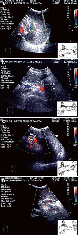
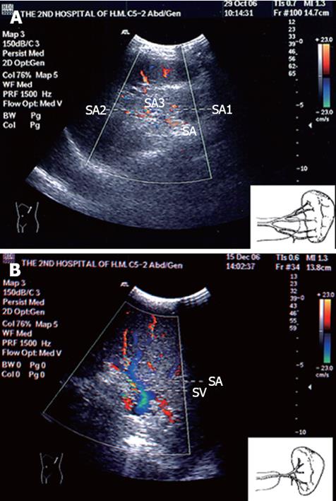
By adopting cross section below left costal margin, CDFI detected four different adjacent relations between three-fourths of the distal segments of spleen vessels and pancreas and two anatomic types of spleen pedicle end. The number and constituent ratio of the different anatomic parameters are listed in Table 1. The appearance of each anatomic parameter on CDFI was described and classified as types 1-4 (Figure 1A-D) and distributed and magistral types (Figure 2A-B). In type I, the limpid spleen arterial blood stream signal was detected constantly in front of the distal pancreatic body or tail and eventually the signal increased and distributed into spleen like a tree (Figure 1A). In type II, the color blood stream signal put forth from the rear of the pancreatic body passed by the pancreatic tail and went up or down to the hilum of spleen (Figure 1B). In type III, the blood stream signal was detected in front of the pancreatic body and suddenly disappeared in the rear of pancreatic tail (Figure 1C). In type IV, the obvious blood stream signal was not detected in front of pancreas although the probe was completely removed. Nevertheless, the vague color signal was found leading to spleen behind pancreas (Figure 1D). In distributed type, two or three color signals of scattering blood stream were detected leading to the hilum of spleen beside the pancreatic tail without signal of main blood stream (Figure 2A). In magistral type, the limpid color signal of main blood stream led to the hilum of spleen beside the pancreatic tail but the evident signal of branches was hard to be detected (Figure 2B).
| Anatomic parameters | CDFI | Intra-operative telerecording | χ2 | P |
| Anatomic types of the end of spleen vessels | ||||
| Distributed type | 23 (63.9) | 25 (69.4) | 0.25 | 0.617 |
| Magistral type | 13 (36.1) | 11 (30.6) | ||
| Adjacent relationships between the distal three-fourths segments of spleen vessels and pancreas | ||||
| Type I | 16 (44.5) | 17 (47.2) | 0.677 | 0.879 |
| Type II | 8 (22.2) | 6 (16.7) | ||
| Type III | 5 (13.9) | 4 (11.1) | ||
| Type IV | 7 (19.4) | 9 (25.0) | ||
The diameters of spleen artery and vein detected by CDFI were 0.46 ± 0.09 cm (range, 0.40-0.60 cm) and 0.85 ± 0.35 cm (range, 0.50-1.60 cm), respectively.
To compare the number and constituent ratio of the parameters recorded by CDFI and telerecording during LS; there was no significant statistical difference between them (Table 1).
CDFI is the latest diagnostic technique for the detection of heart and blood vessels without trauma by using Doppler principle combined with B model imaging and M model echocardiogram. Furthermore, CDFI can display the direction and relative velocity of blood stream and provide temporal and spatial information of blood vessels[7]. With a higher sensitivity and accuracy of diagnosis, CDFI can display 2-dimentional blood stream images directly and quickly[78]. CDFI has been used to prospectively assess resectable carcinoma in the head of pancreas and peri-ampulla since it can detect the tumor and blood vessels around it[9–11].
The spleen artery originates from the celiac artery and is divided into a segment upon the pancreas, a segment in the pancreas, a segment in front of the pancreas and a segment before the hilum of the spleen. One-fourth of the sub-terminal spleen artery segment keeps away from the pancreas. On the contrary, three-fourths of the distal pancreatic and spleen vessel segments are close to each[12–14]. In this study, the adjacent relations between spleen vessels and the pancreas were detected by preoperative CDFI. Four types of segment were found on CDFI. In type I, three-fourths of the distal segments walked along the superior border of the pancreas constantly from celiac artery to hilum of spleen. In type II, two-fourths of the middle segments were located behind or in the pancreas while one-fourth of the distal segments were located at the antero-superior border of the pancreas. In type III, two-fourths of the distal segments walked behind or in the pancreas. In type IV, three-fourths of the distal segments were located behind or in the pancreas. Most adjacent relations belonged to type I (approximately 45%), but the other types are consistent with those reported in the latest literature[1314].
The anatomy of the spleen artery can be divided into magistral type and distributed type according to Michels’ classification[1516]. In this study, CDFI revealed that the anatomy of the spleen pedicle could also be divided into magistral type and distributed type. About 70% belonged to distributed type and 30% belonged to magistral type. The overall categorization accuracy of spleen arterial geography was perfect according to Michels’ classification.
Since hemorrhage is the major reason of conversion during LS, which is closely related to trauma of spleen pedicle and segment[17–20], operation can be performed safely and effectively if the surgeon fully understands the anatomic information of the spleen vessels[21]. In this study, the anatomic types of spleen pedicle and the adjacent relations between spleen vessels and pancreas were detected by preoperative CDFI and compared with the actual anatomy during LS. The results showed that their constituent ratio had no significant statistical difference and the coincidence rate almost reached 92% (23/25), suggesting that preoperative CDFI provides significant anatomic information of the spleen, thus operative procedures can be performed for type I and type II spleen artery segments, the trunk of spleen artery can be ligated to control intra-operative hemorrhage and megalosplenia[522–24]. For type III and type IV spleen artery segments, surgeons should elevate the anus perineum of spleen, separate anadesma tissue around the pancreatic tail and reveal the spleen pedicle.
In regard to the anatomic types of spleen pedicle, CDFI may lead surgeons to hilar approach strategy, particularly during the learning curve time. Surgeons may use the vascular stapler to ligate the trunk of spleen pedicle directly when a magisterial type of vascularization is present, and use clips to separate and ligate the secondary vessels of spleen pedicle when the distributed type is present, which prevents damage to the pancreatic tail and decreases the incidence rate of spleen fever[25]. In addition, surgeons can judge the trunk or the branches of spleen vessels to be ligated according to the diameters of spleen vessels shown by CDFI.
In our study, CDFI was limited to the body corresponded to the caudal extension of the spleen, which prevented a complete assessment of the remote spleen vessels.
Individual operation procedure can be formed according to the anatomic parameters of spleen provided by preoperative CDFI which can increase the safety and shorten intra-operative hemorrhage rate.
Laparoscopic splenectomy can be performed safely and effectively only when surgeons are familiar with the anatomy of spleen vessels. Color Doppler imaging plays an important role in location of target vessels. The value of color Doppler flow imaging (CDFI) for spleen micro-invasive operation was explored in this study.
Conversion often occurs during laparoscopic splenectomy which is closely related to trauma of pancreas and intra-operative hemorrhage. Since methods such as magnetic resonance angiography may result in additional traumas and heavy economic burden on the patients, they are often not used. However, color Doppler flow imaging (CDFI) can provide anatomic information of spleen vessels not leading to trauma, thus make laparoscopic splenectomy more safe and effective.
Individual operative procedures can be formed according to the anatomic parameters of spleen provided by preoperative CDFI. As a commonly used examination, CDFI may have an application prospect in micro-invasive surgery of spleen.
CDFI is the latest diagnostic technique for the detection of heart and blood vessels without trauma by use of Doppler principle combined with B model imaging and M model echocardiogram. Furthermore, CDFI can display the direction and relative velocity of blood stream and provide temporal and spatial information of blood vessels.
In this manuscript, the authors evaluated and classified the anatomy of spleen vessels into four types and two large categories based on the preoperative CDFI and compared the results with intra-operative recordings. The results are acceptable and helpful. The statistical analysis and all figures are adequate.
| 1. | Li SL. Application of laparoscopic splenectomy in hepatic cirrhosis portal hypertension with hypersplenia. Zhonghua Gandan Waike Zazhi. 2004;10:203-204. |
| 2. | Kucuk C, Sozuer E, Ok E, Altuntas F, Yilmaz Z. Laparoscopic versus open splenectomy in the management of benign and malign hematologic diseases: a ten-year single-center experience. J Laparoendosc Adv Surg Tech A. 2005;15:135-139. |
| 3. | Pomp A, Gagner M, Salky B, Caraccio A, Nahouraii R, Reiner M, Herron D. Laparoscopic splenectomy: a selected retrospective review. Surg Laparosc Endosc Percutan Tech. 2005;15:139-143. |
| 4. | Patel AG, Parker JE, Wallwork B, Kau KB, Donaldson N, Rhodes MR, O'Rourke N, Nathanson L, Fielding G. Massive splenomegaly is associated with significant morbidity after laparoscopic splenectomy. Ann Surg. 2003;238:235-240. |
| 5. | Machado MA, Makdissi FF, Herman P, Montagnini AL, Sallum RA, Machado MC. Exposure of splenic hilum increases safety of laparoscopic splenectomy. Surg Laparosc Endosc Percutan Tech. 2004;14:23-25. |
| 6. | Patel AG, Parker JE, Wallwork B, Kau KB, Donaldson N, Rhodes MR, O'Rourke N, Nathanson L, Fielding G. Massive splenomegaly is associated with significant morbidity after laparoscopic splenectomy. Ann Surg. 2003;238:235-240. |
| 7. | Li FX, Jin BR, Wang CJ. The principle and quality control of color Doppler flow imaging. Yiyong Wuli Zazhi. 2007;20:396-397. |
| 8. | Modrzejewska M, Pienkowska-Machoy E, Grzesiak W, Karczewicz D, Wilk G. Predictive value of color Doppler imaging in an evaluation of retrobulbar blood flow perturbation in young type-1 diabetic patients with regard to dyslipidemia. Med Sci Monit. 2008;14:MT47-MT52. |
| 9. | Li FS, Huo XL, Ma SR, Li J, Chen LX. CDFI Assessment of Resectable Carcinoma in pancreatic Head and Peri-ampulla. Zhongguo Chaosheng Yixue Zazhi. 2002;18:621-624. |
| 10. | Janssen J. [(E)US elastography: current status and perspectives]. Z Gastroenterol. 2008;46:572-579. |
| 11. | Janssen J, Schlorer E, Greiner L. EUS elastography of the pancreas: feasibility and pattern description of the normal pancreas, chronic pancreatitis, and focal pancreatic lesions. Gastrointest Endosc. 2007;65:971-978. |
| 12. | Li SL. Pediatric laparoscopic splenectomy with splenic pedicle ligation. Zhonghua Xiandai Erke Zazhi. 2005;2:989-991. |
| 13. | Xia SS. The anatomy of spleen. Modern Splenic Surgery. Jiangsu Science and Technique Publishing Company: Nanking 2000; 4-12. |
| 14. | Pandey SK, Bhattacharya S, Mishra RN, Shukla VK. Anatomical variations of the splenic artery and its clinical implications. Clin Anat. 2004;17:497-502. |
| 15. | Poulin EC, Thibault C. The anatomical basis for laparoscopic splenectomy. Can J Surg. 1993;36:484-488. |
| 17. | Romano F, Caprotti R, Franciosi C, De Fina S, Colombo G, Uggeri F. Laparoscopic splenectomy using Ligasure. Preliminary experience. Surg Endosc. 2002;16:1608-1611. |
| 18. | Torelli P, Cavaliere D, Casaccia M, Panaro F, Grondona P, Rossi E, Santini G, Truini M, Gobbi M, Bacigalupo A. Laparoscopic splenectomy for hematological diseases. Surg Endosc. 2002;16:965-971. |
| 19. | Hashimoto M, Matsuda M, Watanabe G. Simple method of laparoscopic splenectomy. Surg Endosc. 2008;22:2524-2526. |
| 20. | Rescorla FJ, West KW, Engum SA, Grosfeld JL. Laparoscopic splenic procedures in children: experience in 231 children. Ann Surg. 2007;246:683-687; discussion 687-688. |
| 21. | Kercher KW, Matthews BD, Walsh RM, Sing RF, Backus CL, Heniford BT. Laparoscopic splenectomy for massive splenomegaly. Am J Surg. 2002;183:192-196. |
| 22. | Xu WL, Li SL, Shi BJ, Yu ZW, Zhong ZY, Li ZD. Laparoscopic resection of massive splenomegaly for hereditary spherocytosis in children: Report of 7 cases. Zhonguo Weichuang Waike Zazhi. 2005;5:694-707. |
| 23. | Yuney E, Hobek A, Keskin M, Yilmaz O, Kamali S, Oktay C, Bender O. Laparoscopic splenectomy and LigaSure. Surg Laparosc Endosc Percutan Tech. 2005;15:212-215. |
| 24. | Asoglu O, Ozmen V, Gorgun E, Karanlik H, Kecer M, Igci A, Unal ES, Parlak M. Does the early ligation of the splenic artery reduce hemorrhage during laparoscopic splenectomy? Surg Laparosc Endosc Percutan Tech. 2004;14:118-121. |
| 25. | Ying FM, Feng XF. Application of secondary splenic pedicle ligation in laparoscopic splenectomy. Zhongguo Neijing Zazhi. 2004;10:83-84. |


