Published online Aug 14, 2009. doi: 10.3748/wjg.15.3831
Revised: September 16, 2008
Accepted: July 3, 2009
Published online: August 14, 2009
We present a rare case of a 45-year-old woman who presented with epigastric pain associated with early satiety and weight loss. Imaging revealed a large intra-abdominal mass in the epigastrium. Despite intensive investigations, including ultrasound scanning, computed tomography, upper gastrointestinal endoscopy, and percutaneous biopsy, a diagnosis could not be obtained. A histological diagnosis of cavernous hemangioma arising from the gastro-splenic ligament was confirmed after laparoscopic excision and histological examination of the intra-abdominal epigastric mass.
- Citation: Chin KF, Khair G, Babu PS, Morgan DR. Cavernous hemangioma arising from the gastro-splenic ligament: A case report. World J Gastroenterol 2009; 15(30): 3831-3833
- URL: https://www.wjgnet.com/1007-9327/full/v15/i30/3831.htm
- DOI: https://dx.doi.org/10.3748/wjg.15.3831
Cavernous hemangiomas are congenital hamartomatous lesions that originate from the mesodermal tissues, which are composed of large dilated blood vessels and contain large blood-filled spaces that are caused by dilation and thickening of the walls of the capillary loops. They have been reported in different organs, including the liver, colon, retroperitoneum, spleen, adrenal glands, soft tissues, bone, central nervous system, and mediastinum[1]. However, it is extremely rare for these tumors to originate from the mesentery[2] or omentum[3]. In this case report, we present the clinical presentation, diagnosis and treatment of a cavernous haemangioma that arose from the gastro-splenic ligament.
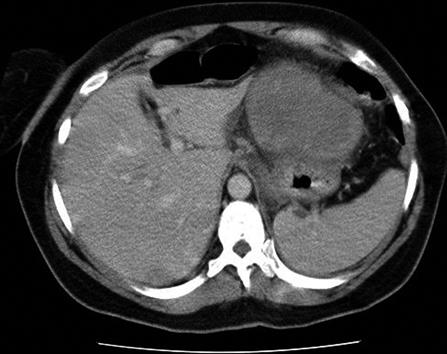
A 45-year-old woman presented with sudden-onset epigastric pain of 2 h duration after eating a meal. She described her pain as a dull ache that radiated to the back, which was associated with early satiety, retching, vomiting and recent weight loss of approximately 6.35 kg. Her past medical history included laparoscopic cholecystectomy and uterine fibroid embolization. She was taking regular omeprazole for dyspepsia. Upon examination, she had neither jaundice nor anemia, and her vital signs were within normal limits. She had no supraclavicular lymphadenopathy. The abdominal examination revealed epigastric tenderness with no guarding or rigidity, and fullness in the left upper quadrant. Her initial laboratory blood results including liver function tests, amylase and full blood count were within normal limits. An abdominal ultrasound scan revealed a large 10.5 cm × 7.5 cm × 6.5 cm solid mass in the epigastrium anterior and to the left of the tail of the pancreas, and a mildly enlarged spleen, normal liver, pancreas and kidneys, and a fibroid uterus with an intrauterine device in situ. The ultrasonic appearance of the epigastric mass was not typical of a gastrointestinal stromal tumor (GIST) or lymphoma. Subsequent computed tomography (CT) of the chest and abdomen showed a well-circumscribed mass of mixed attenuation in the left upper abdomen, which was probably related to the stomach, and measured 10 cm in maximum diameter (Figure 1). In order to rule out gastric cancer, she underwent upper gastrointestinal endoscopy that showed mild reflux oesophagitis and a normal stomach. The diagnosis remained unclear despite an ultrasound-guided biopsy of the epigastric mass, which showed ectatic lymphovascular vessels with a fibrous stroma, with no evidence of malignancy upon histology.
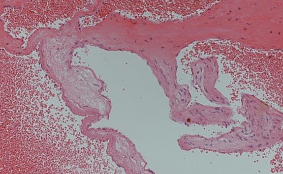
Laparoscopic excision of the intra-abdominal epigastric mass was performed. The intraoperative findings showed that the encapsulated mass, which was related to the omentum and gastro-splenic ligament, was adherent to but not directly invading the anterior wall of the stomach. The mass was dissected off the anterior wall of the stomach, and was mobilized from the omentum and gastro-splenic ligament with laparoscopic LigaSure AdvanceTM device (Covidien Ltd). The mass with an intact capsule was delivered through a Pfannenstiel incision in the suprapubic region. The microscopic specimen showed a hemorrhagic lesion that consisted of numerous thick-walled blood vessels filled with blood, surrounded by fibrosis and a few adjacent, benign-looking simple ductules, which were characteristic of cavernous hemangioma (Figure 2).
The patient was discharged 2 d postoperatively with no complications.
Cavernous hemangiomas of the omentum and mesentery are extremely rare. These tumors frequently are asymptomatic and discovered incidentally at imaging, surgery or autopsy, but can have serious consequences. The clinical presentation of such a lesion varies depending on its location. Our patient presented with symptoms of abdominal pain, vomiting, early satiety and weight loss. In other case reports, these lesions presented as intermittent abdominal pain and dyspepsia[3], palpable abdominal mass[24], or acute abdomen caused by ruptured omental cavernous hemangioma[5], ruptured mesoappendix cavernous hemangioma[6], or an infected greater omental cavernous hemangioma[7].
The diagnosis of such lesions can be challenging, and ultrasonography and CT have been used. Ultrasound scanning reveals a solid mass with a heterogeneous multinodular appearance, whereas CT reveals a homogenous or a heterogeneous mass[6], or a mixed picture[4].
Magnetic resonance imaging of cavernous hemangioma typically shows a uniform high signal intensity on T2-weighted images[34], but occasionally shows heterogeneous signal intensity on T2-weighted images, as a result of fibrosis, hemorrhage or calcification[8], or low signal intensity in T2-weighted images because the tumor is mainly occupied with old blood[2]. In hindsight, this resectable lesion should not be biopsied percutaneously, as it may spread the tumor through other anatomical planes, as in the case of malignant GIST. In addition, a small needle biopsy may produce a poor cellular yield, and the quality of the sample may not be adequate to make an accurate diagnosis. In this case of cavernous hemangioma, a biopsy may potentially cause a rupture or bleeding of the tumor.
In patients with gastrointestinal bleeding of obscure origin who are transfusion-dependent, wireless capsule endoscopy or double-balloon enteroscopy may allow examination of the entire small bowel and successful identification of hemangioma within the bowel lumen. Willert et al[9] have reported successful endoscopic treatment of multiple cavernous hemangioma during double-balloon enteroscopy of the small bowel. However, in our case report of cavernous hemangioma located at the gastro-splenic ligament, capsule endoscopy and double-balloon enteroscopy were unlikely to have been beneficial, as the lesion was located extraluminally. On the other hand, endoscopic ultrasound may help to characterize the extent of vascularity of the hemangioma, and allow selection of cases for endoscopic mucosal resection of cavernous hemangioma in the wall of the esophagus[10] or stomach[11].
The treatment for cavernous hemangioma is surgical excision, and recurrence after complete resection has never been reported. Laparoscopic excision of this vascular lesion is feasible, and has the advantages of less pain and quicker recovery following surgery.
In conclusion, cavernous hemangioma of the omentum or mesentery is a rare tumor that can be very difficult to diagnose preoperatively, despite advanced imaging techniques. As a result, it should be included in the differential diagnosis of any mass that arises in the mesentery or omentum. Surgical excision and histological examination may offer the only means of diagnosis.
| 1. | Kinoshita T, Naganuma H, Yajima Y. Venous hemangioma of the mesocolon. AJR Am J Roentgenol. 1997;169:600-601. |
| 2. | Takamura M, Murakami T, Kurachi H, Kim T, Enomoto T, Narumi Y, Nakamura H. MR imaging of mesenteric hemangioma: a case report. Radiat Med. 2000;18:67-69. |
| 3. | Chung J, Kim M, Lee JT, Yoo HS. Cavernous hemangioma arising from the lesser omentum: MR findings. Abdom Imaging. 2000;25:542-544. |
| 4. | Chateil JF, Saragne-Feuga C, Pérel Y, Brun M, Neuenschwander S, Vergnes P, Diard F. Capillary haemangioma of the greater omentum in a 5-month-old female infant: a case report. Pediatr Radiol. 2000;30:837-839. |
| 5. | Ritossa C, Ferri M, Destefano I, De Giuli P. [Hemoperitoneum caused by cavernous angioma of the omentum]. Minerva Chir. 1989;44:907-908. |
| 6. | Hanatate F, Mizuno Y, Murakami T. Venous hemangioma of the mesoappendix: report of a case and a brief review of the Japanese literature. Surg Today. 1995;25:962-964. |
| 7. | Slizovskiĭ GV, Timashov BA. [An infected cavernous hemangioma of the greater omentum in a child]. Vestn Khir Im I I Grek. 1995;154:69. |
| 8. | Ros PR, Lubbers PR, Olmsted WW, Morillo G. Hemangioma of the liver: heterogeneous appearance on T2-weighted images. AJR Am J Roentgenol. 1987;149:1167-1170. |
| 9. | Willert RP, Chong AK. Multiple cavernous hemangiomas with iron deficiency anemia successfully treated with double-balloon enteroscopy. Gastrointest Endosc. 2008;67:765-767. |
| 10. | Sogabe M, Taniki T, Fukui Y, Yoshida T, Okamoto K, Okita Y, Hayashi H, Kimuara E, Kimura Y, Onose Y. A patient with esophageal hemangioma treated by endoscopic mucosal resection: a case report and review of the literature. J Med Invest. 2006;53:177-182. |
| 11. | Arafa UA, Fujiwara Y, Shiba M, Higuchi K, Wakasa K, Arakawa T. Endoscopic resection of a cavernous haemangioma of the stomach. Dig Liver Dis. 2002;34:808-811. |


