Published online May 28, 2009. doi: 10.3748/wjg.15.2547
Revised: April 14, 2009
Accepted: April 21, 2009
Published online: May 28, 2009
A 78-year-old woman presented with fever, severe abdominal pain, and distension. She had been institutionalized for depression and senile dementia. Laboratory examinations disclosed a leucocytosis (WBC: 12 500/&mgr;L) and elevated levels of serum C-reactive protein (2.8 mEq/L). Diagnosis of acute cecal volvulus was made from a “coffee bean sign” on an abdominal computed tomography and a “beak sign” on a gastrographin enema. An emergent laparotomy confirmed the diagnosis and an ileo-colectomy with primary anastomosis was carried out. The patient recovered after intensive respiratory care and fluid therapy, and then returned to her former institution. A review of Japanese literature disclosed that: (1) a marked increase of aged patients with mental disability presenting with cecal volvulus, (2) adoption of ileo-colectomy as the standard surgical procedure, and (3) improved survival of the patients, were observed in the last decade.
- Citation: Katoh T, Shigemori T, Fukaya R, Suzuki H. Cecal volvulus: Report of a case and review of Japanese literature. World J Gastroenterol 2009; 15(20): 2547-2549
- URL: https://www.wjgnet.com/1007-9327/full/v15/i20/2547.htm
- DOI: https://dx.doi.org/10.3748/wjg.15.2547
Cecal volvulus is axial twisting that occurs involving the cecum, terminal ileum, and ascending colon. Rarely, it may take the form of upward and anterior folding of the ascending colon (“cecal bascule”)[1]. Cecal volvulus is a rare condition, and its incidence is reported to range from 2.8 to 7.1 per million people per year[1].
Clinical presentation is highly variable, ranging from intermittent episodes of abdominal pain to abdominal catastrophe[23]. In this paper, we report a case of cecal volvulus seen in a 78-year-old woman. Ages at presentation, causative factors, treatment and outcome of 40 cases reported in Japan between 1999 and 2008 were also reviewed, and compared with those in cases reported before 1988[4] .
A 78-year-old woman was admitted with acute abdominal pain and distension. She had an operation for gastric cancer at the age of 73, and had been institutionalized for depression and dementia.
On admission, she had a high fever of 38.8°C. Her abdomen was diffusely distended with rebound tenderness. Laboratory examinations disclosed a leucocytosis (WBC: 12 500/&mgr;L) and elevated serum C-reactive protein levels (2.8 mEQ/L).
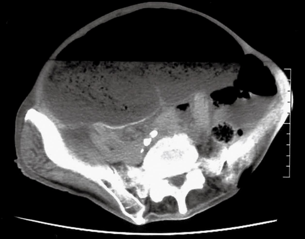
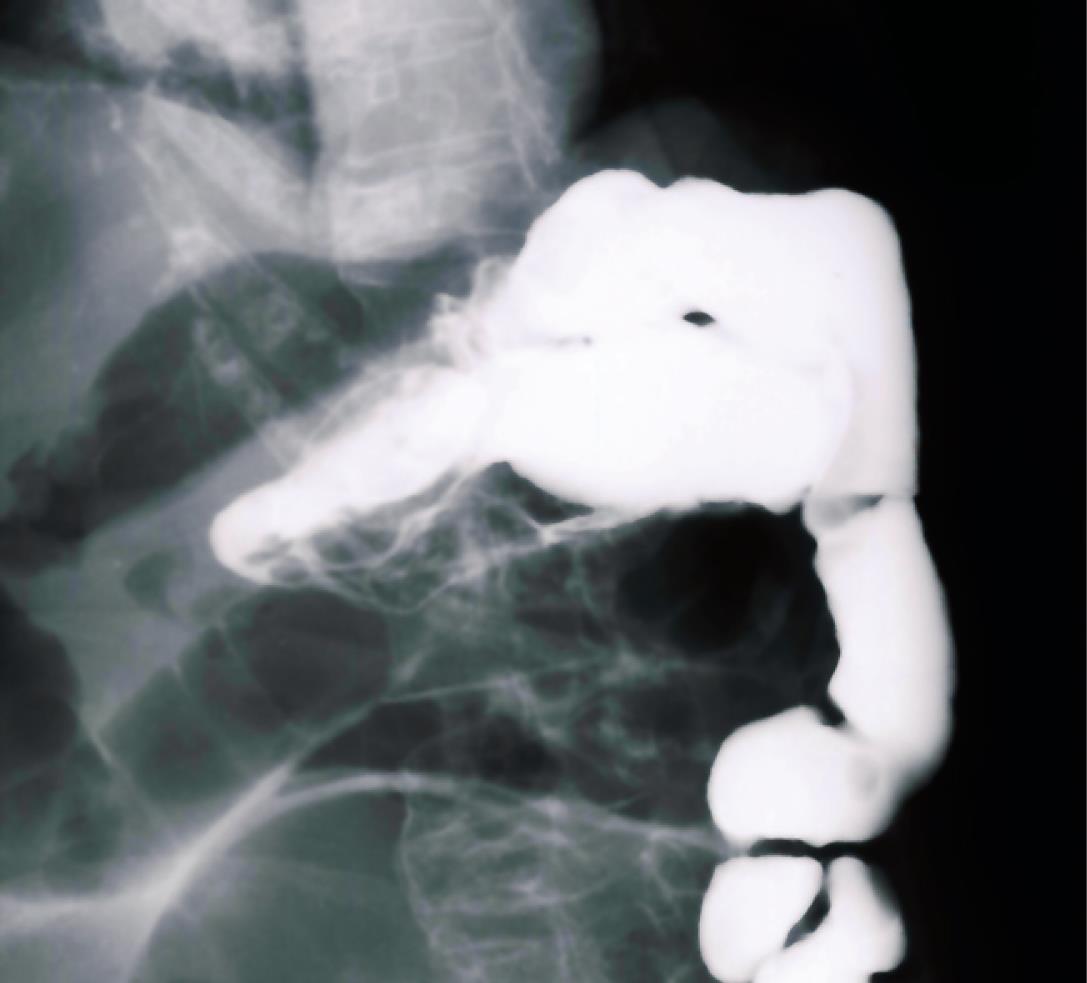
Plain radiographs of the abdomen showed a markedly dilated loop of the intestine, occupying most of the abdomen, but the pathology was not certain. A “coffee-bean sign”, on an abdominal CT (Figure 1), together with “a beak sign” on a gastrogrephin enema (Figure 2), confirmed the diagnosis of cecal volvulus[35].
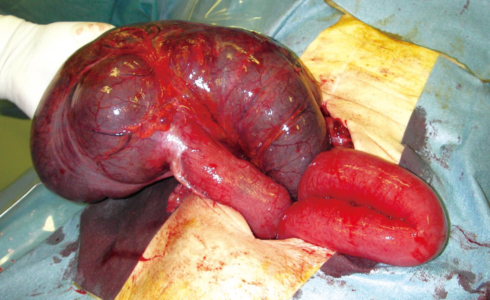
An emergent laparotomy disclosed the axial twisting of the cecum, involving the terminal ileum and the ascending colon. Ischemia of the bowel seemed to be irreversible, but there was no perforation (Figure 3).
An ileo-colectomy with primary anastomosis was carried out. Postoperatively, the patient was placed under close monitoring and managed by an intensive respiratory care and fluid therapy until stabilized. Then she was discharged and returned to her former institution.
The incidence of cecal volvulus is reported to range from 2.8 to 7.1 per million people per year[1]. Patients’ ages at presentation, the treatment of choice, and survival differ by reports chronologically as well as geographically[23].
We compared ages at presentation, treatment of choice, and outcome of 46 patients reported in Japan before 1988[4] with reports of 40 patients collected from a search of Japan Centra Revus Medicina in the last 10 years.
Patients’ age at the presentation are said to be affected by cultural and dietary influences. Gupta and Gupta[6] reported that the average age at presentation in Western countries was 53 years, whereas it was 33 years in India. In Japan, patients treated before 1988 showed two peaks in ages at presentation; one in the 10-29 years range and another in the 60-79 years range. On the other hand, there was only one peak in ages of the patients treated between 1999 and 2008; 70-89 years of age (Table 1). In the young age group, the volvulus was caused by mesenterium commune, other intestinal malformation, or excessive exercise. In contrast, volvulus in the aged patients was associated with chronic constipation, distal colon obstruction, or senile dementia.
| Age | Patients treated before 1988 | Patients treated between 1999 and 2008 |
| 0 to 9 | 3 | 1 |
| 10 to 19 | 9 | 4 |
| 20 to 29 | 5 | 3 |
| 30 to 39 | 5 | 2 |
| 40 to 49 | 3 | 2 |
| 50 to 59 | 4 | 3 |
| 60 to 69 | 10 | 3 |
| 70 to 79 | 50 | 12 |
| 80 to 89 | 2 | 8 |
| Over 80 | 0 | 2 |
Surgical options include cecopexy, cecostomy, and ileo-colectomy by open or laparoscopic approaches. Review of Japanese literature disclosed that resectional surgery was done in 32.6% (15/46) of patients who were treated before 1988, whereas 70% (28/40) of patients underwent ileo-colectomy between 1999 and 2008 (Table 2). This significant increase of the patients treated by resectional surgery (P < 0.0001 by χ2 test) was brought about by advances in surgical techniques and perioperative supportive measures.
| Patients treated before 1988 | Patients treated 1999 to 2008 | |
| Endoscopic detorsion | 0 | 4 |
| Surgery | 45 | 36 |
| Detorsion | 9 | 0 |
| Cecopexy | 14 | 8 |
| Cecostomy | 7 | 0 |
| Ileo-Colectomy | 15 | 28 |
| Unknown | 1 | 0 |
Flexible colonoscopy is commonly used for the diagnosis and the treatment of sigmoid volvulus, but the utility of endoscopy in acute cecal volvulus is limited, because of technical problems and higher rates of recurrence[1]. Our review also showed that reports on colonoscopic detorsion are just emerging (Table 2).
Mortality, morbidity, and recurrence rates were also lower in recent years. O’Mara et al[7] reported in 1979 that they treated 14 patients with cecal volvulus by ileo-colectomy and lost 2 of 7 patients who developed gangrenous bowel, while there were no deaths amongst 7 patients with non-gangrenous bowel. Gupta et al[6] reported that they encountered operative death in 2 of 13 patients who underwent resectional surgery. However, Majeski et al[8] reported in 2005 that there was no operative deaths in 10 patients treated by ileo-colectomy. Our review of Japanese literature disclosed that the outcome after surgical treatment has markedly improved in the last 10 years. Before1988, there were five postoperative death in 45 patients who underwent surgery (15 resectional, and 30 non-resectional), whereas only one of 36 patients treated surgically (8 resectional and 28 non-resectional) between 1999 and 2008 was lost.
As the mortality and the morbidity after resectional surgery for acute cecal volvulus, whether gangrenous or non-gangrenous, have been markedly improved and almost no recurrence is observed, an ileo-colectomy with primary anastomosis should be the preferred surgical option rather than cecopexy or a cecostomy.
| 1. | Consorti ET, Liu TH. Diagnosis and treatment of caecal volvulus. Postgrad Med J. 2005;81:772-776. |
| 2. | Madiba TE, Thomson SR. The management of cecal volvulus. Dis Colon Rectum. 2002;45:264-267. |
| 4. | Masuda R, Isoyama T, Bandou T, Toyoshima H. Cecal volvulus: Report of a case and review of cases in Japan. Nihondaichoukoumonnbyoukaishi. J Jpn Soc Coloproctol. 1988;41:34-38. |
| 5. | Moore CJ, Corl FM, Fishman EK. CT of cecal volvulus: unraveling the image. AJR Am J Roentgenol. 2001;177:95-98. |
| 6. | Gupta S, Gupta SK. Acute caecal volvulus: report of 22 cases and review of literature. Ital J Gastroenterol. 1993;25:380-384. |


