Copyright
©2009 The WJG Press and Baishideng.
World J Gastroenterol. Jan 14, 2009; 15(2): 160-168
Published online Jan 14, 2009. doi: 10.3748/wjg.15.160
Published online Jan 14, 2009. doi: 10.3748/wjg.15.160
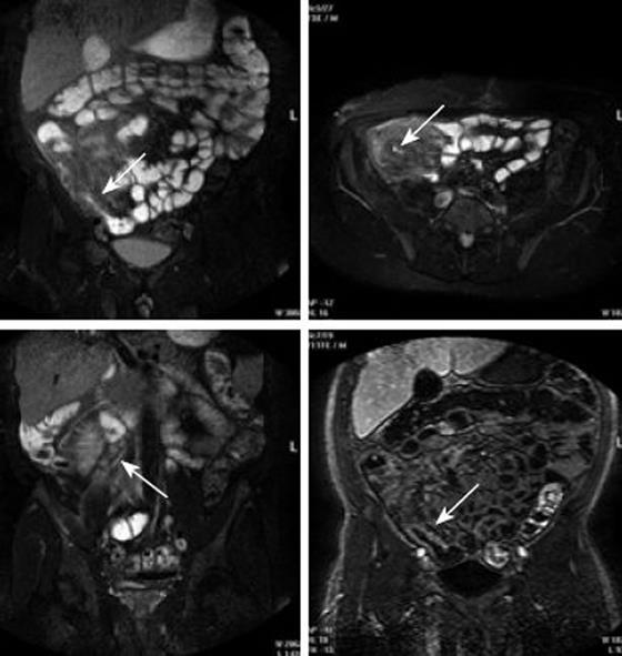
Figure 6 MRI of Crohn’s disease.
The coronal and axial (upper panel) fat-saturated T2-weighted MRI display marked wall thickening, mucosal irregularity and stenosis of the terminal ileum (arrows). Advanced mesenteric inflammation with hypervascularity and enlarged lymph nodes (arrow) are visualised on coronal fat-saturated T2-weighted MRI (lower left). Coronal T1-weighted MRI (lower right) shows clear wall enhancement (arrow). Modified from [36].
- Citation: Frøkjær JB, Drewes AM, Gregersen H. Imaging of the gastrointestinal tract-novel technologies. World J Gastroenterol 2009; 15(2): 160-168
- URL: https://www.wjgnet.com/1007-9327/full/v15/i2/160.htm
- DOI: https://dx.doi.org/10.3748/wjg.15.160


