Review Open Access
Copyright ©2009 The WJG Press and Baishideng. All rights reserved.
World J Gastroenterol. Apr 14, 2009; 15(14): 1702-1707
Published online Apr 14, 2009. doi: 10.3748/wjg.15.1702
Reactive oxygen species: A double-edged sword in oncogenesis
Jin-Shui Pan, Jian-Lin Ren, Division of Gastroenterology, Zhongshan Hospital Xiamen University, Xiamen 361004, Fujian Province, China; Gastroenterology Institute of Xiamen University, Xiamen 361004, Fujian Province, China; Gastroenterology Center of Xiamen, Xiamen 361004, Fujian Province, China
Mei-Zhu Hong, Division of Infectious Diseases, the 174th Hospital of PLA, Xiamen 361003, Fujian Province, China
Author contributions: Pan JS and Hong MZ contributed equally to this work; Pan JS and Hong MZ generated the idea, wrote the paper; Ren JL proofread the paper.
Correspondence to: Jian-Lin Ren, Professor, Division of Gastroenterology, Zhongshan Hospital Xiamen University, Xiamen 361004, Fujian Province, China. jianlinr@msn.com
Telephone: +86-592-2993170
Fax: +86-592-2993170
Received: February 3, 2009
Revised: March 17, 2009
Accepted: March 24, 2009
Published online: April 14, 2009

Abstract

Reactive oxygen species (ROS) are molecules or ions formed by the incomplete one-electron reduction of oxygen. Of interest, it seems that ROS manifest dual roles, cancer promoting or cancer suppressing, in tumorigenesis. ROS participate simultaneously in two signaling pathways that have inverse functions in tumorigenesis, Ras-Raf-MEK1/2-ERK1/2 signaling and the p38 mitogen-activated protein kinases (MAPK) pathway. It is well known that Ras-Raf-MEK1/2-ERK1/2 signaling is related to oncogenesis, while the p38 MAPK pathway contributes to cancer suppression, which involves oncogene-induced senescence, inflammation-induced cellular senescence, replicative senescence, contact inhibition and DNA-damage responses. Thus, ROS may not be an absolute carcinogenic factor or cancer suppressor. The purpose of the present review is to discuss the dual roles of ROS in the pathogenesis of cancer, and the signaling pathway mediating their role in tumorigenesis.

Key Words: p38 mitogen-activated protein kinases; Reactive oxygen species; Signal transduction; Tumorigenesis


INTRODUCTION

Reactive oxygen species (ROS) are molecules or ions formed by the incomplete one-electron reduction of oxygen. These reactive oxygen intermediates include singlet oxygen, superoxides, peroxides, hydroxyl radical, and hypochlorous acid. They contribute to the microbicidal activity of phagocytes, regulation of signal transduction and gene expression, and induce oxidative damage to nucleic acids, proteins, and lipids. Peroxidation by ROS alters the amounts of unsaturated fatty acids and proteins in the cell membrane and thus affects membrane fluidity. In addition, with aging, humans tend to show an increased affectability of lipid peroxides caused by ROS[1]. Recent research has indicated that ROS also play a critical role in the energy dysfunction of mitochondria caused by ethanol-induced gastric mucosa injury[2]. In addition, oxidative damage caused by ROS and other free radicals is involved in a number of pathological conditions including cancer. Data presented herein is consistent with this opinion. Yagoda et al[3] found erastin interacted with voltage-dependent anion channel proteins to induce mitochondrial dysfunction, release of oxidative species and, ultimately, non-apoptotic, oxidative cell death. This process has a degree of selectivity for cells with activated Ras-Raf-MEK signaling. ROS production also involves the induction of autophagy, which contributes to caspase-independent macrophage cell death[4]. ROS, produced in the redox cycle, contribute to p53 mutations, which are dominated by G-to-T transversions. These mutations are suppressed by ROS attenuators[56]. The mutations of p53, a well characterized tumor suppressor, are believed to relate to carcinogenesis. Since oxidative stress comprehensively damages cells and tissues, it is reasonable that factors which induce ROS would contribute to the occurrence and development of tumors, while antioxidant agents that scavenge ROS may inhibit this process. To the best of our knowledge, the former inference is consistent with previous reports[78], however, there is little supportive evidence for the latter[910]. Vitamins C and E, two reducing agents and antioxidants, show no additional benefit in the chemoprevention of gastric cancer[1112]. Inadequate dose, heterogeneous research, poor compliance and multiple effects of antioxidants may lead to this paradox. Is there anything more to be elucidated on this subject?

REGULATION OF ROS PRODUCTION BY RAS

Oxidative stress and Ras activation lead to the production of ROS[13]. Introduction of ROS by Ras may occur at the transcription level. GATA-6 is a component of the specific protein-DNA complexes at the nicotinamide adenine dinucleotide phosphate oxidase (Nox) 1 promoter, and is able to trans-activate the Nox1 promoter. GATA-6 is phosphorylated at serine residues by MEK-activated extracellular signal-regulated kinase (ERK), which enhances GATA-6 DNA binding. The site-directed mutation of the consensus ERK phosphorylation site (PYS(120)P to PYA(120)P) of GATA-6 abolishes its trans-activation activity, suppressing the growth of CaCo-2 cells. By MEK-ERK-dependent phosphorylation of GATA-6, oncogenic Ras signaling enhances the transcription of Nox1[14]. A regulatory subunit, Rac, of the NADPH oxidase complex also involves the regulation of ROS[15]. Other factors that regulate the production of ROS will not be discussed here.

ROS INVOLVE TUMORIGENESIS THAT RELATES TO THE RAS-RAF-MEK-ERK PATHWAY

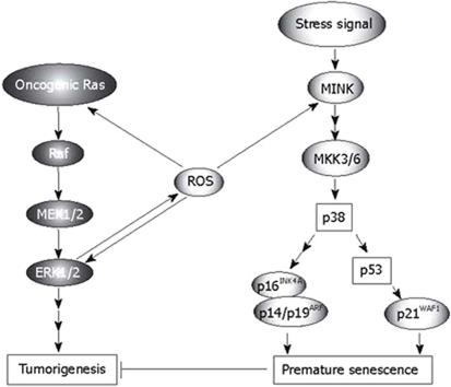
Growth factors, cellular stress, and γ radiation stimulate oncogenic Ras-Raf-MEK signaling, which plays a crucial role in tumorigenesis. As an important mediator of physiological and pathological signal-transduction pathways, ROS is also involved in Ras-Raf-MEK signaling (Figure 1). The functions of ROS in tumorigenesis relating to this pathway include the following. (1) In cells with activated Ras-Raf-MEK signaling, released ROS cause non-apoptotic, oxidative cell death, as previously mentioned[3], and the Ras-ERK pathway is critical in mediating protection against apoptotic cell death induced by increased oxidative stress[16]. (2) The activity of the ROS-generating enzyme Nox1 is required for vascular endothelial growth factor (VEGF), a potent stimulator of tumor angiogenesis. However, if extracellular signal-regulated kinase (ERK)-dependent phosphorylation of the transcription factor Sp1 and Sp1 binding to a VEGF promoter is inhibited, this activity does not occur. Nox1 mediates oncogenic Ras-induced upregulation of VEGF and angiogenesis by activating Sp1 through Ras-ERK-dependent phosphorylation of Sp1[8]. (3) Ras (p19) interaction with p73β, a structural and functional homolog of p53, amplifies p73β-induced apoptotic signaling responses including Bax mitochondrial translocation, cytochrome c release, increased production of ROS and loss of mitochondrial transmembrane potential. After taxol treatment, endogenous expression of Ras and p73β significantly increase, and taxol-enhanced endogenous p73β transcriptional activities are further amplified by p19, which markedly increases cellular apoptosis in the p53-null SAOS2 cancer cell line[17]. (4) In human U937 monocytes, hydrogen peroxide (H2O2) evokes Ca2+ influx through TRPM2 to activate Ca2+-dependent tyrosine kinase Pyk2 and amplify ERK signaling via Ras GTPase. TRPM2 Ca2+ influx controls the ROS-induced signaling cascade responsible for chemokine production, which aggravates inflammation[18].

Figure 1
Figure 1 Roles of ROS in the Ras-Raf-MEK-ERK signaling and p38 pathway (Modified from Ref. 31, with permission).

In contrast, the released ROS have complicated effects on Ras-Raf-MEK signaling, which may occur on several levels (Figure 1). ROS directly enhance the activation of Ras[19], and augment ERK1/2[20]. Melatonin, a natural antioxidant, inhibits the activation of Ras in H4IIE hepatoma cells[21]. Generation of ROS is required for Ras transformation phenotypes including anchorage-independent growth, morphological transformation, and tumorigenesis[22]. In diabetes-related angiogenesis of the retina, activation of H-Ras and its downstream signaling pathway may be under the control of superoxide, and H-Ras activation in diabetes can be prevented by inhibiting superoxide accumulation[23]. H2O2 activates H-Ras and its downstream signaling pathway, including Raf-1 and phosphorylation of p38 MAP kinase. Inhibition of superoxide significantly attenuates glucose-induced activation of H-Ras, Raf-1 and p38 MAP kinase[23]. PI3K is a mediator in the E-Ras-PI3K-Akt signaling pathway, which leads to tumor-like properties in embryonic stem cells. AKR1C2 and AKR1C3 mediated prostaglandin D(2) metabolism augments the PI3K/Akt proliferative signaling pathway in human prostate cancer cells[24]. Activated Ras is usually associated with cancer, but it also produces paradoxical premature senescence in primary cells by inducing ROS followed by the accumulation of tumor suppressors p53 and p16INK4A[25].

ROS INVOLVE TUMOR SUPPRESSION VIA THE P38 PATHWAY

Oncogenic Ras sequentially activates MEK, p38 and two p38 downstream kinases, MAPK-activated protein kinase2 (MK2) and p38 regulated/activated protein kinase (PRAK), which in turn suppress Ras-induced cell proliferation by blocking activation of Jun N-terminal kinase (JNK). Increased intracellular levels of ROS, induced by the Ras-Raf-MEK-ERK signaling cascade, may mediate the activation of the p38 pathway and act as an intermediate signal between the MEK-ERK and MKK3/6-p38 pathways (Figure 1). On the one hand, the activation of p38 mitogen-activated protein kinase (MAPK) is a prerequisite for ROS-mediated functions such as apoptotic cell death in cancer cells[26], and adrenal steroidogenesis[27]. On the other hand, inhibiting or scavenging ROS may attenuate the activation of p38-dependent pathways[2829]. Since Ras induces the production of ROS and the latter activates p38, a conclusion can be derived theoretically that the inhibition of Ras may weaken the tension of p38. This inference is supported by research which involved H4IIE hepatoma cells[21]. However, in some cases, it is not certain that increased intracellular ROS should enhance the activation of p38[30]. The p38 MAPK pathway negatively regulates cell proliferation and tumorigenesis. The involvement of the p38 pathway in the regulation of cellular processes that directly contribute to tumor suppression includes oncogene-induced senescence (OIS), replicative senescence, contact inhibition and DNA-damage responses, which have been discussed in detail[31]. Recently, we found that p38 also plays an important role in inflammation-induced cellular senescence[32], which is believed to be a process related to tumor suppression. Several reports have shown that ROS mediate OIS via p38-dependent pathways[3335]. The accumulation of intracellular ROS induced by oncogenic Ras is ERK-dependent during the activation of p38 and the induction of senescence. After sensing the oxidative stress induced by activated Ras, p38 directs cells to undergo apoptosis[36]. Human cancer cell lines with high ROS levels display enhanced tumorigenicity and impaired p38α activation by ROS. p38α has also been reported to antagonize oncogenic transformation induced by activated N-Ras in murine fibroblasts[37] and by activated K-Ras in colon cancer cell lines[38]. Activated components of the p38 pathway phosphorylate multiple residues on p53, including Ser33 and Ser46 (by p38), Ser37 (by PRAK), and possibly others, leading to increased transcriptional activity of p53 and induction of a transcriptional target of p53 and p21WAF1[31]. Through an unknown mechanism, activated p38 also induces the expression of p16INK4A and p14/p19ARF, which, together with the p53-p21WAF1 cascade, cause premature senescence that serves as a tumor-suppressing defense mechanism both in cell culture and in vivo[31]. Ras also involves senescence, in which Seladin-1 acts as a key mediator of oxidative stress[39]. Seladin-1 has previously been implicated in Alzheimer’s disease and cholesterol metabolism. Following oncogenic and oxidative stress, Seladin-1 binds to the p53 amino terminus and displaces E3 ubiquitin ligase Mdm2 from p53, thus resulting in p53 accumulation. Ablation of Seladin-1 causes the bypass of Ras-induced senescence in rodent and human fibroblasts, and allows Ras to transform these cells. Wild-type Seladin-1, but not mutants that disrupt its association with either p53 or Mdm2, suppresses the transformed phenotype. The same mutants are also inactive in directing the p53-dependent oxidative stress response[39]. p38 related replicative senescence, contact inhibition and DNA-damage responses will not be discussed here, refer to Ref. 31.

ROS INVOLVE APOPTOSIS THAT RELATES TO THE P38 PATHWAY

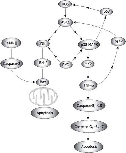
Numerous researchers have shown that ROS relate to apoptosis that is processed through the mitochondrial pathway, which depends on the activation of p38 (Figure 2)[4044]. Apoptosis signal-regulating kinase 1 (ASK1) is an evolutionarily conserved mitogen-activated protein 3-kinase that activates both JNK and p38 MAPK, which may also be triggered by ROS[4549]. However, activation of MAPKs (JNK, p38, ERK) is differentially regulated by cleavage size (40 kDa and 36 kDa) of mammalian sterile 20-like kinase 1, which is controlled by caspase-7 and -3[50]. ASK1-induced and ROS-dependent activation of MAPKs is crucial for apoptosis[4351], and for TLR4-mediated mammalian innate immunity[52]. In the case of oxidative stress, a positive feedback may form in the ASK1-p38-TNF-α pathway, which enhances ROS-mediated apoptosis (Figure 2). Ask1 activates both JNK and p38 MAPK, then the activated p38 translocates into the nucleus and stimulates the expression of MK2. After moving out of the nucleus, MK2 increases TNF-α production. On the other hand, enhanced TNF-α and ROS activate ASK1 activity[46], which leads to the activation of JNK. JNK abrogates Bcl-2, which is believed to be a protector away from mitochondria-related apoptosis, although Bcl-2 may manifest opposing phenotypes in text of interacting with other proteins[53]. In addition, this positive feedback is required for ROS-mediated apoptosis (Figure 2). Functional analyses have revealed that the initial ROS-independent activations of JNK, Bax, and caspase-3 are not sufficient for cell death, and thus, should be re-activated by ROS in order to kill the cells[54]. ROS do not simply mediate the lethal action of γ radiation, but actually amplify it by forming a feedback loop between a downstream effector (caspase) and the upstream initiation signals leading to the activation of JNK. This role of ROS appears to allow Bcl-2 to block the signaling events, which are initially induced upstream[54]. p38α MAPK contributes to the further activation of p53, which also leads to a positive feedback loop, p38α MAPK/p53. The p53/ROS/p38α MAPK cascade is essential for cisplatin-induced cell death in HCT116 cells, and the subsequent p38α/p53 positive feedback loop strongly enhances the initial p53 activation[55]. Of interest, p38 may stimulate indirectly the production of ROS via p53. Glioma pathogenesis-related protein 1 (GLIPR1), a novel p53 target gene, is down-regulated by methylation in prostate cancer and has p53-dependent and -independent pro-apoptotic activities in tumor cells. Overexpression of GLIPR1 in cancer cells leads to suppression of colony growth and induction of apoptosis. Mechanistic analysis indicates that GLIPR1 up-regulation increases the production of ROS, leading to apoptosis through activation of the JNK signaling cascade[56]. However, in p38-related apoptosis that is independent of ROS generation, JNK seems to execute a reverse function. Inhibition of JNK by SP600125 significantly enhanced apoptosis[57].

Figure 2
Figure 2 ROS-mediated apoptosis.

Via regulation of MAPKs, the protein kinase C (PKC)δ-mediated pathway also involves ROS related apoptosis. As a tentative stimulator of p38, JNK1/2 and MEK/ERK signaling[58], PKC-δ regulates cell apoptosis and survival in diverse cellular systems. Knock down of PKC-δ suppresses p38 MAPK phosphorylation. Via p38 MAPK, activated PKC-δ regulates the phosphorylation of heat shock protein (HSP) 27. Attenuated phosphorylation of HSP27 correlates with tumor progression in patients with hepatic cell cancer[59]. PKC-δ translocates to different subcellular sites in response to apoptotic stimuli. The localization of PKC-δ differentially affects the activation of downstream signaling pathways. PKC-δ-cytosol increases the phosphorylation of p38, whereas PKC-δ-nucleus increases c-JNK phosphorylation. Moreover, p38 phosphorylation plays a role in the apoptotic effect of PKC-δ-cyto, whereas c-JNK activation mediates the apoptotic effect of PKC-δ-Nuc[60]. Recent evidence has shown that calcium/calmodulin (Ca2+/CaM)-dependent protein kinase II (CaMKII) activity is also enhanced by pro-oxidant conditions. CaMKII is activated by angiotensin II-induced oxidation, leading to apoptosis in cardiomyocytes both in vitro and in vivo (Figure 2)[61].

Besides apoptosis, ROS also relate to proliferation. In mice lacking Nrf2 transcription factor, oxidative stress-mediated activation of p38, Akt kinase and downstream targets is impaired, resulting in enhanced death and delayed proliferation of hepatocytes[62]. p38 MAPK, p53, and p21 also act as molecular mediators on the way from increased ROS levels to the observed growth arrest[63].

CONCLUSION

Besides their well-known roles, recent studies have demonstrated additional functions of ROS in tumorigenesis. However, the evidence comes from studies performed in cell culture, in addition to data from human tumors. In addition, these cell lines are generally kept in room air, whereas hyperoxic oxygen levels may favor enhanced ROS formation, which is well known. The relevance of ROS in all these events in vivo, especially in humans, is not clear. ROS seem to have dual roles in tumorigenesis, cancer promoting and cancer suppressing. ROS participate in both Ras-Raf-MEK1/2-ERK1/2 signaling and the p38 MAPK pathway. However, these two pathways may have inverse functions in tumorigenesis. The former is related to cancer promotion, whereas the latter is associated with a variety of cellular responses such as OIS, replicative senescence, contact inhibition and DNA-damage responses. Thus, regarding ROS as an absolute “carcinogenic factor” or “cancer suppressor” seems to be inappropriate. It seems that more extensive investigations are needed to determine the integrity of ROS in human cancer development. Two aspects of research remain to be carried out in the future. Firstly, we should determine whether ROS directly mediate the Ras-Raf-MEK1/2-ERK1/2 and p38 MAPK signaling pathways, or whether other mediators are needed. Secondly, the definite shunting mechanism, which controls the steering from triggering Ras-Raf-MEK1/2-ERK1/2 signaling to triggering p38 MAPK signaling and vice versa, should be determined. The relationship between ROS and p38-pathway-mediated OIS is of particular interest because several reports indicate that part of the OIS pathway is intact at least in certain cancer cells, and that senescence responses improve the outcome of chemotherapy. Drugs which artificially trigger senescence in tumor cells will thus improve cancer treatment[31]. Studies on the shunting mechanism would facilitate research on the roles of ROS in tumorigenesis, and could shed light on drug discovery.

Footnotes

Supported by National Natural Science Foundation of China, No. 30750013 and Key Science Research Project Natural Science Foundation of Xiamen, No. WKZ0501

References
1.  Yildirim Z, Kiliç N, Ozer C, Babul A, Take G, Erdogan D. Effects of taurine in cellular responses to oxidative stress in young and middle-aged rat liver. Ann N Y Acad Sci. 2007;1100:553-561.  [PubMed]  [DOI]  [Cited in This Article: ]
2.  Pan JS, He SZ, Xu HZ, Zhan XJ, Yang XN, Xiao HM, Shi HX, Ren JL. Oxidative stress disturbs energy metabolism of mitochondria in ethanol-induced gastric mucosa injury. World J Gastroenterol. 2008;14:5857-5867.  [PubMed]  [DOI]  [Cited in This Article: ]
3.  Yagoda N, von Rechenberg M, Zaganjor E, Bauer AJ, Yang WS, Fridman DJ, Wolpaw AJ, Smukste I, Peltier JM, Boniface JJ. RAS-RAF-MEK-dependent oxidative cell death involving voltage-dependent anion channels. Nature. 2007;447:864-868.  [PubMed]  [DOI]  [Cited in This Article: ]
4.  Xu Y, Kim SO, Li Y, Han J. Autophagy contributes to caspase-independent macrophage cell death. J Biol Chem. 2006;281:19179-19187.  [PubMed]  [DOI]  [Cited in This Article: ]
5.  Yu D, Berlin JA, Penning TM, Field J. Reactive oxygen species generated by PAH o-quinones cause change-in-function mutations in p53. Chem Res Toxicol. 2002;15:832-842.  [PubMed]  [DOI]  [Cited in This Article: ]
6.  Morgan C, Jenkins GJ, Ashton T, Griffiths AP, Baxter JN, Parry EM, Parry JM. Detection of p53 mutations in precancerous gastric tissue. Br J Cancer. 2003;89:1314-1319.  [PubMed]  [DOI]  [Cited in This Article: ]
7.  Lee RA, Kim HA, Kang BY, Kim KH. Hemoglobin induces colon cancer cell proliferation by release of reactive oxygen species. World J Gastroenterol. 2006;12:5644-5650.  [PubMed]  [DOI]  [Cited in This Article: ]
8.  Komatsu D, Kato M, Nakayama J, Miyagawa S, Kamata T. NADPH oxidase 1 plays a critical mediating role in oncogenic Ras-induced vascular endothelial growth factor expression. Oncogene. 2008;27:4724-4732.  [PubMed]  [DOI]  [Cited in This Article: ]
9.  Hung JH, Lu YS, Wang YC, Ma YH, Wang DS, Kulp SK, Muthusamy N, Byrd JC, Cheng AL, Chen CS. FTY720 induces apoptosis in hepatocellular carcinoma cells through activation of protein kinase C delta signaling. Cancer Res. 2008;68:1204-1212.  [PubMed]  [DOI]  [Cited in This Article: ]
10.  Kumar B, Koul S, Khandrika L, Meacham RB, Koul HK. Oxidative stress is inherent in prostate cancer cells and is required for aggressive phenotype. Cancer Res. 2008;68:1777-1785.  [PubMed]  [DOI]  [Cited in This Article: ]
11.  Alkhenizan A, Hafez K. The role of vitamin E in the prevention of cancer: a meta-analysis of randomized controlled trials. Ann Saudi Med. 2007;27:409-414.  [PubMed]  [DOI]  [Cited in This Article: ]
12.  Plummer M, Vivas J, Lopez G, Bravo JC, Peraza S, Carillo E, Cano E, Castro D, Andrade O, Sánchez V. Chemoprevention of precancerous gastric lesions with antioxidant vitamin supplementation: a randomized trial in a high-risk population. J Natl Cancer Inst. 2007;99:137-146.  [PubMed]  [DOI]  [Cited in This Article: ]
13.  Heyworth PG, Knaus UG, Settleman J, Curnutte JT, Bokoch GM. Regulation of NADPH oxidase activity by Rac GTPase activating protein(s). Mol Biol Cell. 1993;4:1217-1223.  [PubMed]  [DOI]  [Cited in This Article: ]
14.  Adachi Y, Shibai Y, Mitsushita J, Shang WH, Hirose K, Kamata T. Oncogenic Ras upregulates NADPH oxidase 1 gene expression through MEK-ERK-dependent phosphorylation of GATA-6. Oncogene. 2008;27:4921-4932.  [PubMed]  [DOI]  [Cited in This Article: ]
15.  Kadara H, Tahara E, Kim HJ, Lotan D, Myers J, Lotan R. Involvement of Rac in fenretinide-induced apoptosis. Cancer Res. 2008;68:4416-4423.  [PubMed]  [DOI]  [Cited in This Article: ]
16.  Jiang H, Zhang L, Koubi D, Kuo J, Groc L, Rodriguez AI, Hunter TJ, Tang S, Lazarovici P, Gautam SC. Roles of Ras-Erk in apoptosis of PC12 cells induced by trophic factor withdrawal or oxidative stress. J Mol Neurosci. 2005;25:133-140.  [PubMed]  [DOI]  [Cited in This Article: ]
17.  Kim JW, Kim WH, Jeong MH, Jang SM, Song KH, Park SI, Song PI, Kang KH, Choi KH. p19(ras) amplifies p73beta-induced apoptosis through mitochondrial pathway. Biochem Biophys Res Commun. 2008;373:146-150.  [PubMed]  [DOI]  [Cited in This Article: ]
18.  Yamamoto S, Shimizu S, Kiyonaka S, Takahashi N, Wajima T, Hara Y, Negoro T, Hiroi T, Kiuchi Y, Okada T. TRPM2-mediated Ca2+influx induces chemokine production in monocytes that aggravates inflammatory neutrophil infiltration. Nat Med. 2008;14:738-747.  [PubMed]  [DOI]  [Cited in This Article: ]
19.  Abe J, Okuda M, Huang Q, Yoshizumi M, Berk BC. Reactive oxygen species activate p90 ribosomal S6 kinase via Fyn and Ras. J Biol Chem. 2000;275:1739-1748.  [PubMed]  [DOI]  [Cited in This Article: ]
20.  Aikawa R, Komuro I, Yamazaki T, Zou Y, Kudoh S, Tanaka M, Shiojima I, Hiroi Y, Yazaki Y. Oxidative stress activates extracellular signal-regulated kinases through Src and Ras in cultured cardiac myocytes of neonatal rats. J Clin Invest. 1997;100:1813-1821.  [PubMed]  [DOI]  [Cited in This Article: ]
21.  Kimball SR, Abbas A, Jefferson LS. Melatonin represses oxidative stress-induced activation of the MAP kinase and mTOR signaling pathways in H4IIE hepatoma cells through inhibition of Ras. J Pineal Res. 2008;44:379-386.  [PubMed]  [DOI]  [Cited in This Article: ]
22.  Shinohara M, Shang WH, Kubodera M, Harada S, Mitsushita J, Kato M, Miyazaki H, Sumimoto H, Kamata T. Nox1 redox signaling mediates oncogenic Ras-induced disruption of stress fibers and focal adhesions by down-regulating Rho. J Biol Chem. 2007;282:17640-17648.  [PubMed]  [DOI]  [Cited in This Article: ]
23.  Kowluru V, Kowluru RA. Increased oxidative stress in diabetes regulates activation of a small molecular weight G-protein, H-Ras, in the retina. Mol Vis. 2007;13:602-610.  [PubMed]  [DOI]  [Cited in This Article: ]
24.  Wang S, Yang Q, Fung KM, Lin HK. AKR1C2 and AKR1C3 mediated prostaglandin D2 metabolism augments the PI3K/Akt proliferative signaling pathway in human prostate cancer cells. Mol Cell Endocrinol. 2008;289:60-66.  [PubMed]  [DOI]  [Cited in This Article: ]
25.  Serrano M, Lin AW, McCurrach ME, Beach D, Lowe SW. Oncogenic ras provokes premature cell senescence associated with accumulation of p53 and p16INK4a. Cell. 1997;88:593-602.  [PubMed]  [DOI]  [Cited in This Article: ]
26.  Kang YH, Lee SJ. The role of p38 MAPK and JNK in Arsenic trioxide-induced mitochondrial cell death in human cervical cancer cells. J Cell Physiol. 2008;217:23-33.  [PubMed]  [DOI]  [Cited in This Article: ]
27.  Abidi P, Zhang H, Zaidi SM, Shen WJ, Leers-Sucheta S, Cortez Y, Han J, Azhar S. Oxidative stress-induced inhibition of adrenal steroidogenesis requires participation of p38 mitogen-activated protein kinase signaling pathway. J Endocrinol. 2008;198:193-207.  [PubMed]  [DOI]  [Cited in This Article: ]
28.  Wang LX, Zeng JP, Wei XB, Wang FW, Liu ZP, Zhang XM. Effects of scutellarin on apoptosis induced by cobalt chloride in PC12 cells. Chin J Physiol. 2007;50:301-307.  [PubMed]  [DOI]  [Cited in This Article: ]
29.  Takahashi M, Suzuki E, Takeda R, Oba S, Nishimatsu H, Kimura K, Nagano T, Nagai R, Hirata Y. Angiotensin II and tumor necrosis factor-alpha synergistically promote monocyte chemoattractant protein-1 expression: roles of NF-kappaB, p38, and reactive oxygen species. Am J Physiol Heart Circ Physiol. 2008;294:H2879-H2888.  [PubMed]  [DOI]  [Cited in This Article: ]
30.  Zhang Y, Qi X, Gong L, Li Y, Liu L, Xue X, Xiao Y, Wu X, Ren J. Roles of reactive oxygen species and MAP kinases in the primary rat hepatocytes death induced by toosendanin. Toxicology. 2008;249:62-68.  [PubMed]  [DOI]  [Cited in This Article: ]
31.  Han J, Sun P. The pathways to tumor suppression via route p38. Trends Biochem Sci. 2007;32:364-371.  [PubMed]  [DOI]  [Cited in This Article: ]
32.  Ren JL, Pan JS, Lu YP, Sun P, Han J. Inflammatory signaling and cellular senescence. Cell Signal. 2009;21:378-383.  [PubMed]  [DOI]  [Cited in This Article: ]
33.  Colavitti R, Finkel T. Reactive oxygen species as mediators of cellular senescence. IUBMB Life. 2005;57:277-281.  [PubMed]  [DOI]  [Cited in This Article: ]
34.  Nicke B, Bastien J, Khanna SJ, Warne PH, Cowling V, Cook SJ, Peters G, Delpuech O, Schulze A, Berns K. Involvement of MINK, a Ste20 family kinase, in Ras oncogene-induced growth arrest in human ovarian surface epithelial cells. Mol Cell. 2005;20:673-685.  [PubMed]  [DOI]  [Cited in This Article: ]
35.  Zdanov S, Debacq-Chainiaux F, Remacle J, Toussaint O. Identification of p38MAPK-dependent genes with changed transcript abundance in H2O2-induced premature senescence of IMR-90 hTERT human fibroblasts. FEBS Lett. 2006;580:6455-6463.  [PubMed]  [DOI]  [Cited in This Article: ]
36.  Dolado I, Swat A, Ajenjo N, De Vita G, Cuadrado A, Nebreda AR. p38alpha MAP kinase as a sensor of reactive oxygen species in tumorigenesis. Cancer Cell. 2007;11:191-205.  [PubMed]  [DOI]  [Cited in This Article: ]
37.  Wolfman JC, Palmby T, Der CJ, Wolfman A. Cellular N-Ras promotes cell survival by downregulation of Jun N-terminal protein kinase and p38. Mol Cell Biol. 2002;22:1589-1606.  [PubMed]  [DOI]  [Cited in This Article: ]
38.  Qi X, Tang J, Pramanik R, Schultz RM, Shirasawa S, Sasazuki T, Han J, Chen G. p38 MAPK activation selectively induces cell death in K-ras-mutated human colon cancer cells through regulation of vitamin D receptor. J Biol Chem. 2004;279:22138-22144.  [PubMed]  [DOI]  [Cited in This Article: ]
39.  Wu C, Miloslavskaya I, Demontis S, Maestro R, Galaktionov K. Regulation of cellular response to oncogenic and oxidative stress by Seladin-1. Nature. 2004;432:640-645.  [PubMed]  [DOI]  [Cited in This Article: ]
40.  Jantová S, Repický A, Letasiová S, Cipák L. 4-Amino-3-acetylquinoline-induced apoptosis of murine L1210 leukemia cells involves ROS-mitochondrial-mediated death signaling and activation of p38 MAPK. Cell Biochem Funct. 2008;26:609-619.  [PubMed]  [DOI]  [Cited in This Article: ]
41.  Lee KB, Lee JS, Park JW, Huh TL, Lee YM. Low energy proton beam induces tumor cell apoptosis through reactive oxygen species and activation of caspases. Exp Mol Med. 2008;40:118-129.  [PubMed]  [DOI]  [Cited in This Article: ]
42.  Lee SJ, Kim MS, Park JY, Woo JS, Kim YK. 15-Deoxy-delta 12,14-prostaglandin J2 induces apoptosis via JNK-mediated mitochondrial pathway in osteoblastic cells. Toxicology. 2008;248:121-129.  [PubMed]  [DOI]  [Cited in This Article: ]
43.  Noguchi T, Ishii K, Fukutomi H, Naguro I, Matsuzawa A, Takeda K, Ichijo H. Requirement of reactive oxygen species-dependent activation of ASK1-p38 MAPK pathway for extracellular ATP-induced apoptosis in macrophage. J Biol Chem. 2008;283:7657-7665.  [PubMed]  [DOI]  [Cited in This Article: ]
44.  Nusuetrong P, Pengsuparp T, Meksuriyen D, Tanitsu M, Kikuchi H, Mizugaki M, Shimazu K, Oshima Y, Nakahata N, Yoshida M. Satratoxin H generates reactive oxygen species and lipid peroxides in PC12 cells. Biol Pharm Bull. 2008;31:1115-1120.  [PubMed]  [DOI]  [Cited in This Article: ]
45.  Hong HY, Kim BC. Mixed lineage kinase 3 connects reactive oxygen species to c-Jun NH2-terminal kinase-induced mitochondrial apoptosis in genipin-treated PC3 human prostate cancer cells. Biochem Biophys Res Commun. 2007;362:307-312.  [PubMed]  [DOI]  [Cited in This Article: ]
46.  Kuo PL, Chen CY, Hsu YL. Isoobtusilactone A induces cell cycle arrest and apoptosis through reactive oxygen species/apoptosis signal-regulating kinase 1 signaling pathway in human breast cancer cells. Cancer Res. 2007;67:7406-7420.  [PubMed]  [DOI]  [Cited in This Article: ]
47.  Fürst R, Zahler S, Vollmar AM. Dexamethasone-induced expression of endothelial mitogen-activated protein kinase phosphatase-1 involves activation of the transcription factors activator protein-1 and 3’,5’-cyclic adenosine 5’-monophosphate response element-binding protein and the generation of reactive oxygen species. Endocrinology. 2008;149:3635-3642.  [PubMed]  [DOI]  [Cited in This Article: ]
48.  Kim MH, Kim MO, Heo JS, Kim JS, Han HJ. Acetylcholine inhibits long-term hypoxia-induced apoptosis by suppressing the oxidative stress-mediated MAPKs activation as well as regulation of Bcl-2, c-IAPs, and caspase-3 in mouse embryonic stem cells. Apoptosis. 2008;13:295-304.  [PubMed]  [DOI]  [Cited in This Article: ]
49.  Zhou J, Chen Y, Lang JY, Lu JJ, Ding J. Salvicine inactivates beta 1 integrin and inhibits adhesion of MDA-MB-435 cells to fibronectin via reactive oxygen species signaling. Mol Cancer Res. 2008;6:194-204.  [PubMed]  [DOI]  [Cited in This Article: ]
50.  Song JJ, Lee YJ. Differential cleavage of Mst1 by caspase-7/-3 is responsible for TRAIL-induced activation of the MAPK superfamily. Cell Signal. 2008;20:892-906.  [PubMed]  [DOI]  [Cited in This Article: ]
51.  Nakao N, Kurokawa T, Nonami T, Tumurkhuu G, Koide N, Yokochi T. Hydrogen peroxide induces the production of tumor necrosis factor-alpha in RAW 264.7 macrophage cells via activation of p38 and stress-activated protein kinase. Innate Immun. 2008;14:190-196.  [PubMed]  [DOI]  [Cited in This Article: ]
52.  Matsuzawa A, Saegusa K, Noguchi T, Sadamitsu C, Nishitoh H, Nagai S, Koyasu S, Matsumoto K, Takeda K, Ichijo H. ROS-dependent activation of the TRAF6-ASK1-p38 pathway is selectively required for TLR4-mediated innate immunity. Nat Immunol. 2005;6:587-592.  [PubMed]  [DOI]  [Cited in This Article: ]
53.  Lin B, Kolluri SK, Lin F, Liu W, Han YH, Cao X, Dawson MI, Reed JC, Zhang XK. Conversion of Bcl-2 from protector to killer by interaction with nuclear orphan receptor Nur77/TR3. Cell. 2004;116:527-540.  [PubMed]  [DOI]  [Cited in This Article: ]
54.  Kim EM, Yang HS, Kang SW, Ho JN, Lee SB, Um HD. Amplification of the gamma-irradiation-induced cell death pathway by reactive oxygen species in human U937 cells. Cell Signal. 2008;20:916-924.  [PubMed]  [DOI]  [Cited in This Article: ]
55.  Bragado P, Armesilla A, Silva A, Porras A. Apoptosis by cisplatin requires p53 mediated p38alpha MAPK activation through ROS generation. Apoptosis. 2007;12:1733-1742.  [PubMed]  [DOI]  [Cited in This Article: ]
56.  Li L, Abdel Fattah E, Cao G, Ren C, Yang G, Goltsov AA, Chinault AC, Cai WW, Timme TL, Thompson TC. Glioma pathogenesis-related protein 1 exerts tumor suppressor activities through proapoptotic reactive oxygen species-c-Jun-NH2 kinase signaling. Cancer Res. 2008;68:434-443.  [PubMed]  [DOI]  [Cited in This Article: ]
57.  Torres F, Quintana J, Díaz JG, Carmona AJ, Estévez F. Trifolin acetate-induced cell death in human leukemia cells is dependent on caspase-6 and activates the MAPK pathway. Apoptosis. 2008;13:716-728.  [PubMed]  [DOI]  [Cited in This Article: ]
58.  Yacoub D, Théorêt JF, Villeneuve L, Abou-Saleh H, Mourad W, Allen BG, Merhi Y. Essential role of protein kinase C delta in platelet signaling, alpha IIb beta 3 activation, and thromboxane A2 release. J Biol Chem. 2006;281:30024-30035.  [PubMed]  [DOI]  [Cited in This Article: ]
59.  Takai S, Matsushima-Nishiwaki R, Tokuda H, Yasuda E, Toyoda H, Kaneoka Y, Yamaguchi A, Kumada T, Kozawa O. Protein kinase C delta regulates the phosphorylation of heat shock protein 27 in human hepatocellular carcinoma. Life Sci. 2007;81:585-591.  [PubMed]  [DOI]  [Cited in This Article: ]
60.  Gomel R, Xiang C, Finniss S, Lee HK, Lu W, Okhrimenko H, Brodie C. The localization of protein kinase Cdelta in different subcellular sites affects its proapoptotic and antiapoptotic functions and the activation of distinct downstream signaling pathways. Mol Cancer Res. 2007;5:627-639.  [PubMed]  [DOI]  [Cited in This Article: ]
61.  Erickson JR, Joiner ML, Guan X, Kutschke W, Yang J, Oddis CV, Bartlett RK, Lowe JS, O’Donnell SE, Aykin-Burns N. A dynamic pathway for calcium-independent activation of CaMKII by methionine oxidation. Cell. 2008;133:462-474.  [PubMed]  [DOI]  [Cited in This Article: ]
62.  Beyer TA, Xu W, Teupser D, auf dem Keller U, Bugnon P, Hildt E, Thiery J, Kan YW, Werner S. Impaired liver regeneration in Nrf2 knockout mice: role of ROS-mediated insulin/IGF-1 resistance. EMBO J. 2008;27:212-223.  [PubMed]  [DOI]  [Cited in This Article: ]
63.  Heiss EH, Schilder YD, Dirsch VM. Chronic treatment with resveratrol induces redox stress- and ataxia telangiectasia-mutated (ATM)-dependent senescence in p53-positive cancer cells. J Biol Chem. 2007;282:26759-26766.  [PubMed]  [DOI]  [Cited in This Article: ]