Published online Feb 14, 2008. doi: 10.3748/wjg.14.948
Revised: December 10, 2007
Published online: February 14, 2008
Foreign bodies in the colon are encountered with increasing frequency, but only sporadic reports concerning their management have appeared in the literature. While most ingested foreign bodies usually pass through the gastrointestinal tract uneventfully, sharp foreign bodies such as toothpicks infrequently cause intestinal perforation and may even result in death. We report our experience with a patient with a sigmoid colon pseudodiverticulum formation, a complication of accidental ingestion of a toothpick that was diagnosed and successfully managed colonoscopically.
- Citation: Chung YS, Chung YW, Moon SY, Yoon SM, Kim MJ, Kim KO, Park CH, Hahn T, Yoo KS, Park SH, Kim JH, Park CK. Toothpick impaction with sigmoid colon pseudodiverticulum formation successfully treated with colonoscopy. World J Gastroenterol 2008; 14(6): 948-950
- URL: https://www.wjgnet.com/1007-9327/full/v14/i6/948.htm
- DOI: https://dx.doi.org/10.3748/wjg.14.948
If an ingested foreign body successfully navigates the esophagus, it will frequently pass through the entire gastrointestinal tract. Opposition to passage beyond the esophagus, however, can be caused by anatomic conditions or factors specific to the foreign body. Impaction generally occurs at sites of narrowing, such as the pylorus, the ligament of Treitz, the ileocecal valve, or the rectosigmoid junction[1]. Furthermore, long, narrow, or pointed foreign bodies are associated with a higher risk of impaction, as well as complications such as perforation.
We describe here a case of abdominal pain caused by a toothpick impacting the sigmoid colon, which resulted in a pseudodiverticulum formation. The foreign body was diagnosed and managed successfully with colonoscopy. To our knowledge, colonic pseudodiverticulum caused by toothpick impaction has not been reported.
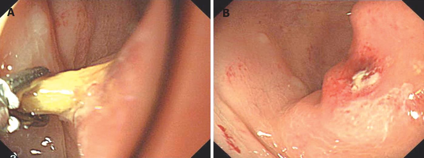
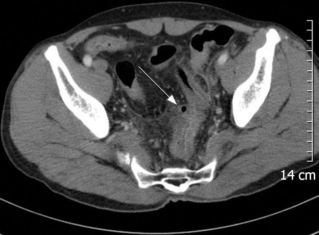
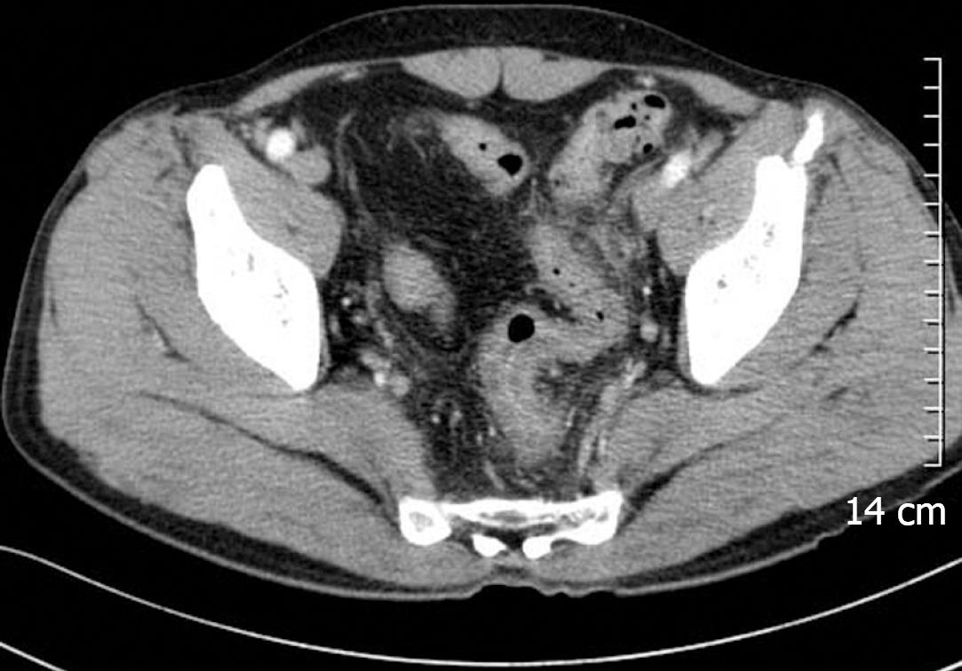
A 52-year-old man presented with lower abdominal pain of 1-mo duration. The pain began in the epigastric area and migrated to the lower abdominal area. He had a history of traumatic subdural hematoma 3 mo ago and has been treated with an anticonvulsant drug. He was not a smoker and his use of alcohol was described as only social. Physical examination revealed mild lower abdominal tenderness without rebound tenderness. Laboratory and endoscopic examinations were recommended, but he refused further evaluation. Seven days later, he revisited our hospital with severe lower abdominal pain. On physical examination, aggravated tenderness was noted in the lower abdominal region without rebound tenderness. Laboratory examination revealed only mild leukocytosis of 11 000/mm3 with elevated C-reactive protein (38.90 mg/L). The next day, colonoscopy was performed and revealed a fixed sigmoid loop with a toothpick of 6 cm length that had impacted the distal sigmoid colon with surrounding mucosal erythema and edema. The toothpick was cautiously removed using foreign-body extraction forceps (Figure 1A). There were some yellowish discharge and hyperemia on the surface of impacted colonic mucosa, but no bleeding was observed (Figure 1B). Subsequent questioning yielded a history of accidental toothpick ingestion during an afternoon nap 2 wk ago. Abdominal CT revealed severe wall thickening, pericolic fat infiltration and peritoneal thickening in the distal sigmoid colon. In addition, a small air-containing cavity lined by thin epithelium was noted in the serosal surface of the distal sigmoid colon, which was consistent with pseudodiverticulum (Figure 2). There was no evidence of free air or pneumoperitoneum. Treatment with broad spectrum parenteral antibiotics was started and continued for 7 d. The patient had an uneventful hospital course; the white blood cell count and C-reactive protein returned to normal within 48 h. Follow-up abdominal CT 4 d later revealed interval improvement with disappearance of the pseudodiverticulum (Figure 3). The patient did well after discharge.
About 80%-93% of ingested foreign bodies entering the stomach will be passed through the gastrointestinal tract uneventfully[2]. However, if the ingested foreign bodies are long, narrow, sharp such as toothpicks, the incidence of perforation could be as high as 15%-35%[3]. Patients may manifest symptoms due to bowel wall penetration, peritonitis, or an obstructive process. Toothpick ingestion appears to be commonly implicated in intestinal perforations due to the length and bilateral pointed ends of this foreign body[1]. The most frequent site of injury from ingested toothpicks is duodenum, followed by sigmoid colon[4]. Toothpicks seem to be benign materials from its wooden composition comparing to metallic pin, but surprisingly high mortality rate of toothpick injury (18%) was reported in one study[4]. Moreover, the mortality of patients presenting in shock or with enteric-vascular fistulas was extremely higher (70% and 80%, respectively)[4].
Until recently, toothpick impaction in the lower gastrointestinal tract has been managed by early surgery. One possible explanation for this fact is that most toothpicks are made of wood and may not apparent on imaging studies. Li et al reported that toothpicks were apparent on imaging studies only in 14% of the cases[4]. The definite diagnosis was most commonly made at laparotomy (53%), followed by endoscopy (12%)[4]. Another possible explanation is that most patients do not remember swallowing a toothpick. This may lead the physicians to hardly consider toothpick impaction as the cause of patients’ symptoms. However, as the skills and devices of endoscopist improve, this approach to management should be reconsidered. Obviously, operative removal of a frankly perforating toothpick is prudent, but it appears that in some cases endoscopic intervention may be accomplished at acceptable rates of morbidity and mortality. In several cases, endoscopic removal of toothpicks injury was described in the literature[15–9]. Preceding 5 cases presented with colonic perforation and the last one presented with toothpick impaction in the transverse colon without evidence of perforation. By contrast, severe sigmoid colonic inflammation with pseudodiverticulum formation was noted in our case. Free air or pneumoperitoneum, which is a sign of perforation, was not evident. To our knowledge, colonic pseudodiverticulum associated with toothpick impaction has not been reported. Colonoscopic removal of the foreign body and antibiotics treatment led to an uneventful recovery.
In summary, we report a case of toothpick impaction with sigmoid colon pseudodiverticulum formation successfully managed with colonoscopy. This case emphasizes the uncommon but possibly mortal hazards associated with the ingestion of toothpicks. Such sharp objects may cause acute inflammation without obvious perforation and present with signs of an acute abdomen. If this unusual but important cause of abdominal pain is taken into account and diagnosed in time, endoscopic management is possible.
| 1. | Reddy SK, Griffith GS, Goldstein JA, Stollman NH. Toothpick impaction with localized sigmoid perforation: successful colonoscopic management. Gastrointest Endosc. 1999;50:708-709. |
| 2. | Henderson CT, Engel J, Schlesinger P. Foreign body ingestion: review and suggested guidelines for management. Endoscopy. 1987;19:68-71. |
| 3. | Webb WA. Management of foreign bodies of the upper gastrointestinal tract. Gastroenterology. 1988;94:204-216. |
| 4. | Li SF, Ender K. Toothpick injury mimicking renal colic: case report and systematic review. J Emerg Med. 2002;23:35-38. |
| 5. | Tenner S, Wong RC, Carr-Locke D, Davis SK, Farraye FA. Toothpick ingestion as a cause of acute and chronic duodenal inflammation. Am J Gastroenterol. 1996;91:1860-1862. |
| 6. | Meltzer SJ, Goldberg MD, Meltzer RM, Claps F. Appendiceal obstruction by a toothpick removed at colonoscopy. Am J Gastroenterol. 1986;81:1107-1108. |
| 7. | Callon RA Jr, Brady PG. Toothpick perforation of the sigmoid colon: an unusual case associated with Erysipelothrix rhusiopathiae septicemia. Gastrointest Endosc. 1990;36:141-143. |
| 8. | Mohr HH, Dierkes-Globisch A. Endoscopic removal of a perforating toothpick. Endoscopy. 2001;33:295. |
| 9. | Monkemuller KE, Patil R, Marino CR. Endoscopic removal of a toothpick from the transverse colon. Am J Gastroenterol. 1996;91:2438-2439. |


