Review Open Access
Copyright ©2008 The WJG Press and Baishideng. All rights reserved.
World J Gastroenterol. Oct 28, 2008; 14(40): 6115-6121
Published online Oct 28, 2008. doi: 10.3748/wjg.14.6115
Ste20-related proline/alanine-rich kinase: A novel regulator of intestinal inflammation
Yutao Yan, Didier Merlin, Department of Medicine, Division of Digestive Diseases, Emory University School of Medicine, 615 Michael Street, Atlanta, GA 30322, United States
Author contributions: Yan Y and Merlin D contributed equally to this work.
Correspondence to: Didier Merlin, PhD, Associate Professor, Emory University Department of Medicine Division of Digestive Diseases 615 Michael Street Atlanta GA 30322, United States. dmerlin@emory.edu
Telephone: +01-404-7276454 Fax: +01-404-727-5767
Received: May 14, 2008
Revised: July 28, 2008
Accepted: August 3, 2008
Published online: October 28, 2008

Abstract

Recently, inflammatory bowel disease (IBD) has been the subject of considerable research, with increasing attention being paid to the loss of intestinal epithelial cell barrier function as a mechanism of pathogenesis. Ste20-related proline/alanine-rich kinase (SPAK) is involved in regulating barrier function. SPAK is known to interact with inflammation-related kinases (such as p38, JNK, NKCC1, PKCtheta;, WNK and MLCK), and with transcription factor AP-1, resulting in diverse biological phenomena, including cell differentiation, cell transformation and proliferation, cytoskeleton rearrangement, and regulation of chloride transport. This review examines the involvement of Ste20-like kinases and downstream mitogen-activated protein kinases (MAPKs) pathways in the pathogenesis and control of intestinal inflammation. The primary focus will be on the molecular features of intestinal inflammation, with an emphasis on the interaction between SPAK and other molecules, and the effect of these interactions on homeostatic maintenance, cell volume regulation and increased cell permeability in intestinal inflammation.

Key Words: Inflammatory bowel diseases; WNK; NKCC1; Barrier function; Ste20-related proline/alanine-rich kinase


INTRODUCTION
Figure 1
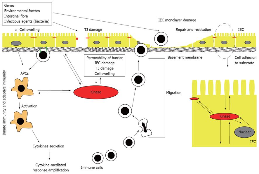
Figure 1 Pathogenesis of IBD. Many different factors, such as genetic factors, environmental factors, and intestinal non-pathogenic or pathogenic bacteria can damage the mucus, epithelium, or the tight junction, to initiate the inappropriate regulation or deregulation of the immune response, leading to the secretion of pro-inflammatory cytokines, decrease in epithelial barrier function and initiation of the inflammation-related signaling pathways. IEC: Intestinal epithelial cell; APC: Antigen presenting cell; TJ: Tight junction.
Figure 2
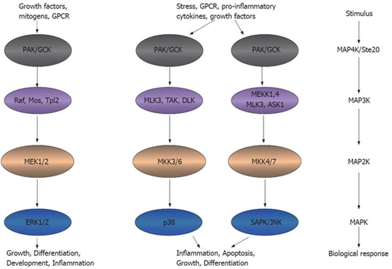
Figure 2 Ste20 kinases participate in inflammation. Ste20 kinases that function as an MAP4K can activate MAP3K, MAP2K and MAPK, leading to the inflammatory functions. This model adapted from the model presented in http://www.cellsignal.com/pathways/map-kinase.jsp. MAPK: Mitogen-activated protein kinase. GPCR: G-protein coupled receptor; PAK: p21 activated kinase; GCK: Germinal central kinase; MLK: Multiple lineage kinase; TAK: Tat-associated kinase; DLK: Dual leucine zipper-bearing kinase; MEK: MAPK/Erk kinase; MEKK: MEK kinase; ASK: Aspartate kinase; MKK: MAPK kinase; Erk: Extracellular signal-regulated kinase; SAPK: Stress-activated protein kinase; JNK: Jun-amino-terminal kinase.
Figure 3
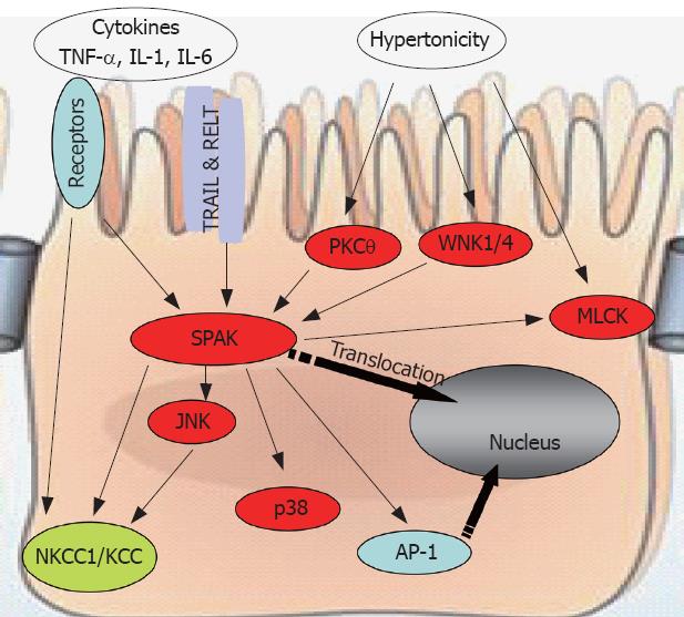
Figure 3 SPAK interacts with other molecules to maintain cellular homeostasis. SPAK can be a substrate, indirectly or directly, for pro-inflammatory cytokines, environmental stress including hypertonicity, some other kinases such as PKCtheta;, WNK1/4, or other receptors, for example TRAIL & RELT. Also SPAK can function as upstream kinase to JNK, p38, or ion transport NKCC1/KCC, transcription factor AP-1, as well as MLCK. WNK: With no lysine kinase 1/4; TRIL: TNF-related apoptosis-inducing ligand; RELT: Receptor expressed in lymphoid tissues; MLCK: Myosin II regulatory light chain kinase; NKCC1: Sodium potassium chloride chloride transporter 1; KCC: Potassium chloride chloride transporter; AP-1: Activating protein 1.

Inflammatory bowel diseases (IBD), primarily ulcerative colitis (UC) and Crohn’s disease (CD), are chronic idiopathic inflammatory disorders of the gastrointestinal tract that are thought to arise as a result of an interplay of genetic and environmental factors. The mechanisms implicated in the pathogenesis of IBD (Figure 1) include: (1) inappropriate regulation of the innate immune response at the level of the intestinal mucosa; (2) deregulation of the adaptive immune system stemming from an imbalance between regulatory and effector-cell immune responses to luminal antigens; and (3) increased permeability across the mucosal epithelial barrier due to loss of structural integrity and/or abnormal transepithelial transport[1,2]. The loss of barrier function, in particular, has gained increasing support as an IBD pathogenic mechanism because the epithelium represents a potential intersection of both genetic and environmental influences. The intestinal mucosa is composed of a single layer of polarized intestinal epithelial cells (IECs) that protects against direct contact with enteric antigens, bacteria and other pathogens (Figure 1). The integrity of the epithelium is maintained primarily through a combination of intercellular adhesion structures and specialized junctions. In addition, other factors such as the presence of mucins, rapid turnover of epithelial cells, and peristaltic movement of the gastrointestinal tract, all help to protect against colonization and invasion of the intestinal mucosa by pathogens[3]. Moreover, epidemiological and genetic linkage studies have confirmed a strong link between modulation of the barrier function and IBD; these include, for example, the loci IBD1-9, corresponding to regions on chromosomes 16, 12, 6, 14, 5, 19, 1, 16 and 3, respectively[4-13], and a new IBD locus on chromosome 2[14].

MITOGEN-ACTIVATED PROTEIN KINASES (MAPKs) ARE INVOLVED IN INTESTINAL INFLAMMATION

Intracellular signaling cascades are the main route of communication between the plasma membrane and regulatory targets in various intracellular compartments. The evolutionarily conserved MAPK signaling pathway plays an important role in transducing signals from diverse extra-cellular stimuli (including growth factors, cytokines and environmental stresses) to the nucleus in order to affect a wide range of cellular processes, such as proliferation, differentiation, development, stress responses and apoptosis. MAPK signaling cascades, which comprise up to five levels of protein kinases that are sequentially activated by phosphorylation, are also involved in intestinal inflammation[15-17] (Figure 2).

MAPK signaling pathways are involved in regulating crucial inflammatory mediators and could thus serve as molecular targets for anti-inflammatory therapy. At least six distinct MAPK pathways have been identified in multicellular organisms, of which three, the extra-cellular signal-regulated kinase (ERK), Jun N-terminal kinase (JNK) and p38 cascades, are significantly activated and directly involved in inflammatory diseases such as IBD (Figure 2). In this context, cross-talk between these pathways and other inflammatory signaling pathways, including the NF-κB and Janus kinase/signal transducers, and activation of transcription (STAT) cascades[18-20], is also relevant to the action of MAPK pathways.

The involvement of some MAPK members in IBD is suggested by linkage studies. For example, the ERK1 gene is located in a major IBD susceptibility region on chromosome 16[4], and the p38α gene is located in a major IBD susceptibility region on chromosome 6[9]. Activation of p38 MAPK is also known to induce the production and secretion of pro-inflammatory cytokines, such as interleukin (IL)-1β and tumor necrosis factor-α (TNF-α)[21], and increased activity of p38 MAPK has been observed in patients with IBD[18,22]. Inhibition of p38 has been well documented to suppress IBD[17], and the guanylhydrazone compound, CNI-1493, which inhibits both JNK and p38, strongly reduces clinical disease activity in CD patients. In addition, inhibition of either ERK or p38 kinase pathway decreases lipopolysaccharide (LPS)-induced production of the cytokines, IL-6 and TNF-α[23]. The involvement of JNK pathways in intestinal inflammation has been intensively studied both in patients with IBD and in an experimental colitis model[18,24,25]. JNK inhibitors, which affect either JNK signaling pathway indirectly (e.g. CEP1347) or block the catalytic domain of JNK (e.g. SP 600125), have been tested for their potential value in treating IBD. Collectively, these observations demonstrate a very important role for MAPK pathways in the control and therapy of IBD.

STE20-LIKE KINASES ACT UPSTREAM OF MAPK PATHWAYS

The various MAPK pathways share a common family of upstream mediators: the Ste20 kinases. Ste20 was originally identified as a component of the pheromone-response pathway in budding yeast, and has also been shown to participate in the signaling pathways that regulate osmotic responses, including those to high osmolarity glycerol (HOG)[26]. Several mammalian Ste20 homologs have been identified. The Ste20 family includes two subfamilies that share basic structural and functional properties. The first subfamily includes the p21-activated kinases (PAKs), which are characterized by a C-terminal catalytic domain and an N-terminal binding site for the small G proteins, Rac1 and Cdc42. The second family comprises of the germinal center kinases (GCKs), which contain an N-terminal kinase domain and a C-terminal regulatory domain.

Ste20-like kinases function as MAP4Ks, triggering activation of MAPK cascade[27-29] and transmitting signals from extra-cellular stimuli that activate transcription factors (Figure 2). The resulting changes in gene expression, in turn, regulate cellular functions[27-31] that are important in the maintenance of epithelial barrier function, apoptosis, growth, morphogenesis, cell permeability, and rearrangements of the cytoskeleton that lead to changes in cell shape and motility. For example, members of the PAK subfamily of Ste20 kinases have been shown to increase endothelial permeability[32,33]. The pro-inflammatory cytokine, TNF-α, stimulates expression of the yeast Ste20 homolog, Map4k4, through TNF-α-receptor-1-mediated signaling to c-Jun[34], the chemokine CXCL12 and the complement factor C5a. The resulting increase in Map4k4 activity triggers cell migration via a PAK1/2-p38α MAPK-MAPKAP-K2-HSP27 pathway[35]. Other relevant examples include: (1) Ste20-like kinase (SLK)[36], Ste20-like oxidant stress-activated kinase (SOK)[37] and prostate-derived Ste20-like kinase 1-α (PSK1-α)[38], which induce apoptosis by activating the JNK pathway; (2) lymphocyte-oriented kinase (LOK)[39] and SLK[40], which regulate Rac1-mediated actin reorganization during cell adhesion and spreading; (3) mixed lineage kinase-3 (MLK-3)[41], which activates the SAPK/JNK and p38/RK pathways via SEK1 and MKK3/6; and (4) hematopoietic progenitor kinase 1 (HPK1), which is activated by prostaglandin E2 (PGE2) through a G-protein coupled receptor (GPCR) pathway, and negatively regulates transcription of the fos gene[42].

Ste20-like kinases has been reported to be activated by at least three pathogen-associated molecular patterns (PAMPs)-lipopolysaccharide, peptidoglycan, and flagellin-produced by invading microbial pathogens, and has been shown to initiate innate immune responses by binding to pattern recognition receptors (PRRs)[43]. PAMPs activate GCKs (Ste-20 family of kinases), which signal through MLK-2 and -3 to recruit JNK, p38 and their effectors[43]. These findings indicate an important role for GCKs and MLKs in PAMP-stimulated MAPK pathway activation, and therefore in stimulating the expression of pro-inflammatory genes involved in intestinal inflammation.

STE20-RELATED PROLINE/ALANINE-RICH KINASE (SPAK) IS A STE20-LIKE KINASES INVOLVED IN INTESTINAL INFLAMMATION

The GCKs may be divided into eight subfamilies based on homologies in their C-terminal domains (GCKI-VII). The Ste20-like kinase SPAK[44], PASK (the rat SPAK homolog)[45,46] and OSR1[47] share GCK VI homologies. Among these, SPAK and OSR1 are ubiquitously expressed. PASK is also expressed in most rat tissues, but its expression is particularly notable in cells with high ion-transport activity[45,48]. Both SPAK and PASK are highly expressed in epithelia and neurons[49]. On the other hand, PASK is found only in negligible levels in the liver and skeletal muscle[50]. SPAK, OSR1 and PASK contain a series of proline and alanine repeats (PAPA box) at the extreme N-terminus, followed by a serine/threonine kinase domain, a nuclear localization signal, a consensus caspase cleavage recognition motif, and a C-terminal regulatory region. However, the colonic SPAK isoform is unique in that it lacks the PAPA box and N-terminal F-alpha helix loop, due to the presence of a 5' splice junction-like sequence within exon-1[51]. Given its ubiquitous expression and diverse functional domains, the SPAK protein may be associated with diverse biological roles. It has been shown that under hyper-osmotic (but not hypo-osmotic) stress conditions, SPAK translocates from the cytosolic pool to a Triton X-100-insoluble fraction; although present in both fractions, SPAK associated with the Triton X-100-insoluble pool is dephosphorylated[52]. Our laboratory has observed that upon SPAK over-expression[51] or under TNF-α stress conditions (unpublished data), SPAK is cleaved and the N-terminal fragment is translocated to the nucleus.

The Na+-K+-2Cl- cotransporter 1 (NKCC1), a member of the Slc12 family of solute carriers and target of SPAK, plays a crucial role in cell volume regulation, cell proliferation and survival, and epithelial transport[53]. The activity and expression of NKCC1 can be regulated by cell volume[53] and intracellular chloride concentration[54], which act through NKCC1’s N-terminal (R/K) FX (V/I) binding motif. The pro-inflammatory cytokines IL-1β, TNF-α[55] and IL-6[56] also regulate NKCC1 activity. In addition, NKCC1 can be activated by α- and β-adrenergic stimulation via the cAMP/PKA-dependent pathway[57-59] and can be stimulated by PKC in a cell-specific manner[60,61]. Notably, NKCC1 can be phosphorylated by hyperosmolarity and, in vitro, by JNK, which can also be activated by hyperosmolarity[62,63]. As an upstream kinase to NKCC1, SPAK can associate through its conserved C-terminal domain with the (R/K) FX (V/I) motif of NKCC1 and phosphorylate Thr203, Thr207, and Thr212 residues on NKCC1, thereby playing an important role in inflammation[45,64,65]. However, SPAK alone is unable to activate NKCC1. SPAK is a substrate of WNK1/4, which are serine threonine kinases lacking a lysine in subdomain I of the catalytic domain[66]. SPAK physically associates through its conserved C-terminal domain with the C-terminus of WNK, resulting in phosphorylation and activation of SPAK by WNK. WNK is also unable to activate NKCC1 in the absence of SPAK, indicating that this association of SPAK with WNK is required for SPAK-dependent phosphorylation and activation of NKCC1. A mutation of WNK1 is involved in the pathogenesis of pseudohypoaldosteronism type II (PHAII), characterized by hypertension and hyperkalemia[67].

SPAK can also activate p38 pathways in different cell types[51,68,69] to play a role in cell differentiation; an observation that may be relevant in the context of the known relationship between the p38 pathway and inflammation[17,70-74]. Interestingly, p38 activation has been noted in damaged corneal epithelial tissue and in an in vitro intestinal epithelial restitution model[75-78], suggesting that under some circumstances p38 may be involved in regulating cell motility and wound healing. Protein kinase C θ (PKCθ) is known to be an intestinal inflammation-related kinase[79]. By associating with Rho GTPases, PKCθ migrates from the cytosol to the membrane and the actin cytoskeleton[80], where SPAK may act as both a substrate and target of PKCθ in a TCR/CD28-induced signaling pathway that leads selectively to AP-1 activation, T-cell transformation and proliferation, and IL-2 production[81]. SPAK is also known to associate with F-actin under conditions of stress, which, along with the activation and phosphorylation of myosin light chain kinase (MLCK), leads to cytoskeleton rearrangement[47,52]. Fray, the Drosophila orthologue of mammalian SPAK, has been shown to participate in the activation of the JNK pathway by sorbitol[47]. Fray probably functions by activating MAP3K, leading to activation of MAP2K/MEK4 and MEK7, and ultimately, JNK activation.

Accumulating evidence points to the important role that SPAK plays in the physiology and pathogenesis of intestinal inflammation (Figure 3). First, by activating and phosphorylating p38, Ap-1, NKCC1, as well as p21-activated protein kinase 1 (PAK1, another Ste20 line kinase), SPAK induces the transcription of inflammation-related genes or modulates the function of inflammation-related proteins. Second, SPAK is activated and phosphorylated by WNK1/4, PKCtheta; and MLCK. In addition, SPAK has been reported to associate with the heat shock protein HSP105, the cytoskeleton protein gelsolin, and the apoptosis-associated tyrosine kinase AATYK. We have observed that SPAK can increase the permeability of Caco2-BBE cells (unpublished observations). Additional unpublished data indicate that colonic epithelial SPAK expression is increased in IBD patients and in mice with experimentally induced colitis. Importantly, we have also found that the pro-inflammatory cytokine, TNF-α, increases colonic SPAK expression, an observation that underscores the importance of SPAK in the pathogenesis of intestinal inflammation.

PERSPECTIVE

Increased permeability across the mucosal epithelial barrier resulting from loss of structural integrity and/or abnormal transepithelial transport is thought to be one of the main functional changes that lead to IBD. Numerous studies have focused on epithelial barrier function, measuring transepithelial electrical resistance (TER), which is known to be decreased in intestinal epithelium by over-expression of SPAK[51]. Other studies have assessed cell adhesion and migration, providing a measure of wound healing. The pro-inflammatory cytokine TNF-α is both necessary and sufficient to trigger the onset of IBD. In fact, nearly half of the drugs used for the treatment of IBD target TNF-α. In in vitro studies, we have found that TNF-α increases SPAK expression in intestinal epithelial cells in a dose- and time- dependent manner (unpublished data). It is therefore reasonable to speculate that the regulation of SPAK by TNF-α could account for TNF-α-mediated alterations of barrier function and inflammation in intestinal epithelial cells. Additional studies on the role of SPAK in intestinal barrier function would likely substantially advance the field of IBD.

Intestinal inflammation is usually associated with hyper-osmotic status in the lumen. The WNK1/4-SPAK-NKCC1 pathway has been highlighted in this context as a molecular mechanism that may contribute to ion transport and cell volume changes. This pathway, together with its interactions with other related molecules, such as MLCK, claudin and zo-1, may play an important role in maintaining cell shape, since the epithelial cell tight junctions that play a dominant role in TER would collapse in IBD. In short, more attention should be paid to tight junction and cell volume regulation as important contributing factors in IBD.

It should be evident from this review that SPAK occupies an important intracellular position, integrating extra-cellular pro-inflammatory signals and converting them into pro-inflammatory cellular responses. Given its unique position at the crossroads of multiple pathways, SPAK appears to represent an attractive target for developing effective and efficient strategies to treat IBD. Continuing work along the lines suggested above could make important contributions to the effort to realize the potential of this therapeutic approach.

Footnotes

Peer reviewer: Alessandro Fichera, MD, FACS, FASCRS, Assistant Professor, Department of Surgery-University of Chicago, 5841 S. Maryland Ave, MC 5031, Chicago, IL 60637, United States

S- Editor Tian L L- Editor Anand BS E- Editor Lin YP

References
1.  Gaudier E, Michel C, Segain JP, Cherbut C, Hoebler C. The VSL# 3 probiotic mixture modifies microflora but does not heal chronic dextran-sodium sulfate-induced colitis or reinforce the mucus barrier in mice. J Nutr. 2005;135:2753-2761.  [PubMed]  [DOI]  [Cited in This Article: ]
2.  Goyette P, Labbe C, Trinh TT, Xavier RJ, Rioux JD. Molecular pathogenesis of inflammatory bowel disease: genotypes, phenotypes and personalized medicine. Ann Med. 2007;39:177-199.  [PubMed]  [DOI]  [Cited in This Article: ]
3.  Lievin-Le Moal V, Servin AL. The front line of enteric host defense against unwelcome intrusion of harmful microorganisms: mucins, antimicrobial peptides, and microbiota. Clin Microbiol Rev. 2006;19:315-337.  [PubMed]  [DOI]  [Cited in This Article: ]
4.  Hugot JP, Laurent-Puig P, Gower-Rousseau C, Olson JM, Lee JC, Beaugerie L, Naom I, Dupas JL, Van Gossum A, Orholm M. Mapping of a susceptibility locus for Crohn's disease on chromosome 16. Nature. 1996;379:821-823.  [PubMed]  [DOI]  [Cited in This Article: ]
5.  Satsangi J, Parkes M, Louis E, Hashimoto L, Kato N, Welsh K, Terwilliger JD, Lathrop GM, Bell JI, Jewell DP. Two stage genome-wide search in inflammatory bowel disease provides evidence for susceptibility loci on chromosomes 3, 7 and 12. Nat Genet. 1996;14:199-202.  [PubMed]  [DOI]  [Cited in This Article: ]
6.  van Heel DA, Fisher SA, Kirby A, Daly MJ, Rioux JD, Lewis CM. Inflammatory bowel disease susceptibility loci defined by genome scan meta-analysis of 1952 affected relative pairs. Hum Mol Genet. 2004;13:763-770.  [PubMed]  [DOI]  [Cited in This Article: ]
7.  Peltekova VD, Wintle RF, Rubin LA, Amos CI, Huang Q, Gu X, Newman B, Van Oene M, Cescon D, Greenberg G. Functional variants of OCTN cation transporter genes are associated with Crohn disease. Nat Genet. 2004;36:471-475.  [PubMed]  [DOI]  [Cited in This Article: ]
8.  Hampe J, Franke A, Rosenstiel P, Till A, Teuber M, Huse K, Albrecht M, Mayr G, De La Vega FM, Briggs J. A genome-wide association scan of nonsynonymous SNPs identifies a susceptibility variant for Crohn disease in ATG16L1. Nat Genet. 2007;39:207-211.  [PubMed]  [DOI]  [Cited in This Article: ]
9.  Hampe J, Shaw SH, Saiz R, Leysens N, Lantermann A, Mascheretti S, Lynch NJ, MacPherson AJ, Bridger S, van Deventer S. Linkage of inflammatory bowel disease to human chromosome 6p. Am J Hum Genet. 1999;65:1647-1655.  [PubMed]  [DOI]  [Cited in This Article: ]
10.  Stoll M, Corneliussen B, Costello CM, Waetzig GH, Mellgard B, Koch WA, Rosenstiel P, Albrecht M, Croucher PJ, Seegert D. Genetic variation in DLG5 is associated with inflammatory bowel disease. Nat Genet. 2004;36:476-480.  [PubMed]  [DOI]  [Cited in This Article: ]
11.  Cho JH, Nicolae DL, Gold LH, Fields CT, LaBuda MC, Rohal PM, Pickles MR, Qin L, Fu Y, Mann JS. Identification of novel susceptibility loci for inflammatory bowel disease on chromosomes 1p, 3q, and 4q: evidence for epistasis between 1p and IBD1. Proc Natl Acad Sci USA. 1998;95:7502-7507.  [PubMed]  [DOI]  [Cited in This Article: ]
12.  Hampe J, Frenzel H, Mirza MM, Croucher PJ, Cuthbert A, Mascheretti S, Huse K, Platzer M, Bridger S, Meyer B. Evidence for a NOD2-independent susceptibility locus for inflammatory bowel disease on chromosome 16p. Proc Natl Acad Sci USA. 2002;99:321-326.  [PubMed]  [DOI]  [Cited in This Article: ]
13.  Duerr RH, Barmada MM, Zhang L, Achkar JP, Cho JH, Hanauer SB, Brant SR, Bayless TM, Baldassano RN, Weeks DE. Evidence for an inflammatory bowel disease locus on chromosome 3p26: linkage, transmission/disequilibrium and partitioning of linkage. Hum Mol Genet. 2002;11:2599-2606.  [PubMed]  [DOI]  [Cited in This Article: ]
14.  Paavola-Sakki P, Ollikainen V, Helio T, Halme L, Turunen U, Lahermo P, Lappalainen M, Farkkila M, Kontula K. Genome-wide search in Finnish families with inflammatory bowel disease provides evidence for novel susceptibility loci. Eur J Hum Genet. 2003;11:112-120.  [PubMed]  [DOI]  [Cited in This Article: ]
15.  Dubuquoy L, Dharancy S, Nutten S, Pettersson S, Auwerx J, Desreumaux P. Role of peroxisome proliferator-activated receptor gamma and retinoid X receptor heterodimer in hepatogastroenterological diseases. Lancet. 2002;360:1410-1418.  [PubMed]  [DOI]  [Cited in This Article: ]
16.  Arulampalam V, Pettersson S. Uncoupling the p38 MAPK kinase in IBD: a double edged sword? Gut. 2002;50:446-447.  [PubMed]  [DOI]  [Cited in This Article: ]
17.  Hollenbach E, Neumann M, Vieth M, Roessner A, Malfertheiner P, Naumann M. Inhibition of p38 MAP kinase- and RICK/NF-kappaB-signaling suppresses inflammatory bowel disease. FASEB J. 2004;18:1550-1552.  [PubMed]  [DOI]  [Cited in This Article: ]
18.  Waetzig GH, Seegert D, Rosenstiel P, Nikolaus S, Schreiber S. p38 mitogen-activated protein kinase is activated and linked to TNF-alpha signaling in inflammatory bowel disease. J Immunol. 2002;168:5342-5351.  [PubMed]  [DOI]  [Cited in This Article: ]
19.  Brinkman BM, Telliez JB, Schievella AR, Lin LL, Goldfeld AE. Engagement of tumor necrosis factor (TNF) receptor 1 leads to ATF-2- and p38 mitogen-activated protein kinase-dependent TNF-alpha gene expression. J Biol Chem. 1999;274:30882-30886.  [PubMed]  [DOI]  [Cited in This Article: ]
20.  Hoffmeyer A, Grosse-Wilde A, Flory E, Neufeld B, Kunz M, Rapp UR, Ludwig S. Different mitogen-activated protein kinase signaling pathways cooperate to regulate tumor necrosis factor alpha gene expression in T lymphocytes. J Biol Chem. 1999;274:4319-4327.  [PubMed]  [DOI]  [Cited in This Article: ]
21.  Yoshinari D, Takeyoshi I, Koibuchi Y, Matsumoto K, Kawashima Y, Koyama T, Ohwada S, Morishita Y. Effects of a dual inhibitor of tumor necrosis factor-alpha and interleukin-1 on lipopolysaccharide-induced lung injury in rats: involvement of the p38 mitogen-activated protein kinase pathway. Crit Care Med. 2001;29:628-634.  [PubMed]  [DOI]  [Cited in This Article: ]
22.  Hommes D, van den Blink B, Plasse T, Bartelsman J, Xu C, Macpherson B, Tytgat G, Peppelenbosch M, Van Deventer S. Inhibition of stress-activated MAP kinases induces clinical improvement in moderate to severe Crohn's disease. Gastroenterology. 2002;122:7-14.  [PubMed]  [DOI]  [Cited in This Article: ]
23.  Carter AB, Monick MM, Hunninghake GW. Both Erk and p38 kinases are necessary for cytokine gene transcription. Am J Respir Cell Mol Biol. 1999;20:751-758.  [PubMed]  [DOI]  [Cited in This Article: ]
24.  Mitsuyama K, Suzuki A, Tomiyasu N, Tsuruta O, Kitazaki S, Takeda T, Satoh Y, Bennett BL, Toyonaga A, Sata M. Pro-inflammatory signaling by Jun-N-terminal kinase in inflammatory bowel disease. Int J Mol Med. 2006;17:449-455.  [PubMed]  [DOI]  [Cited in This Article: ]
25.  Moon DO, Jin CY, Lee JD, Choi YH, Ahn SC, Lee CM, Jeong SC, Park YM, Kim GY. Curcumin decreases binding of Shiga-like toxin-1B on human intestinal epithelial cell line HT29 stimulated with TNF-alpha and IL-1beta: suppression of p38, JNK and NF-kappaB p65 as potential targets. Biol Pharm Bull. 2006;29:1470-1475.  [PubMed]  [DOI]  [Cited in This Article: ]
26.  Raitt DC, Posas F, Saito H. Yeast Cdc42 GTPase and Ste20 PAK-like kinase regulate Sho1-dependent activation of the Hog1 MAPK pathway. EMBO J. 2000;19:4623-4631.  [PubMed]  [DOI]  [Cited in This Article: ]
27.  Bagrodia S, Cerione RA. Pak to the future. Trends Cell Biol. 1999;9:350-355.  [PubMed]  [DOI]  [Cited in This Article: ]
28.  Sells MA, Chernoff J. Emerging from the Pak: the p21-activated protein kinase family. Trends Cell Biol. 1997;7:162-167.  [PubMed]  [DOI]  [Cited in This Article: ]
29.  Kyriakis JM. Signaling by the germinal center kinase family of protein kinases. J Biol Chem. 1999;274:5259-5262.  [PubMed]  [DOI]  [Cited in This Article: ]
30.  Widmann C, Gibson S, Jarpe MB, Johnson GL. Mitogen-activated protein kinase: conservation of a three-kinase module from yeast to human. Physiol Rev. 1999;79:143-180.  [PubMed]  [DOI]  [Cited in This Article: ]
31.  Ip YT, Davis RJ. Signal transduction by the c-Jun N-terminal kinase (JNK)--from inflammation to development. Curr Opin Cell Biol. 1998;10:205-219.  [PubMed]  [DOI]  [Cited in This Article: ]
32.  Stockton RA, Schaefer E, Schwartz MA. p21-activated kinase regulates endothelial permeability through modulation of contractility. J Biol Chem. 2004;279:46621-46630.  [PubMed]  [DOI]  [Cited in This Article: ]
33.  Stockton R, Reutershan J, Scott D, Sanders J, Ley K, Schwartz MA. Induction of vascular permeability: beta PIX and GIT1 scaffold the activation of extracellular signal-regulated kinase by PAK. Mol Biol Cell. 2007;18:2346-2355.  [PubMed]  [DOI]  [Cited in This Article: ]
34.  Tesz GJ, Guilherme A, Guntur KV, Hubbard AC, Tang X, Chawla A, Czech MP. Tumor necrosis factor alpha (TNFalpha) stimulates Map4k4 expression through TNFalpha receptor 1 signaling to c-Jun and activating transcription factor 2. J Biol Chem. 2007;282:19302-19312.  [PubMed]  [DOI]  [Cited in This Article: ]
35.  Rousseau S, Dolado I, Beardmore V, Shpiro N, Marquez R, Nebreda AR, Arthur JS, Case LM, Tessier-Lavigne M, Gaestel M. CXCL12 and C5a trigger cell migration via a PAK1/2-p38alpha MAPK-MAPKAP-K2-HSP27 pathway. Cell Signal. 2006;18:1897-1905.  [PubMed]  [DOI]  [Cited in This Article: ]
36.  Sabourin LA, Rudnicki MA. Induction of apoptosis by SLK, a Ste20-related kinase. Oncogene. 1999;18:7566-7575.  [PubMed]  [DOI]  [Cited in This Article: ]
37.  Pombo CM, Bonventre JV, Molnar A, Kyriakis J, Force T. Activation of a human Ste20-like kinase by oxidant stress defines a novel stress response pathway. EMBO J. 1996;15:4537-4546.  [PubMed]  [DOI]  [Cited in This Article: ]
38.  Zihni C, Mitsopoulos C, Tavares IA, Baum B, Ridley AJ, Morris JD. Prostate-derived sterile 20-like kinase 1-alpha induces apoptosis. JNK- and caspase-dependent nuclear localization is a requirement for membrane blebbing. J Biol Chem. 2007;282:6484-6493.  [PubMed]  [DOI]  [Cited in This Article: ]
39.  Endo J, Toyama-Sorimachi N, Taya C, Kuramochi-Miyagawa S, Nagata K, Kuida K, Takashi T, Yonekawa H, Yoshizawa Y, Miyasaka N. Deficiency of a STE20/PAK family kinase LOK leads to the acceleration of LFA-1 clustering and cell adhesion of activated lymphocytes. FEBS Lett. 2000;468:234-238.  [PubMed]  [DOI]  [Cited in This Article: ]
40.  Wagner S, Flood TA, O'Reilly P, Hume K, Sabourin LA. Association of the Ste20-like kinase (SLK) with the microtubule. Role in Rac1-mediated regulation of actin dynamics during cell adhesion and spreading. J Biol Chem. 2002;277:37685-37692.  [PubMed]  [DOI]  [Cited in This Article: ]
41.  Tibbles LA, Ing YL, Kiefer F, Chan J, Iscove N, Woodgett JR, Lassam NJ. MLK-3 activates the SAPK/JNK and p38/RK pathways via SEK1 and MKK3/6. EMBO J. 1996;15:7026-7035.  [PubMed]  [DOI]  [Cited in This Article: ]
42.  Sawasdikosol S, Russo KM, Burakoff SJ. Hematopoietic progenitor kinase 1 (HPK1) negatively regulates prostaglandin E2-induced fos gene transcription. Blood. 2003;101:3687-3689.  [PubMed]  [DOI]  [Cited in This Article: ]
43.  Zhong J, Kyriakis JM. Dissection of a signaling pathway by which pathogen-associated molecular patterns recruit the JNK and p38 MAPKs and trigger cytokine release. J Biol Chem. 2007;282:24246-24254.  [PubMed]  [DOI]  [Cited in This Article: ]
44.  Johnston AM, Naselli G, Gonez LJ, Martin RM, Harrison LC, DeAizpurua HJ. SPAK, a STE20/SPS1-related kinase that activates the p38 pathway. Oncogene. 2000;19:4290-4297.  [PubMed]  [DOI]  [Cited in This Article: ]
45.  Dowd BF, Forbush B. PASK (proline-alanine-rich STE20-related kinase), a regulatory kinase of the Na-K-Cl cotransporter (NKCC1). J Biol Chem. 2003;278:27347-27353.  [PubMed]  [DOI]  [Cited in This Article: ]
46.  Ushiro H, Tsutsumi T, Suzuki K, Kayahara T, Nakano K. Molecular cloning and characterization of a novel Ste20-related protein kinase enriched in neurons and transporting epithelia. Arch Biochem Biophys. 1998;355:233-240.  [PubMed]  [DOI]  [Cited in This Article: ]
47.  Chen W, Yazicioglu M, Cobb MH. Characterization of OSR1, a member of the mammalian Ste20p/germinal center kinase subfamily. J Biol Chem. 2004;279:11129-11136.  [PubMed]  [DOI]  [Cited in This Article: ]
48.  Moriguchi T, Urushiyama S, Hisamoto N, Iemura S, Uchida S, Natsume T, Matsumoto K, Shibuya H. WNK1 regulates phosphorylation of cation-chloride-coupled cotransporters via the STE20-related kinases, SPAK and OSR1. J Biol Chem. 2005;280:42685-42693.  [PubMed]  [DOI]  [Cited in This Article: ]
49.  Piechotta K, Garbarini N, England R, Delpire E. Characterization of the interaction of the stress kinase SPAK with the Na+-K+-2Cl- cotransporter in the nervous system: evidence for a scaffolding role of the kinase. J Biol Chem. 2003;278:52848-52856.  [PubMed]  [DOI]  [Cited in This Article: ]
50.  Miao N, Fung B, Sanchez R, Lydon J, Barker D, Pang K. Isolation and expression of PASK, a serine/threonine kinase, during rat embryonic development, with special emphasis on the pancreas. J Histochem Cytochem. 2000;48:1391-1400.  [PubMed]  [DOI]  [Cited in This Article: ]
51.  Yan Y, Nguyen H, Dalmasso G, Sitaraman SV, Merlin D. Cloning and characterization of a new intestinal inflammation-associated colonic epithelial Ste20-related protein kinase isoform. Biochim Biophys Acta. 2007;1769:106-116.  [PubMed]  [DOI]  [Cited in This Article: ]
52.  Tsutsumi T, Ushiro H, Kosaka T, Kayahara T, Nakano K. Proline- and alanine-rich Ste20-related kinase associates with F-actin and translocates from the cytosol to cytoskeleton upon cellular stresses. J Biol Chem. 2000;275:9157-9162.  [PubMed]  [DOI]  [Cited in This Article: ]
53.  Russell JM. Sodium-potassium-chloride cotransport. Physiol Rev. 2000;80:211-276.  [PubMed]  [DOI]  [Cited in This Article: ]
54.  Breitwieser GE, Altamirano AA, Russell JM. Osmotic stimulation of Na(+)-K(+)-Cl- cotransport in squid giant axon is [Cl-]i dependent. Am J Physiol. 1990;258:C749-C753.  [PubMed]  [DOI]  [Cited in This Article: ]
55.  Topper JN, Wasserman SM, Anderson KR, Cai J, Falb D, Gimbrone MA Jr. Expression of the bumetanide-sensitive Na-K-Cl cotransporter BSC2 is differentially regulated by fluid mechanical and inflammatory cytokine stimuli in vascular endothelium. J Clin Invest. 1997;99:2941-2949.  [PubMed]  [DOI]  [Cited in This Article: ]
56.  Sun D, Lytle C, O'Donnell ME. IL-6 secreted by astroglial cells regulates Na-K-Cl cotransport in brain microvessel endothelial cells. Am J Physiol. 1997;272:C1829-C1835.  [PubMed]  [DOI]  [Cited in This Article: ]
57.  Liedtke CM. Bumetanide-sensitive NaCl uptake in rabbit tracheal epithelial cells is stimulated by neurohormones and hypertonicity. Am J Physiol. 1992;262:L621-L627.  [PubMed]  [DOI]  [Cited in This Article: ]
58.  Paulais M, Turner RJ. Beta-adrenergic upregulation of the Na(+)-K(+)-2Cl- cotransporter in rat parotid acinar cells. J Clin Invest. 1992;89:1142-1147.  [PubMed]  [DOI]  [Cited in This Article: ]
59.  Andersen GO, Enger M, Thoresen GH, Skomedal T, Osnes JB. Alpha1-adrenergic activation of myocardial Na-K-2Cl cotransport involving mitogen-activated protein kinase. Am J Physiol. 1998;275:H641-H652.  [PubMed]  [DOI]  [Cited in This Article: ]
60.  Homma T, Burns KD, Harris RC. Agonist stimulation of Na+/K+/Cl- cotransport in rat glomerular mesangial cells. Evidence for protein kinase C-dependent and Ca2+/calmodulin-dependent pathways. J Biol Chem. 1990;265:17613-17620.  [PubMed]  [DOI]  [Cited in This Article: ]
61.  Torchia J, Yi Q, Sen AK. Carbachol-stimulated phosphorylation of the Na-K-Cl cotransporter of avian salt gland. Requirement for Ca2+ and PKC Activation. J Biol Chem. 1994;269:29778-29784.  [PubMed]  [DOI]  [Cited in This Article: ]
62.  O'Neill WC, Klein JD. Regulation of vascular endothelial cell volume by Na-K-2Cl cotransport. Am J Physiol. 1992;262:C436-C444.  [PubMed]  [DOI]  [Cited in This Article: ]
63.  Klein JD, Lamitina ST, O'Neill WC. JNK is a volume-sensitive kinase that phosphorylates the Na-K-2Cl cotransporter in vitro. Am J Physiol. 1999;277:C425-C431.  [PubMed]  [DOI]  [Cited in This Article: ]
64.  Piechotta K, Lu J, Delpire E. Cation chloride cotransporters interact with the stress-related kinases Ste20-related proline-alanine-rich kinase (SPAK) and oxidative stress response 1 (OSR1). J Biol Chem. 2002;277:50812-50819.  [PubMed]  [DOI]  [Cited in This Article: ]
65.  Gagnon KB, England R, Delpire E. Characterization of SPAK and OSR1, regulatory kinases of the Na-K-2Cl cotransporter. Mol Cell Biol. 2006;26:689-698.  [PubMed]  [DOI]  [Cited in This Article: ]
66.  Xu B, English JM, Wilsbacher JL, Stippec S, Goldsmith EJ, Cobb MH. WNK1, a novel mammalian serine/threonine protein kinase lacking the catalytic lysine in subdomain II. J Biol Chem. 2000;275:16795-16801.  [PubMed]  [DOI]  [Cited in This Article: ]
67.  Wilson FH, Disse-Nicodeme S, Choate KA, Ishikawa K, Nelson-Williams C, Desitter I, Gunel M, Milford DV, Lipkin GW, Achard JM. Human hypertension caused by mutations in WNK kinases. Science. 2001;293:1107-1112.  [PubMed]  [DOI]  [Cited in This Article: ]
68.  Johnston AM, Naselli G, Gonez LJ, Martin RM, Harrison LC, DeAizpurua HJ. SPAK, a STE20/SPS1-related kinase that activates the p38 pathway. Oncogene. 2000;19:4290-4297.  [PubMed]  [DOI]  [Cited in This Article: ]
69.  Dan I, Watanabe NM, Kusumi A. The Ste20 group kinases as regulators of MAP kinase cascades. Trends Cell Biol. 2001;11:220-230.  [PubMed]  [DOI]  [Cited in This Article: ]
70.  Chen S, Supakar PC, Vellanoweth RL, Song CS, Chatterjee B, Roy AK. Functional role of a conformationally flexible homopurine/homopyrimidine domain of the androgen receptor gene promoter interacting with Sp1 and a pyrimidine single strand DNA-binding protein. Mol Endocrinol. 1997;11:3-15.  [PubMed]  [DOI]  [Cited in This Article: ]
71.  Guan Z, Buckman SY, Pentland AP, Templeton DJ, Morrison AR. Induction of cyclooxygenase-2 by the activated MEKK1 --> SEK1/MKK4 --> p38 mitogen-activated protein kinase pathway. J Biol Chem. 1998;273:12901-12908.  [PubMed]  [DOI]  [Cited in This Article: ]
72.  Badger AM, Cook MN, Lark MW, Newman-Tarr TM, Swift BA, Nelson AH, Barone FC, Kumar S. SB 203580 inhibits p38 mitogen-activated protein kinase, nitric oxide production, and inducible nitric oxide synthase in bovine cartilage-derived chondrocytes. J Immunol. 1998;161:467-473.  [PubMed]  [DOI]  [Cited in This Article: ]
73.  Craxton A, Shu G, Graves JD, Saklatvala J, Krebs EG, Clark EA. p38 MAPK is required for CD40-induced gene expression and proliferation in B lymphocytes. J Immunol. 1998;161:3225-3236.  [PubMed]  [DOI]  [Cited in This Article: ]
74.  Pietersma A, Tilly BC, Gaestel M, de Jong N, Lee JC, Koster JF, Sluiter W. p38 mitogen activated protein kinase regulates endothelial VCAM-1 expression at the post-transcriptional level. Biochem Biophys Res Commun. 1997;230:44-48.  [PubMed]  [DOI]  [Cited in This Article: ]
75.  Sharma GD, He J, Bazan HE. p38 and ERK1/2 coordinate cellular migration and proliferation in epithelial wound healing: evidence of cross-talk activation between MAP kinase cascades. J Biol Chem. 2003;278:21989-21997.  [PubMed]  [DOI]  [Cited in This Article: ]
76.  Lu L, Reinach PS, Kao WW. Corneal epithelial wound healing. Exp Biol Med (Maywood). 2001;226:653-664.  [PubMed]  [DOI]  [Cited in This Article: ]
77.  Yu CF, Sanders MA, Basson MD. Human caco-2 motility redistributes FAK and paxillin and activates p38 MAPK in a matrix-dependent manner. Am J Physiol Gastrointest Liver Physiol. 2000;278:G952-G966.  [PubMed]  [DOI]  [Cited in This Article: ]
78.  Frey MR, Golovin A, Polk DB. Epidermal growth factor-stimulated intestinal epithelial cell migration requires Src family kinase-dependent p38 MAPK signaling. J Biol Chem. 2004;279:44513-44521.  [PubMed]  [DOI]  [Cited in This Article: ]
79.  Hokama A, Mizoguchi E, Sugimoto K, Shimomura Y, Tanaka Y, Yoshida M, Rietdijk ST, de Jong YP, Snapper SB, Terhorst C. Induced reactivity of intestinal CD4(+) T cells with an epithelial cell lectin, galectin-4, contributes to exacerbation of intestinal inflammation. Immunity. 2004;20:681-693.  [PubMed]  [DOI]  [Cited in This Article: ]
80.  Sedwick CE, Altman A. Perspectives on PKCtheta in T cell activation. Mol Immunol. 2004;41:675-686.  [PubMed]  [DOI]  [Cited in This Article: ]
81.  Li Y, Hu J, Vita R, Sun B, Tabata H, Altman A. SPAK kinase is a substrate and target of PKCtheta in T-cell receptor-induced AP-1 activation pathway. EMBO J. 2004;23:1112-1122.  [PubMed]  [DOI]  [Cited in This Article: ]