Published online Aug 28, 2008. doi: 10.3748/wjg.14.5096
Revised: July 18, 2008
Accepted: July 25, 2008
Published online: August 28, 2008
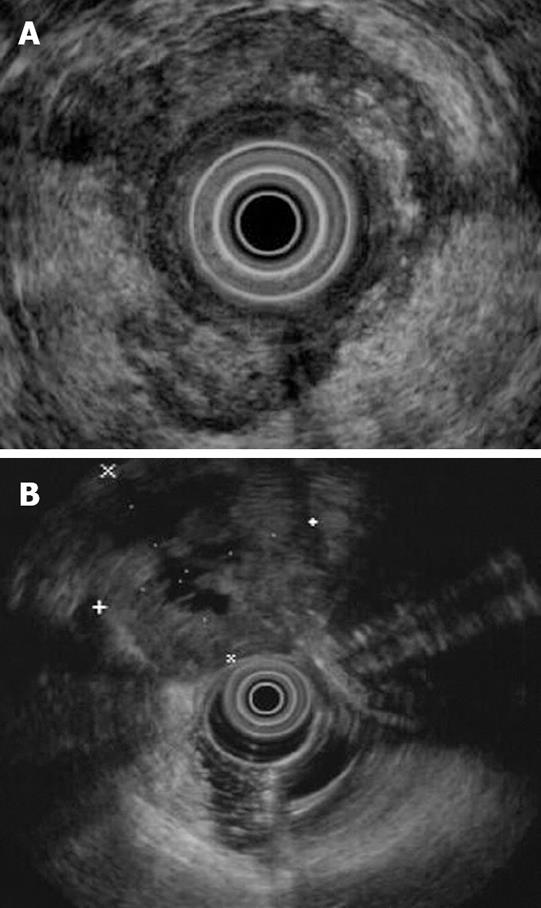
A case is presented of rectal carcinoma in which during staging by endoscopic ultrasound (EUS) a second large extrarectal mass was seen not otherwise visualized on computer tomography (CT) that was a solitary ovarian metastasis. The surgeon was alerted to the EUS finding prior to the planned laparoscopic colectomy. On retrospective review of the CT pelvis after surgery, the radiologist could still not diagnose the ovarian lesion separated from the primary rectal tumor due to their close proximity. However, on EUS we were able to clearly see on real-time imaging that there was a distinct peri-rectal mass apart from the primary rectal tumor.
- Citation: Moparty B, Gomez G, Bhutani MS. Large solitary ovarian metastasis from colorectal cancer diagnosed by endoscopic ultrasound. World J Gastroenterol 2008; 14(32): 5096-5097
- URL: https://www.wjgnet.com/1007-9327/full/v14/i32/5096.htm
- DOI: https://dx.doi.org/10.3748/wjg.14.5096
Approximately 148 000 new cases of colorectal cancer are detected yearly in the USA[1]. Ovarian metastasis has been reported in various studies to occur in 3%-14% of patients[2-5]. Endoscopic ultrasound (EUS) has been shown to have high accuracy in staging rectal cancer[6]. The use of EUS for detecting ovarian metastasis from colorectal cancer has not been established. We describe a case of a patient with ovarian metastasis from a rectal cancer that was not detected on computer tomography (CT) scan but detected as a peri-rectal mass on preoperative EUS.
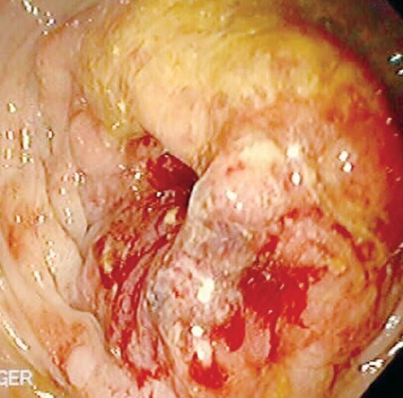
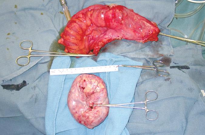
A 46-year-old female had a colonoscopy revealing an obstructing circumferential mass at 15 cm confirmed to be an invasive rectal adenocarcinoma (Figure 1). CT scan demonstrated a large rectal mass. EUS performed for staging showed a hypoechoic mass that was suspicious for a T3 lesion with penetration through the muscularis propria into the adventitia (Figure 2A). An 8 mm oval peri-tumorous lymph node was also visualized which was suspicious for malignant invasion. Inferior to the mass, EUS visualized another extra-rectal hypoechoic mass with anechoic areas. The mass measured about 5 cm and was seen in the peri-rectal area (Figure 2B). Colorectal surgery was performed and revealed a moderately differentiated rectal adenocarcinoma. Another pelvic mass was also noted (consistent with EUS findings) and found to be an ovarian metastasis (Figure 3). EUS was able to demonstrate a second mass not otherwise visualized on CT. The surgeon was alerted to the EUS finding prior to the planned laparoscopic colectomy. Based on this finding, the surrounding area was explored for a second mass and a pelvic tumor was found. On retrospective review of the CT pelvis after surgery, the radiologist could still not diagnose the ovarian lesion separated from the primary rectal tumor due to their close proximity. However, on EUS we were able to clearly see on real-time imaging that there was a distinct peri-rectal mass apart from the primary rectal tumor.
Colorectal cancer with ovarian metastasis has been reported in multiple studies[2-5]. Patients generally present with vague symptoms[7]. Colonoscopy or barium enema can help identify an intrinsic colonic lesion. If a rectal cancer is detected, EUS is performed to stage the cancer by assessing the extent of infiltration, and the presence/absence of lymph nodes[6]. EUS also allows for visualization of adjacent organs such as bladder or prostate. CT abdomen/pelvis is generally performed to evaluate metastasis of the colorectal cancer to the liver and other organs. It may be difficult at times to assess on CT, concomitant adjacent pelvic organ metastasis due to the close proximity to the bowel or to be able to differentiate the origin of a pelvic mass, whether colorectal or perirectal.
Our case demonstrates the utility of EUS in detecting peri-rectal lesion, which in our patient was difficult to detect even on retrospective review of the CT. Combining information from imaging modalities such as CT and EUS may be even more important when minimally invasive surgical techniques are employed for cancer surgery to provide the surgeon with the maximal amount of preoperative information. If a solitary ovarian lesion is noted, oophorectomy is generally performed. There has been debate on whether routine bilateral oophorectomy should be performed routinely in those undergoing surgery for colorectal cancer[8]. Some recommend discussing this option with the patient prior to surgery since reports suggest that ovarian metastasis occurs in 3%-14%[2-5].
In conclusion, for patients who present with a pelvic mass, and are found to have a colorectal cancer, EUS may be performed preoperatively to detect adjacent peri-rectal masses such as ovarian metastatic lesions.
Peer reviewer: Otto Schiueh-Tzang Lin, MD, C3-Gas, Gastroenterology Section, Virginia Mason Medical Center, 1100 Ninth Avenue, Seattle WA 98101, United States
S- Editor Li DL L- Editor Wang XL E- Editor Ma WH
| 1. | Jemal A, Siegel R, Ward E, Murray T, Xu J, Smigal C, Thun MJ. Cancer statistics, 2006. CA Cancer J Clin. 2006;56:106-130. |
| 2. | Banerjee S, Kapur S, Moran BJ. The role of prophylactic oophorectomy in women undergoing surgery for colorectal cancer. Colorectal Dis. 2005;7:214-217. |
| 3. | Koves I, Vamosi-Nagy I, Besznyak I. Ovarian metastases of colorectal tumours. Eur J Surg Oncol. 1993;19:633-635. |
| 4. | Abrams HL, Spiro R, Goldstein N. Metastases in carcinoma; analysis of 1000 autopsied cases. Cancer. 1950;3:74-85. |
| 5. | Demopoulos RI, Touger L, Dubin N. Secondary ovarian carcinoma: a clinical and pathological evaluation. Int J Gynecol Pathol. 1987;6:166-175. |
| 6. | Harewood GC, Wiersema MJ, Nelson H, Maccarty RL, Olson JE, Clain JE, Ahlquist DA, Jondal ML. A prospective, blinded assessment of the impact of preoperative staging on the management of rectal cancer. Gastroenterology. 2002;123:24-32. |
| 7. | Wright JD, Powell MA, Mutch DG, Rader JS, Gibb RK, Huettner PC, Herzog TJ. Synchronous ovarian metastases at the time of laparotomy for colon cancer. Gynecol Oncol. 2004;92:851-855. |
| 8. | Young-Fadok TM, Wolff BG, Nivatvongs S, Metzger PP, Ilstrup DM. Prophylactic oophorectomy in colorectal carcinoma: preliminary results of a randomized, prospective trial. Dis Colon Rectum. 1998;41:277-283; discussion 283-285. |


