Published online May 14, 2008. doi: 10.3748/wjg.14.2932
Revised: March 19, 2008
Published online: May 14, 2008
Hemolymphangioma of the pancreas is a very rare benign tumor. There were only five reports of this disease until March 2008. Herein, we report a case of hemolymphangioma of the pancreas with gastrointestinal bleeding due to duodenal invasion. A 53-year-old man had been admitted a referral hospital because of severe anemia due to gastrointestinal bleeding in December 2005. He was then transferred to our institute with a diagnosis of a tumor of the head of the pancreas with duodenal invasion in January 2006. No abnormalities were revealed except for anemia in laboratory data including CEA and CA19-9. Gastrointestinal endoscopy revealed bleeding at the duodenum. Computed tomography also demonstrated a heterogenous mass at the pancreatic head and suspected invasion to the duodenal wall. Ultrasonography showed a huge mass at the pancreatic head with a mixture of high and low echoic areas. Pylorous-preserving pancreatoduodenectomy was performed. The pancreatic tumor was soft and had invaded to the duodenum. The pathological diagnosis was a hemolymphangioma of the pancreas invaded to the duodenum. His postoperative course was uneventful and he was discharged on the 26th d after surgery. Hemolymphangioma of the pancreas is a very rare benign tumor. In a literature review until March 2008, we found five case reports. Major symptoms are abdominal pain and distension due to the enlarged tumor. However, we experienced a case of hemolymphangioma of the pancreas with gastrointestinal bleeding due to invasion to the duodenum. This disease is a very rare entity, but should be considered when patients have gastrointestinal bleeding.
- Citation: Toyoki Y, Hakamada K, Narumi S, Nara M, Kudoh D, Ishido K, Sasaki M. A case of invasive hemolymphangioma of the pancreas. World J Gastroenterol 2008; 14(18): 2932-2934
- URL: https://www.wjgnet.com/1007-9327/full/v14/i18/2932.htm
- DOI: https://dx.doi.org/10.3748/wjg.14.2932
Hemolymphangioma of the pancreas is an uncommon disease and basically benign tumor. There were only five reports of this tumor of the pancreas until March 2008[1–5]. Vascular tumors of the pancreas are cystic tumors accounting for 0.1% of all pancreatic tumors[6]. The most frequent of these pancreatic tumors is lymphangioma.
Major symptoms in this hemolymphangioma are abdominal pain and distension associated with the enlarged tumor. This tumor is commonly a benign disease and has no invasion ability. Herein, we report a case of hemolymphangioma of the pancreas with gastrointestinal bleeding due to invasion to the duodenum.
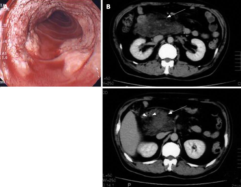
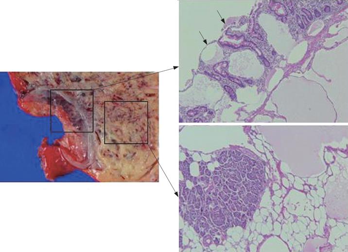
A 53-year-old man was admitted to a referral hospital because of severe anemia due to gastrointestinal bleeding in December 2005. He was then transferred to our institute with a diagnosis of a tumor of the head of the pancreas with duodenal invasion in January 2006. No abnormalities were revealed except for anemia in laboratory data including tumor markers such as CEA and CA19-9. Gastrointestinal endoscopy revealed bleeding at the duodenum from a flat elevated lesion (Figure 1A). Computed tomography also demonstrated a heterogenous mass at the pancreatic head and suspected invasion to the duodenal wall (Figure 1B). Ultrasonography showed a huge mass at the pancreatic head with a mixture of high and low echoic areas. Endoscopic retrograde cholangiopancratography showed no abnormal findings in the bile duct and pancreatic duct. Pylorous-preserving pancreatoduodenectomy was performed. The pancreatic head tumor was soft and had invaded to the duodenum. Final pathological diagnosis of this tumor was a hemolymphangioma of the pancreas with invasion to the duodenum (Figure 2). His postoperative course was uneventful and he was discharged 26 d after surgery. He is currently enjoying normal life without signs of recurrence.
Hemolymphangioma of the pancreas is an uncommon and benign tumor. In a literature review until March 2008 (PubMed), there were only five reports of this tumor occurring in the pancreas[1–5]. The first report by a French group, Couinaud et al reported a giant hemolymphangioma of the pancreas in 1966[5]. Banchini et al considered this tumor a congenital malformation of the vascular system[2]. However, there are no reports that describe invasive features of this tumor.
Most frequent symptoms of this disease in these 5 case reports were abdominal pain and distension associated with enlarged tumor. Other symptoms such as vomiting and nausea are caused by occupied tumor[5]. This tumor is commonly a benign disease and has no invasion ability. In this case, the chief complaint was severe anemia caused by duodenal bleeding because of hemolymphangioma of the pancreas which invaded to the duodenum. This symptom is extremely rare. In generally, this disease is benign, but it is possible that this tumor invaded other organs like our case.
Despite its low frequency, this disease should be considered when gastrointestinal bleeding is seen. Surgery including local resection is a definitive modality. All cases in the literature had good postoperative courses as did our case. The risk of recurrence or metastasis seems very low, but careful follow-up is necessary. Herein, we reported a case of hemolymphangioma of the pancreas head with gastrointestinal bleeding due to duodenal invasion.
| 1. | Balderramo DC, Di Tada C, de Ditter AB, Mondino JC. Hemolymphangioma of the pancreas: case report and review of the literature. Pancreas. 2003;27:197-199. |
| 2. | Banchini E, Bonati L, Villani LG. [A case of hemolym-phangioma of the pancreas]. Minerva Chir. 1987;42:807-813. |
| 3. | Montete P, Marmuse JP, Claude R, Charleux H. [Hemolymphangioma of the pancreas]. J Chir (Paris). 1985;122:659-663. |
| 4. | Couinaud C, Jouan , Prot , Chalut , Favre , Schneiter . [A rare tumor of the head of the pancreas. (Hemolymphangioma weighing 1,500 kg.)]. Presse Med. 1967;75:1955-1956. |
| 5. | Couinaud , Jouan , Prot , Chalut , Schneiter . [Hemolym-phangioma of the head of the pancreas]. Mem Acad Chir (Paris). 1966;92:152-155. |


