Published online May 14, 2008. doi: 10.3748/wjg.14.2915
Revised: March 22, 2008
Published online: May 14, 2008
Developmental anomalies of the pancreas have been reported but dorsal pancreatic agenesis is an extremely rare entity. We report an asymptomatic 62-year-old woman with complete agenesis of the dorsal pancreas. Abdominal computed tomography (CT) revealed a normal pancreatic head, but pancreatic body and tail were not visualized. Magnetic resonance imaging (MRI) findings were similar to CT. At magnetic resonance cholangiopancreatography (MRCP), the major pancreatic duct was short and the dorsal pancreatic duct was not visualized. The final diagnosis was dorsal pancreatic agenesis.
- Citation: Pasaoglu L, Vural M, Hatipoglu HG, Tereklioglu G, Koparal S. Agenesis of the dorsal pancreas. World J Gastroenterol 2008; 14(18): 2915-2916
- URL: https://www.wjgnet.com/1007-9327/full/v14/i18/2915.htm
- DOI: https://dx.doi.org/10.3748/wjg.14.2915
Agenesis of the dorsal pancreas is an extremely rare anomaly which results from defective pancreas formation. A few case reports have been published in the literature about this anomaly. Agenesis of the dorsal pancreas is mostly asymptomatic but abdominal pain, pancreatitis and diabetes mellitus may be associated[1]. We present a case of asymptomatic dorsal pancreatic agenesis.
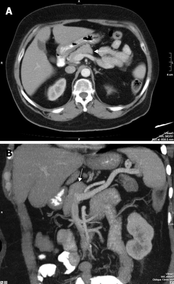
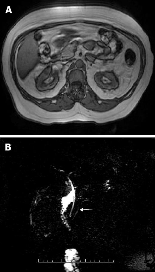
A 62-year-old female patient was admitted to our hospital with a prediagnosis of gastric lymphoma. The patient has a suspicion of lymphoma with gastroscopy. The biochemical evaluation of the patient revealed mild elevation of fasting plasma glucose (126 mg/dL; reference range: 70-115 mg/dL[1]). Pancreatic amylase and lipase levels in serum were within normal limits. On abdominal computed tomography (CT), perigastric lymph nodes were demonstrated ranging between 10 mm and 20 mm. At this examination pancreas body and tail were not seen. Pancreatic head and neck were normal in size and shape (Figure 1A and B). Upper abdominal Magnetic resonance imaging (MRI) and magnetic resonance cholangiopancreatography (MRCP) were carried out with the suspicion of dorsal pancreatic agenesis. At MRCP, the major pancreatic duct was short and the dorsal pancreatic duct was not visualized (Figure 2A and B).
The pancreas develops by ventral and dorsal endodermal buds. The dorsal bud forms the upper part of the head, body and tail of the pancreas which drains through the Santorini duct. The ventral bud gives rise to the major part of the head and uncinate process which drains through Wirsung duct[1]. Agenesis of the ventral pancreas and complete agenesis of the pancreas are lethal conditions[2]. Dorsal pancreatic agenesis is an extremely rare congenital anomaly. In the literature, about 23 cases of partial agenesis of dorsal pancreas were reported between 1913 and 2007. Pancreas divisum and autodigestion secondary to chronic pancreatitis must be considered in the differential diagnosis of the dorsal pancreatic agenesis. Failure of the ventral and dorsal pancreatic ducts to fuse is called pancreas divisum. In this entity, the ventral duct drains into major papilla, while the dorsal duct drains into separate minor papilla[1]. Atrophy of the body and the tail of the pancreas secondary to chronic pancreatitis and sparing of the pancreatic head is called pseudo-agenesis[3]. This situation may mimic dorsal pancreatic agenesis. In the differential diagnosis of pseudo-agenesis, histories of previous abdominal pain, pancreatitis, CT scanning and the serum amylase level may be helpful. The complete absence of the dorsal duct and demonstration of short ventral duct is important in the diagnosis of the dorsal bud agenesis. Abdominal CT may not evaluate the pancreatic duct in a detailed fashion. Therefore ERCP or MRCP is necessary for revealing the major and the accessory duct systems. ERCP is an invasive technique. It might challenge the cannulation of the minor papilla in selected cases. There is also radiation risk to the patient. MRCP can help the diagnosis of the dorsal pancreatic agenesis noninvasively with no radiation risk[4–6].
In our patient, distal pancreas was absent on CT and MRI images. On MRCP, dorsal duct system was not visualized and a short ventral duct was present which supports the diagnosis of dorsal pancreatic agenesis. In summary, this extremely rare congenital anomaly must be kept in mind when the corpus and tail of the pancreas are not seen at routine examinations or as in our case an incidental finding at the examinations for different pathologies.
| 1. | Fukuoka K, Ajiki T, Yamamoto M, Fujiwara H, Onoyama H, Fujita T, Katayama N, Mizuguchi K, Ikuta H, Kuroda Y. Complete agenesis of the dorsal pancreas. J Hepatobiliary Pancreat Surg. 1999;6:94-97. |
| 2. | Voldsgaard P, Kryger-Baggesen N, Lisse I. Agenesis of pancreas. Acta Paediatr. 1994;83:791-793. |
| 3. | Gold RP. Agenesis and pseudo-agenesis of the dorsal pancreas. Abdom Imaging. 1993;18:141-144. |
| 4. | Balakrishnan V, Narayanan VA, Siyad I, Radhakrishnan L, Nair P. Agenesis of the dorsal pancreas with chronic calcific pancreatitis. case report, review of the literature and genetic basis. JOP. 2006;7:651-659. |
| 5. | Uygur-Bayramicli O, Dabak R, Kilicoglu G, Dolapcioglu C, Oztas D. Dorsal pancreatic agenesis. JOP. 2007;8:450-452. |
| 6. | DU J, Xu GQ, Xu P, Jin EY, Liu Q, Li YM. Congenital short pancreas. Chin Med J (Engl). 2007;120:259-262. |


