Published online Mar 7, 2007. doi: 10.3748/wjg.v13.i9.1449
Revised: December 1, 2006
Accepted: February 22, 2007
Published online: March 7, 2007
A case of strangulation of the transverse colon in a traumatic left diaphragmatic hernia manifesting as pericarditis is reported. This is unusual because pericardial signs in traumatic diaphragmatic hernia have been previously described in association with direct pericardial injury. This is the only such case where electrocardiographic changes of pericarditis were seen without direct pericardial trauma. The possibility of internal herniation through a traumatic diaphragmatic hernia must be considered in patients with chest symptoms and a compatible history.
- Citation: Makhija R, Akoh JA. Strangulated diaphragmatic hernia presenting clinically as pericarditis. World J Gastroenterol 2007; 13(9): 1449-1450
- URL: https://www.wjgnet.com/1007-9327/full/v13/i9/1449.htm
- DOI: https://dx.doi.org/10.3748/wjg.v13.i9.1449
We report an unusual case of strangulated diaphragmatic hernia presenting as pericarditis, which was not reported in English literature.
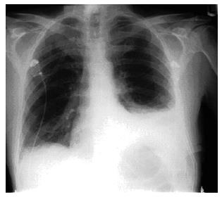
Mr CV, a 60-year old man presented to the casualty department with a history of vomiting and crampy abdominal pain. His previous medical history included a fall 4 years ago during which he sustained multiple rib fractures and a small left haemopneumothorax, which was treated conservatively at that time. Physical examination was unremarkable and he was empirically treated for gastroenteritis and discharged home. He returned the following day, with pleuritic left chest pain, dyspnoea, pyrexia of 38.5°C and tachypnoea. Investigations revealed minimally elevated white cell count, left lower lobe consolidation on chest X-ray (CXR) (Figure 1) and normal electrocardiogram (ECG).
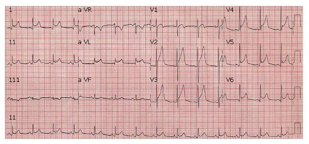
He was admitted to Medical Assessment Unit where he later complained of chest tightness. Repeat ECG revealed generalised ST elevation with upward concavity (Figure 2). A differential diagnosis of acute myocardial infarction and pericarditis was entertained and he was transferred to Coronary Care Unit (CCU) where he had a short episode of supraventricular tachycardia controlled with intravenous adenosine. Echocardiography revealed normal ventricular function with minor apical hypokinesis and no pericardial fluid. Twelve hours later he had signs of acute abdomen. Abdominal radiograph suggested large bowel obstruction, which was confirmed by GastrografinTM enema. The surgical team reviewed him and took him to the operating theatre with a diagnosis of peritonitis. However, its cause still remained unknown.
Laparotomy findings were faecal peritonitis with caecal perforation due to strangulated transverse colon in a left diaphragmatic hernia. Extended right hemicolectomy was performed with formation of ileostomy and a mucous fistula. The diaphragmatic opening was primarily repaired with non-absorbable sutures. Post-operative recovery was protracted with a long spell in the Intensive Care Unit. ECG changes reverted to normal shortly after surgery.
Traumatic diaphragmatic hernia is usually caused by blunt trauma and occurs more frequently on the left side as the liver often protects the right side[1,2]. As in this case, it is a diagnosis, which is often missed at the time of initial injury and only determined at laparotomy much later. This case is unusual because the presenting features so strongly simulated pericarditis that the patient was initially admitted to CCU after assessment by a cardiologist. This type of presentation has not been published in English literature previously. Whenever pericardial symptoms are associated with traumatic diaphragmatic hernia, there is a direct trauma to the pericardial sac[3,4]. However, in this patient, the pleuritic chest pain and ECG changes were presumably caused by pressure effects from the adjacent distended colon along with contaminated faecal fluid in the hernial sac.
In conclusion, all patients who have persistent basal abnormality on their chest X-ray after blunt thoraco-abdominal trauma should be radiologically evaluated to exclude the possibility of a ruptured diaphragm owing to the serious consequences of an undetected diaphragmatic hernia. The preferred radiological investigation is a contrast-enhanced computed tomography scan of the abdomen. In addition, the possibility of an incarceration should be kept in mind when assessing patients presenting with chest symptoms and a history suggestive of previously ruptured diaphragm.
S- Editor Liu Y L- Editor Wang XL E- Editor Zhou T
| 1. | Brown GL, Richardson JD. Traumatic diaphragmatic hernia: a continuing challenge. Ann Thorac Surg. 1985;39:170-173. [RCA] [PubMed] [DOI] [Full Text] [Cited by in Crossref: 40] [Cited by in RCA: 39] [Article Influence: 1.0] [Reference Citation Analysis (0)] |
| 2. | Beauchamp G, Khalfallah A, Girard R, Dube S, Laurendeau F, Legros G. Blunt diaphragmatic rupture. Am J Surg. 1984;148:292-295. [RCA] [PubMed] [DOI] [Full Text] [Cited by in Crossref: 29] [Cited by in RCA: 31] [Article Influence: 0.8] [Reference Citation Analysis (0)] |
| 3. | Aldhoheyan A, Jain SK, Hamdy M, Alsebayel MJ. Traumatic intrapericardial diaphragmatic hernia. Injury. 1992;23:331-332. [RCA] [PubMed] [DOI] [Full Text] [Cited by in Crossref: 9] [Cited by in RCA: 7] [Article Influence: 0.2] [Reference Citation Analysis (0)] |
| 4. | Girzadas DV, Fligner DJ. Delayed traumatic intrapericardial diaphragmatic hernia associated with cardiac tamponade. Ann Emerg Med. 1991;20:1246-1247. [RCA] [PubMed] [DOI] [Full Text] [Cited by in Crossref: 18] [Cited by in RCA: 18] [Article Influence: 0.5] [Reference Citation Analysis (0)] |


