Published online Feb 28, 2007. doi: 10.3748/wjg.v13.i8.1289
Revised: December 29, 2006
Accepted: January 24, 2007
Published online: February 28, 2007
Capsule endoscopy is an easy and painless procedure permitting visualization of the entire small-bowel during its normal peristalsis. However, important problems exist concerning capsule retention in patients at risk of small bowel obstruction. The present report describes a young patient who had recurrent episodes of overt gastrointestinal bleeding of obscure origin, 18 years after small bowel resection in infancy for ileal atresia. Capsule endoscopy was performed, resulting in capsule retention in the distal small bowel. However, this event contributed to patient management by clearly identifying the site of obstruction and can be used to guide surgical intervention, where an anastomotic ulcer is identified.
- Citation: Kalantzis C, Apostolopoulos P, Mavrogiannis P, Theodorou D, Papacharalampous X, Bramis I, Kalantzis N. Capsule endoscopy retention as a helpful tool in the management of a young patient with suspected small-bowel disease. World J Gastroenterol 2007; 13(8): 1289-1291
- URL: https://www.wjgnet.com/1007-9327/full/v13/i8/1289.htm
- DOI: https://dx.doi.org/10.3748/wjg.v13.i8.1289
Capsule endoscopy allows direct visualization of the entire small bowel in a noninvasive manner and has become the gold standard in evaluating suspected disease of the small bowel[1-4]. Capsule retention remains a major concern for physicians performing capsule endoscopy, since it can lead to a need for surgery to remove the capsule in a patient who otherwise might have been treated medically for the same illness. Nevertheless, it has been postulated that capsule endoscopy in certain patients with suspected small bowel obstruction, contributes to patient management by clearly identifying the site of obstruction and can be used to guide surgical intervention[5-7].
A 22-year old patient was admitted to our hospital for evaluation of recurrent episodes of overt gastrointestinal (GI) bleeding of obscure origin. The patient presented at birth with ileal atresia and underwent partial small bowel resection (excision of 30 cm of ileal loop sparing ileocaecal valve). He was reoperated three times during the first years of his life, initially due to anastomotic leak and subsequently twice for postoperative repair of fistulae. Since then, the patient had remained free of symptoms and developed normally. At the age of 18 years (in 2001), he had a severe episode of overt GI bleeding requiring blood transfusion, and two new bleeding episodes during the next two years. Upper and lower GI endoscopy, computed tomography of the abdomen, Meckel scintigraphy and enteroclysis, revealed no remarkable findings. In 2004 he had symptoms of incomplete intestinal obstruction, which resolved after conservative treatment. A new episode of GI bleeding with negative GI endoscopy requiring transfusion a few days before admission was the reason for referral to our unit, in order to carry out capsule endoscopy.
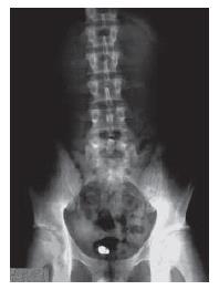
Because of his medical history, a patent capsule (Given Imaging Ltd., Yoqneam, Israel) was given prior to Pillcam SB, in order to avoid capsule retention. The patent capsule was impacted in the distal small bowel, causing temporary abdominal pain, which was confirmed by two abdominal X-rays on the second and forth day, as the patent capsule was detected at the lower abdomen to the middle line. On the sixth day, a new abdominal X-ray showed that the patent capsule was dissolved. Considering that the patent capsule stopped in a stenotic lesion, probably on the level of entero-enteric ileal anastomosis, surgical management was decided. After discussion with the surgeons and based on data supporting that capsule endoscopy in patients with suspected small bowel obstruction can guide surgical intervention by identifying the site of obstruction[5], capsule endoscopy was carried out after a written consent was obtained from the patient. Four sachets of PEG preparation were given the day before capsule endoscopy procedure was performed.
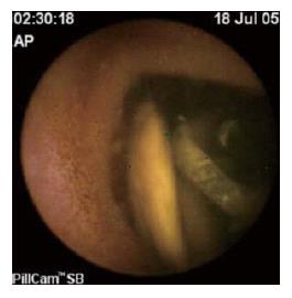
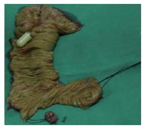
The capsule passed through the stomach, duodenum and jejunum in 2 h and 30 min, with no evidence of disease and remained impacted in an ileal loop, where a food bezoar was observed (Figure 1). Abdominal X-rays after 24 h and on the fifth day confirmed capsule retention (Figure 2). The patient was operated on the sixth day. Loose adhesions were identified and resected. The capsule was palpated 50 cm proximal to the ileocaecal valve, on the level of the entero-enteric anastomosis and an anastomotic ulcer causing stenosis was found after incision. Intraoperative enteroscopy through the incision point was performed in order to examine the ileal segment from the stenosis to the ileocaecal valve that was found normal. The lesion was resected (Figure 3) and histology demonstrated focal ulceration with chronic inflammation, but no evidence of granulomata, crypt abscesses or malignancy. The patient was discharged on the fifth postoperative day and remained asymptomatic 12 mo post-surgery.
Atresia and stenosis are common birth defects affecting the small intestine, but few population-based studies have examined the epidemiology of small intestinal atresia and stenosis. It was reported that the rate of small intestinal atresia and stenosis is 2.9 per 10 000 live births (I 95% CI = 2.3-3.6)[8]. Current operative techniques and contemporary neonatal critical care result in a 5% morbidity and mortality rate, with late complications not uncommon[9]. Protein-losing enteropathy and gastrointestinal bleeding due to anastomotic ulcer after 4-12 years have been reported[10]. Actually, gastrointestinal bleeding occurred in our patient due to anastomotic ulcer 18 years after small bowel resection. For these reasons, follow-up of these patients in their adulthood is recommended and physicians must be aware to identify and address these late occurrences[9].
Capsule endoscopy is currently the preferred test for mucosal imaging of the entire small intestine[4]. When integrated into a global approach to the patient, capsule endoscopy can achieve effective decision-making, concerning subsequent investigations and treatments. Capsule retention remains the major concern for physicians performing capsule endoscopy, since retention could lead to a need for an otherwise unnecessary surgery to remove the capsule. As there is no accepted imaging method for completely avoiding capsule retention, it is clear that obtaining a good medical history is the best single method[11].
Our patient had two risk factors for capsule retention: a history of previous small-bowel resection and a distant history of partial small-bowel obstruction. For these reasons, a patent capsule was given initially to the patient. The retention of patent capsule in the small intestine, confirmed our suspicions. Nevertheless, Cheifetz et al[5] showed that capsule retention in the small intestine could help a pre-decided operation by guiding the surgeons to clearly indentify the site of obstruction in patients with suspected small-bowel obstruction. Even though capsule endoscopy did not help us to diagnose a definite lesion, as we did not take clear images of the site of capsule retention, palpation of the capsule by the surgeons during operation, could lead them directly to the site of stenosis.
In conclusion, capsule retention is not always an adverse event, as in certain patients with suspected small bowel obstruction, the retained capsule can be used as a “guiding point” for a pre-decided surgical intervention.
S- Editor Liu Y L- Editor Zhu LH E- Editor Lu W
| 1. | Iddan G, Meron G, Glukhovsky A, Swain P. Wireless capsule endoscopy. Nature. 2000;405:417. [RCA] [PubMed] [DOI] [Full Text] [Cited by in Crossref: 1994] [Cited by in RCA: 1383] [Article Influence: 55.3] [Reference Citation Analysis (1)] |
| 2. | Appleyard M, Glukhovsky A, Swain P. Wireless-capsule diagnostic endoscopy for recurrent small-bowel bleeding. N Engl J Med. 2001;344:232-233. [RCA] [PubMed] [DOI] [Full Text] [Cited by in Crossref: 191] [Cited by in RCA: 166] [Article Influence: 6.9] [Reference Citation Analysis (0)] |
| 3. | Mishkin DS, Chuttani R, Croffie J, Disario J, Liu J, Shah R, Somogyi L, Tierney W, Song LM, Petersen BT. ASGE Technology Status Evaluation Report: wireless capsule endoscopy. Gastrointest Endosc. 2006;63:539-545. [RCA] [PubMed] [DOI] [Full Text] [Cited by in Crossref: 186] [Cited by in RCA: 169] [Article Influence: 8.9] [Reference Citation Analysis (0)] |
| 4. | Leighton JA, Goldstein J, Hirota W, Jacobson BC, Johanson JF, Mallery JS, Peterson K, Waring JP, Fanelli RD, Wheeler-Harbaugh J. Obscure gastrointestinal bleeding. Gastrointest Endosc. 2003;58:650-655. [RCA] [PubMed] [DOI] [Full Text] [Cited by in Crossref: 86] [Cited by in RCA: 82] [Article Influence: 3.7] [Reference Citation Analysis (0)] |
| 5. | Cheifetz A, Sachar D, Lewis B. Small bowel obstruction: indication or contraindication for capsule endoscopy. Gastrointest Endosc. 2004;59:461. [DOI] [Full Text] |
| 6. | Cheifetz AS, Lewis BS. Capsule endoscopy retention: is it a complication? J Clin Gastroenterol. 2006;40:688-691. [RCA] [PubMed] [DOI] [Full Text] [Cited by in Crossref: 81] [Cited by in RCA: 77] [Article Influence: 4.1] [Reference Citation Analysis (0)] |
| 7. | Baichi MM, Arifuddin RM, Mantry PS. What we have learned from 5 cases of permanent capsule retention. Gastrointest Endosc. 2006;64:283-287. [RCA] [PubMed] [DOI] [Full Text] [Cited by in Crossref: 36] [Cited by in RCA: 35] [Article Influence: 1.8] [Reference Citation Analysis (0)] |
| 8. | Forrester MB, Merz RD. Population-based study of small intestinal atresia and stenosis, Hawaii, 1986-2000. Public Health. 2004;118:434-438. [RCA] [PubMed] [DOI] [Full Text] [Cited by in Crossref: 37] [Cited by in RCA: 35] [Article Influence: 1.7] [Reference Citation Analysis (0)] |
| 9. | Escobar MA, Ladd AP, Grosfeld JL, West KW, Rescorla FJ, Scherer LR, Engum SA, Rouse TM, Billmire DF. Duodenal atresia and stenosis: long-term follow-up over 30 years. J Pediatr Surg. 2004;39:867-871; discussion 867-871. [RCA] [PubMed] [DOI] [Full Text] [Cited by in Crossref: 183] [Cited by in RCA: 142] [Article Influence: 6.8] [Reference Citation Analysis (0)] |
| 10. | Couper RT, Durie PR, Stafford SE, Filler RM, Marcon MA, Forstner GG. Late gastrointestinal bleeding and protein loss after distal small-bowel resection in infancy. J Pediatr Gastroenterol Nutr. 1989;9:454-460. [RCA] [PubMed] [DOI] [Full Text] [Cited by in Crossref: 26] [Cited by in RCA: 27] [Article Influence: 0.8] [Reference Citation Analysis (0)] |
| 11. | Cave D, Legnani P, de Franchis R, Lewis BS. ICCE consensus for capsule retention. Endoscopy. 2005;37:1065-1067. [RCA] [PubMed] [DOI] [Full Text] [Cited by in Crossref: 261] [Cited by in RCA: 255] [Article Influence: 12.8] [Reference Citation Analysis (0)] |


