Published online Feb 7, 2007. doi: 10.3748/wjg.v13.i5.806
Revised: November 29, 2006
Accepted: December 25, 2006
Published online: February 7, 2007
Hydatid disease is an endemic disease in certain areas of the world. It is located mostly in the liver. Spontaneous rupture of the hydatid cyst into the peritoneum is a rare condition, which is accompanied by serious morbidity and mortality generally. We present herein a case with a spontaneous rupture of a hepatic hidatid disease into the peritoneum without any serious symptoms. A 15-year-old boy was admitted to the emergency room with a mild abdominal pain lasting for a day. Physical examination revealed only mild abdominal tenderness. There was no history of trauma or complaints related to hydatid diseases. Ultrasonography showed a large amount of free fluid and a cystic lesion with irregular borders in the liver. He was operated on. Postoperative albendazol therapy was given for 2 mo. No recurrence or secondary hydatidosis was seen on CT investigation in the 3rd, 6th and 12th mo following surgery.
- Citation: Karakaya K. Spontaneous rupture of a hepatic hydatıd cyst into the peritoneum causing only mild abdominal pain: A case report. World J Gastroenterol 2007; 13(5): 806-808
- URL: https://www.wjgnet.com/1007-9327/full/v13/i5/806.htm
- DOI: https://dx.doi.org/10.3748/wjg.v13.i5.806
Hydatid disease is a parasitic infestation caused by Echino-coccus granulosus[1-3]. Hydatid disease is an endemic problem in Turkey as well as in sheep-bearing regions of the world[1,2,4].
Although hepatic hydatid disease may be asymptomatic for many years, it can become symptomatic due to expansion, rupture or pyogenic infection[2,3,5]. Hydatid cyst rupture may cause mild to fatal complications[2,5]. Rupture of the hepatic hydatid disease generally occurs into the biliary tree. Rupture of the cyst in the peritoneal cavity is rare and generally followed by anaphylactic reactions.
In this study, we present a case of a spontaneous rupture of a hepatic hydatid cyst into the peritoneum, in which the patient was admitted to the emergency room for mild abdominal pain without any other symptom. Spontaneous rupture of the hepatic hydatid cyst into peritoneum without any serious symptom is unusual.
A 15-year-old boy perceived sudden abdominal pain in the epigastrium and right upper quadrant a day ago. After a short duration his pain was ameliorated spontaneously. The following day he perceived mild pain in the right lower quadrant. He had no pruritis, erythema or any other symptom except the abdominal pain. No history of blunt trauma was found.
Physical examination revealed a blood pressure of 100/70 mmHg, and a pulse rate of 78 beats/min. The patient was afebrile. Abdominal palpation revealed mild tenderness. He was comfortable on his movements. There was no history of nausea, vomiting or loss of appetite. Laboratory investigations were normal except mild leukocytosis (WBC: 11 000/mm3).
Abdominal ultrasonography showed a large amount of free fluid and a cystic lesion measuring 10 cm × 12 cm with irregular borders in the right lobe of the liver. The anterior border of the cyst was incomplete. Floating membrane image was seen. The biliary ducts were normal in calibration.
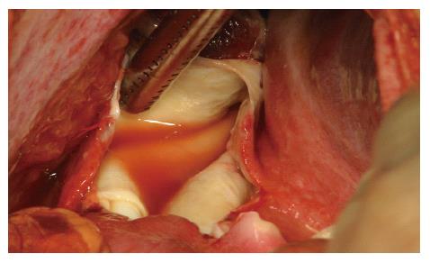
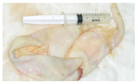
The abdomen was exposed through a right paramedian incision. Approximately 3500 mL clear fluid was aspirated from the intraperitoneal space. There was a hydatid cyst in the right lobe of the liver. The cyst was 13 cm in size and had a ruptured area of 5 cm × 7 cm anteriorly (Figure 1). There were firm adhesions between the liver and right diaphragm. There was no daughter vesicle in the abdominal cavity. The germinative membrane was taken out completely (Figure 2). No biliary leakage was seen. We could not find any other pathological evidence on exploration. The cyst pouch was irrigated with hypertonic saline (20%) and then followed by isotonic saline. The peritoneal cavity was irrigated with isotonic saline. Introflexion procedure was performed. Latex drains were placed into the sub-hepatic and recto-vesicle areas.
The patient was discharged from the hospital on the 9th postoperative day without any complication. Postoperative albendazole therapy (10 mg/kg) was prescribed for 2 mo under the liver enzyme and blood count monitoring. The patient was controlled as an outpatient. Postoperative follow up was uneventful for 13 mo. No recurrence or secondary hydatidosis was seen on CT investigation in the 3rd, 6 th and 12th mo following surgery.
The hydatid cyst is typically filled with clear fluid (hydatid fluid). The cyst consists of an internal cellular layer (germinal layer) and an outer, acellular layer (laminated layer). As the cyst expands gradually, a granulomatous host reaction followed by a fibrous reaction forms a connective tissue layer, which is called a pericyst[1].
Hydatid disease is a serious health problem in endemic areas as well as in Turkey [6,7]. The diagnosis and appropriate surgical therapy is usually delayed because most of the hydatid cysts remain asymptomatic until it is getting complicated [1-3,7,8].
Precautions for the hydatid disease are mostly insufficient. Treatment of dogs with antihelmintic is the main procedure to control the parasite (Echinococcus spp.)[9]. In rural areas of Turkey this treatment is not applied routinely. In the rural area, sheep are home-slaughtered routinely. Dogs can access to the infected viscera. This is true in almost all of the developing or underdeveloped countries in the world[9].
Initially almost all of the hydatid cysts are asympto-matic. Later in time some symptoms depending on the involved organ, localisation of the cyst and pressure effect of the cyst on the surrounding tissues and structures develop[1,7,10]. Abdominal pain is the most commonly encountered symptom. The second one is the symptoms arising from the pressure effect of the cyst[7,8,11]. It is known that rupture of the cyst ameliorates the abdominal pain and the patient feels comfortable to some degree[2]. Later in time rupture of the hydatid cyst produces symptoms and signs of allergy or peritoneal irritation almost all the time[10]. No history of previous abdominal pain or any other symptom relating to the hydatid cyst was present in the case presented here.
Diagnosis of the hydatid cyst is mainly based on ultrasonography and computed tomography (CT)[1-3,7,8,10,11]. CT is especially valuable for the 3-dimensional localisation of the cyst preoperatively[8,11]. MRI, magnetic resonance cholangio pancreatography (MRCP), scintigraphy scans and laparoscopy may be useful for diagnosis of the rare, undiagnosed cases and for its complications. As we made the diagnosis of a ruptured hepatic hydatid cyst by US examination, the other investigational techniques were not used for preoperative evaluation.
Surgery is the preferred treatment for the cure of the hydatid disease[3,6,8]. Surgical procedures may differ from percutaneous aspiration instillation and reaspiration to complete excision of the cyst[1,6,7]. We performed introfleksion procedure on the patient. Rupture of the cyst is usually related to increased intracystic pressure. This may be related to trauma, or over enlargement of the cyst[2,10]. Perforation of the hydatid cyst may cause dissemination of the parasite and increased morbidity and mortality rate[1,2,9]. As cyst size increases, risk of rupture increases[5]. Cyst size measured 13 cm preoperatively in our case. Intraperitoneal rupture of the hydatid cyst can cause abdominal pain, allergy and anaphylaxis[1,2,5]. Mild abdominal tenderness was present in our patient on admission. The patient had no history of trauma or any event that increases intra-abdominal pressure such as coughing or constipation.
Medical treatment with albendazole is described elsewhere[1,6,7,10,11]. The patient was treated with albendazole 10 mg/kg for 2 mo under liver enzyme and blood count monitoring. Postoperative follow up was uneventful.
Intra-peritoneal hydatidosis is a major problem for the patients who have hydatid cyst rupture. The patient presented here had no recurrence or secondary hydatidosis.
In endemic regions, it is useful to consider hydatid cyst disease for patients with abdominal pain admitted to the emergency room. Rupture of the hydatid cyst may be fatal.
S- Editor Liu Y L- Editor Zhu LH E- Editor Ma WH
| 1. | Eckert J, Deplazes P. Biological, epidemiological, and clinical aspects of echinococcosis, a zoonosis of increasing concern. Clin Microbiol Rev. 2004;17:107-135. [RCA] [PubMed] [DOI] [Full Text] [Cited by in Crossref: 1121] [Cited by in RCA: 1184] [Article Influence: 56.4] [Reference Citation Analysis (1)] |
| 2. | Erdogmus B, Yazici B, Akcan Y, Ozdere BA, Korkmaz U, Alcelik A. Latent fatality due to hydatid cyst rupture after a severe cough episode. Tohoku J Exp Med. 2005;205:293-296. [RCA] [PubMed] [DOI] [Full Text] [Cited by in Crossref: 9] [Cited by in RCA: 11] [Article Influence: 0.6] [Reference Citation Analysis (0)] |
| 3. | Sayek I, Yalin R, Sanaç Y. Surgical treatment of hydatid disease of the liver. Arch Surg. 1980;115:847-850. [RCA] [PubMed] [DOI] [Full Text] [Cited by in Crossref: 103] [Cited by in RCA: 109] [Article Influence: 2.4] [Reference Citation Analysis (0)] |
| 4. | Altintaş N. Cystic and alveolar echinococcosis in Turkey. Ann Trop Med Parasitol. 1998;92:637-642. [RCA] [PubMed] [DOI] [Full Text] [Cited by in Crossref: 12] [Cited by in RCA: 9] [Article Influence: 0.3] [Reference Citation Analysis (0)] |
| 5. | Kantarci M, Onbas O, Alper F, Celebi Y, Yigiter M, Okur A. Anaphylaxis due to a rupture of hydatid cyst: imaging findings of a 10-year-old boy. Emerg Radiol. 2003;10:49-50. [PubMed] |
| 6. | Yagci G, Ustunsoz B, Kaymakcioglu N, Bozlar U, Gorgulu S, Simsek A, Akdeniz A, Cetiner S, Tufan T. Results of surgical, laparoscopic, and percutaneous treatment for hydatid disease of the liver: 10 years experience with 355 patients. World J Surg. 2005;29:1670-1679. [RCA] [PubMed] [DOI] [Full Text] [Cited by in Crossref: 126] [Cited by in RCA: 115] [Article Influence: 6.1] [Reference Citation Analysis (0)] |
| 7. | Hatipoglu AR, Coskun I, Karakaya K, Ibis C. Retroperitoneal localization of hydatid cyst disease. Hepatogastroenterology. 2001;48:1037-1039. [PubMed] |
| 8. | Schwartz SI. Liver. 7th ed. Schwartz SI, Shires GT, Spencer FC, Daly JM, Fischer JE, Galloway AC, editors. Principles of Surgery. International Edition: McGraw-Hill Book Company 1999; 1403-1405. |
| 9. | Oku Y, Malgor R, Benavidez U, Carmona C, Kamiya H. Control program against hydatidosis and the decreased prevalence in Uruguay. International Congress Series 1267;. 2004;98-104. [RCA] [DOI] [Full Text] [Cited by in Crossref: 7] [Cited by in RCA: 8] [Article Influence: 0.4] [Reference Citation Analysis (0)] |
| 10. | Derici H, Tansug T, Reyhan E, Bozdag AD, Nazli O. Acute intraperitoneal rupture of hydatid cysts. World J Surg. 2006;30:1879-1883; discussion 1884-1885;. [PubMed] |
| 11. | Langer B, Gallinger S. Cystic disease of the liver. Shackelford's Surgery of the Alimentary Tract. Philadelphia, London, Toronto, Montreal, Sydney, Tokyo: WB Saunders Company 1996; 531-540. |


