Published online Nov 7, 2007. doi: 10.3748/wjg.v13.i41.5512
Revised: July 18, 2007
Accepted: August 19, 2007
Published online: November 7, 2007
Endosonography-guided biliary drainage (ESBD) is a new method enabling internal drainage of an obstructed bile duct. However, the histological conditions associated with fistula development via the duodenum to the bile duct have not been reported. We performed ESBD 14 d preoperatively in a patient with an ampullary carcinoma and histologically confirmed changes in and around the fistula. The female patient developed no complications relevant to ESBD. Levels of serum bilirubin and hepatobiliary enzymes declined quickly, and pancreatoduodenectomy was carried out uneventfully. The resected specimen was sliced and stained with hematoxylin-eosin. Histological evaluation of the puncture site in the duodenum and bile-duct wall, and the sinus tract revealed no hematoma, bile leakage, or abscess in or around the sinus tract. Little sign of granulation, fibrosis, and inflammatory cell infiltration was observed. Although further large-scale confirmatory studies are needed, the findings here may encourage more active use of ESBD as a substitute for percutaneous transhepatic drainage in cases with failed/difficult endoscopic biliary stenting.
- Citation: Fujita N, Noda Y, Kobayashi G, Ito K, Obana T, Horaguchi J, Takasawa O, Nakahara K. Histological changes at an endosonography-guided biliary drainage site: A case report. World J Gastroenterol 2007; 13(41): 5512-5515
- URL: https://www.wjgnet.com/1007-9327/full/v13/i41/5512.htm
- DOI: https://dx.doi.org/10.3748/wjg.v13.i41.5512
The role of endosonography (ES) in digestive diseases is gradually expanding from diagnostic to therapeutic applications. In the mid 1990s, the feasibility of ES-guided cholangiopancreatography was first reported by Harada et al[1] (pancreatography) and Wiersema et al[2] (cholangiography). Several reports on the application of this technique for therapeutic purposes, such as ES as a guide for biliary drainage, have been published.
We have recently applied ES-guided biliary drainage (ESBD) for preoperative decompression of the biliary tree in a patient with cancer of the papilla of Vater. The results of histological evaluation of and around the sinus tract are reported herein.
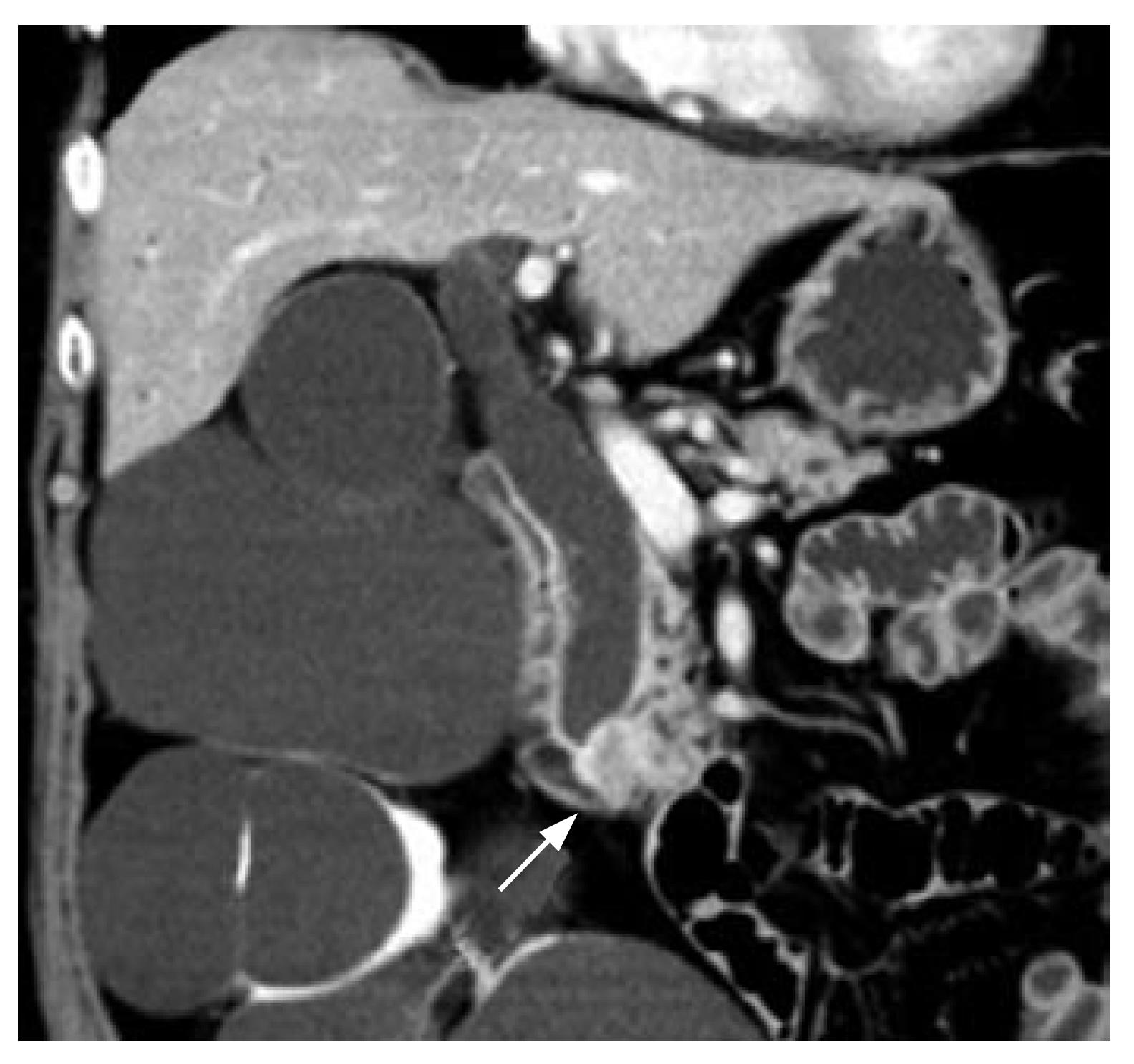
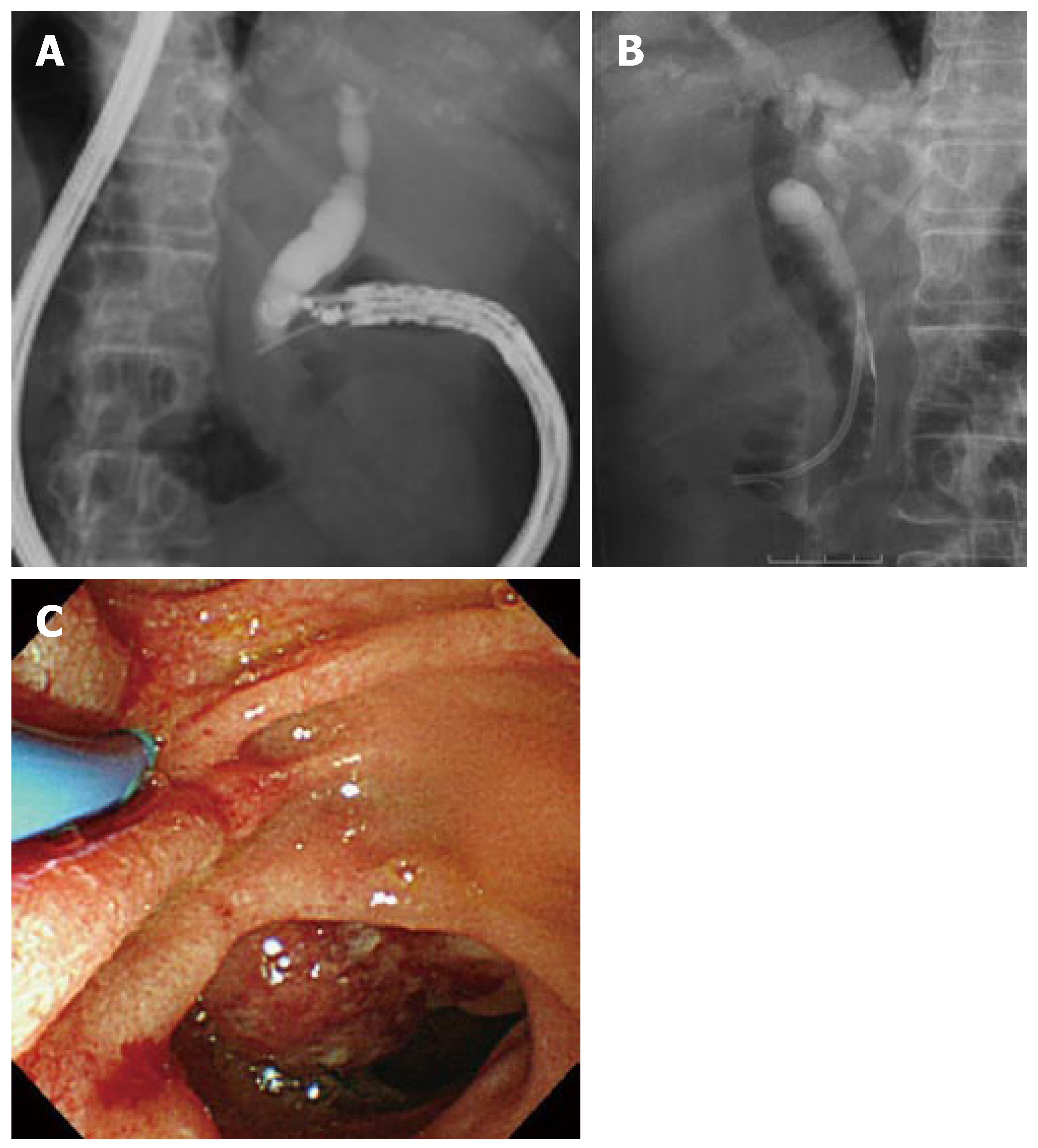
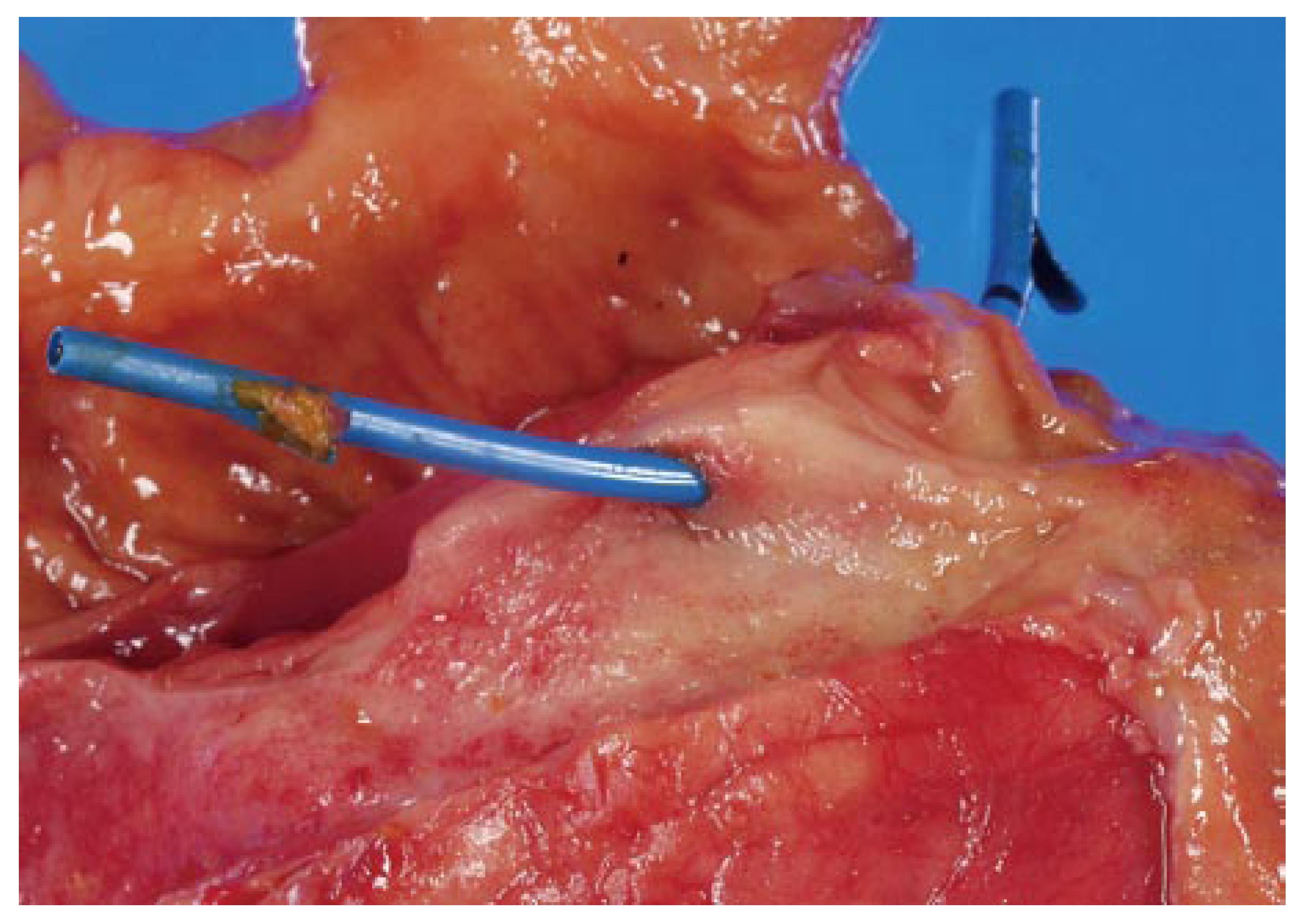
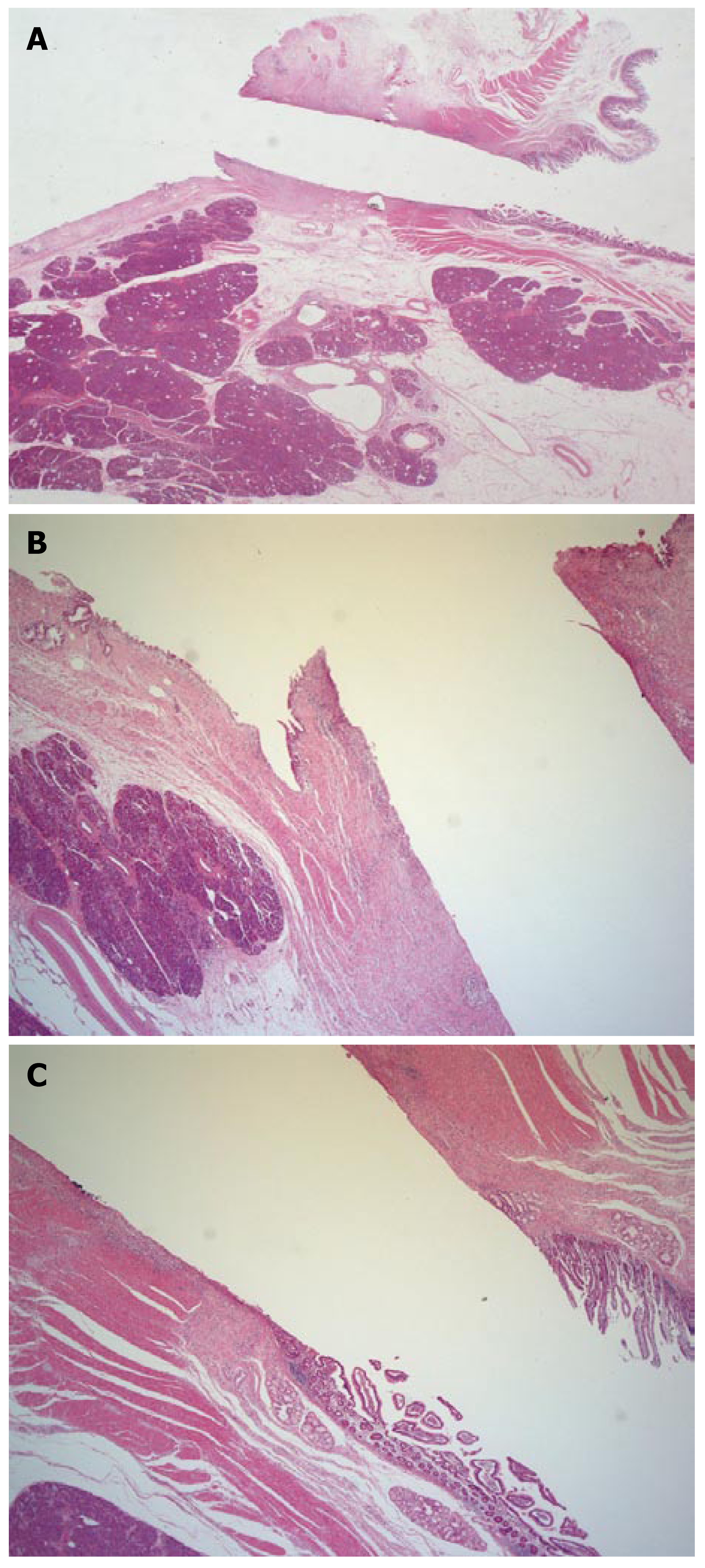
A 76-year-old Japanese woman was admitted to our department, complaining of jaundice and itching. Laboratory data on admission showed the following abnormalities: serum total bilirubin, 11.7 mg/dL; glutamic oxaloacetic transaminase (GOT), 389 IU/L; glutamic pyruvic transaminase (GPT), 285 IU/L; and alkaline phosphatase (ALP), 1487 IU/L. Transabdominal ultrasonography and abdominal computed tomography revealed a mass in the ampullary region, along with dilatation of the bile and pancreatic ducts (Figure 1). To resolve her complaints, endoscopic retrograde cholangiopancreatography (ERCP) with biliary stenting was attempted. However, cannulation of the bile duct was unsuccessful because stenosis of the descending portion of the duodenum, and the presence of a tumor at the papilla of Vater that bled easily when contacted, made it impossible to manipulate the endoscope to identify the orifice. After obtaining written informed consent, ESBD was undertaken. Following visualization of the extrahepatic bile duct with a curved linear array echoendoscope (GF-UC240P; Olympus, Tokyo, Japan), the dilated bile duct was punctured via the upper part of the descending portion of the duodenum with a 19G needle (Olympus) (Figure 2A). Following removal of the core needle, white bile was aspirated. A small amount of contrast agent was then injected via the sheath catheter into the bile duct, to guide stent placement and to confirm the absence of bile leakage or extravasation. A guidewire 0.889 mm in diameter (Jagwire; Boston Scientific, Natik, MA, USA) was introduced into the sheath catheter and inserted into the intrahepatic bile duct. Subsequent to removing the sheath catheter and leaving the guidewire in situ, we dilated the puncture tract with a dilator catheter 7F in diameter, and placed a 7F plastic stent (Figure 2B and C). The patient developed no symptoms related to the procedure and her initial complaints also soon disappeared. One week after the procedure, levels of serum total bilirubin, GOT, GPT, and ALP had declined to 3.2 mg/dL, 33 IU/L, 47 IU/L, and 780 IU/L, respectively. She underwent pancreaticoduodenectomy 14 d after ESBD. Macroscopically, the sites of the puncture in the bile duct and duodenum were clear without infection, hemorrhage or hematoma (Figure 3).Histological examination revealed mild inflammatory cell infiltrate adjacent to the sinus tract in the duodenal and bile duct walls, without hemorrhage (Figure 4). A fistula was formed along the tract of the puncture without significant reactive changes. No evidence of severe inflammation, such as bile peritonitis, was found on the extraluminal side of either the duodenum or the bile duct.
The role of ES in the management of gastrointestinal diseases has evolved from imaging of the gut wall and organs adjacent to the alimentary tract, to its use as a guide for tissue sampling with a fine-needle, as well as in therapeutic applications such as injection of agents into tumors and drainage of pancreatic pseudocysts. Endosonographic approaches to an inaccessible bile duct and pancreatic duct by ERCP were first reported in the mid 1990s. In 1996, Wiersema et al[2] reported the feasibility of ES-guided cholangiopancreatography. Subsequently, several case series have been published on the feasibility and effectiveness of ESBD, the first being by Giovannini et al[3] in 2001, in a pancreatic cancer patient with a history of failed cannulation of the bile duct, even after precutting, who underwent preoperative chemoradiotherapy. They succeeded in the deployment of a 10F stent in a two-step procedure by changing endoscopes. In 2003, Burmester et al[4] reported three cases of successful stent placement among four attempts at ESBD. They achieved biliary drainage by a one-step technique, a modification of the Seifert technique[9] for drainage of pancreatic pseudocysts. They approached the bile duct via the duodenum, stomach and even the jejunum. Mallery et al[5] performed endoscopic ultrasound-guided rendezvous drainage of the bile duct and pancreatic duct after unsuccessful ERCP. They succeeded in stent placement following antegrade traverse of the stenosis with a guidewire in three (two biliary and one pancreatic) of six cases. Puspok et al[6] applied this technique to cases of bile duct stones with difficult cannulation of the bile duct, and reported excellent results. Kahaleh et al[7] recently published their experience of ESBD in 23 patients. They carried out puncture of the bile duct via the stomach, duodenum and jejunum, and concluded that intrahepatic access to the biliary system appears safer than the extrahepatic approach.
As described here, acceptable success rates and incidence of complications were reported for puncture and stent placement under sonographic guidance. However, there have been no reports of the influence of this technique on the gut wall, bile duct, or intervening tissues between them. This is believed to be the first report describing the preoperative performance of ESBD and the histological condition of the sinus tract established by this method. The duodenum, bile duct and sinus tract showed no adverse histological changes. These results are attributable to the use of endosonography as a guide. This technique has a low potential risk of major bleeding as color Doppler evaluation is utilized for determining the route of the puncture. Injury to adjacent organs is also minimized due to clear visualization of the structures in the area of interest, with the use of high-frequency ultrasound. Some authors[4,6,8] have applied a fistulotome with electrocautery for puncture. However, it seems theoretically safer to apply a simple puncture needle to avoid damage to the tissues in and around the pathway of the puncture.
The present study elucidated histological changes at and around the sinus tract, following ESBD. The results that no severe inflammation or hemorrhage occurred should encourage the wider use of this technique, although further large-scale studies will be needed.
In general, the method of choice for biliary obstruction is endoscopic biliary stenting. Percutaneous transhepatic cholangio-drainage (PTCD) is considered a substitute. However, PTCD can result in pain after placement of the drainage tube, and can restrict activities of daily living. In contrast, ESBD is a safe and effective method for biliary drainage and does not cause pain or restriction of daily living, as is the case with endoscopic biliary stenting. It should therefore replace PTCD in a large proportion of those patients with an obstructed biliary tree and with difficult cannulation of the bile duct, duodenal stenosis, or deformity of the papilla of Vater caused by cancer, which hinders detection of the orifice, regardless of the likelihood of successful PTCD.
As is the case with plastic stents in endoscopic biliary stenting, the diameter of the stent available in ESBD is restricted by the diameter of the working channel of the endoscope employed. The endoscope we used had a 2.8-mm diameter working channel, allowing the use of only 7F stents, or smaller.
Dilation of the sinus tract can be achieved with a dilator balloon, following insertion of the guidewire into the bile duct via the lumen of the placed stent and removal of the sent alone. Therefore, once access to the bile duct has been established, it is possible to place a stent with a larger caliber, even a metallic stent, without difficulty in either a one- or two-step procedure. In the long term, it is inevitable that plastic stents will become occluded. Deployment of a metallic stent will prolong patency, as shown in endoscopic transpapillary biliary stenting[10]. Covered metallic stents are deemed to be advantageous in avoiding bile leakage.
Further development of accessory devices specialized for ESBD will help expand its indications.
S- Editor Zhu LH L- Editor Kerr C E-Editor Li JL
| 1. | Harada N, Kouzu T, Arima M, Asano T, Kikuchi T, Isono K. Endoscopic ultrasound-guided pancreatography: a case report. Endoscopy. 1995;27:612-615. [RCA] [PubMed] [DOI] [Full Text] [Cited by in Crossref: 57] [Cited by in RCA: 53] [Article Influence: 1.8] [Reference Citation Analysis (0)] |
| 2. | Wiersema MJ, Sandusky D, Carr R, Wiersema LM, Erdel WC, Frederick PK. Endosonography-guided cholangiopancreatography. Gastrointest Endosc. 1996;43:102-106. [RCA] [PubMed] [DOI] [Full Text] [Cited by in Crossref: 189] [Cited by in RCA: 178] [Article Influence: 6.1] [Reference Citation Analysis (0)] |
| 3. | Giovannini M, Moutardier V, Pesenti C, Bories E, Lelong B, Delpero JR. Endoscopic ultrasound-guided bilioduodenal anastomosis: a new technique for biliary drainage. Endoscopy. 2001;33:898-900. [RCA] [PubMed] [DOI] [Full Text] [Cited by in Crossref: 446] [Cited by in RCA: 480] [Article Influence: 20.0] [Reference Citation Analysis (0)] |
| 4. | Burmester E, Niehaus J, Leineweber T, Huetteroth T. EUS-cholangio-drainage of the bile duct: report of 4 cases. Gastrointest Endosc. 2003;57:246-251. [RCA] [PubMed] [DOI] [Full Text] [Cited by in Crossref: 216] [Cited by in RCA: 196] [Article Influence: 8.9] [Reference Citation Analysis (0)] |
| 5. | Mallery S, Matlock J, Freeman ML. EUS-guided rendezvous drainage of obstructed biliary and pancreatic ducts: Report of 6 cases. Gastrointest Endosc. 2004;59:100-107. [RCA] [PubMed] [DOI] [Full Text] [Cited by in Crossref: 260] [Cited by in RCA: 259] [Article Influence: 12.3] [Reference Citation Analysis (0)] |
| 6. | Püspök A, Lomoschitz F, Dejaco C, Hejna M, Sautner T, Gangl A. Endoscopic ultrasound guided therapy of benign and malignant biliary obstruction: a case series. Am J Gastroenterol. 2005;100:1743-1747. [RCA] [PubMed] [DOI] [Full Text] [Cited by in Crossref: 127] [Cited by in RCA: 111] [Article Influence: 5.6] [Reference Citation Analysis (0)] |
| 7. | Kahaleh M, Hernandez AJ, Tokar J, Adams RB, Shami VM, Yeaton P. Interventional EUS-guided cholangiography: evaluation of a technique in evolution. Gastrointest Endosc. 2006;64:52-59. [RCA] [PubMed] [DOI] [Full Text] [Cited by in Crossref: 193] [Cited by in RCA: 188] [Article Influence: 9.9] [Reference Citation Analysis (0)] |
| 8. | Yamao K, Sawaki A, Takahashi K, Imaoka H, Ashida R, Mizuno N. EUS-guided choledochoduodenostomy for palliative biliary drainage in case of papillary obstruction: report of 2 cases. Gastrointest Endosc. 2006;64:663-667. [RCA] [PubMed] [DOI] [Full Text] [Cited by in Crossref: 77] [Cited by in RCA: 86] [Article Influence: 4.5] [Reference Citation Analysis (0)] |
| 9. | Seifert H, Faust D, Schmitt T, Dietrich C, Caspary W, Wehrmann T. Transmural drainage of cystic peripancreatic lesions with a new large-channel echo endoscope. Endoscopy. 2001;33:1022-1026. [RCA] [PubMed] [DOI] [Full Text] [Cited by in Crossref: 43] [Cited by in RCA: 52] [Article Influence: 2.2] [Reference Citation Analysis (0)] |
| 10. | Isayama H, Komatsu Y, Tsujino T, Sasahira N, Hirano K, Toda N, Nakai Y, Yamamoto N, Tada M, Yoshida H. A prospective randomised study of "covered" versus "uncovered" diamond stents for the management of distal malignant biliary obstruction. Gut. 2004;53:729-734. [RCA] [PubMed] [DOI] [Full Text] [Cited by in Crossref: 460] [Cited by in RCA: 459] [Article Influence: 21.9] [Reference Citation Analysis (0)] |


