Published online Sep 7, 2007. doi: 10.3748/wjg.v13.i33.4473
Revised: April 15, 2007
Accepted: April 18, 2007
Published online: September 7, 2007
AIM: To investigate the role of angiopoietin (Ang) -1, -2 and -4 and its receptors, Tie-1 and -2, in the growth and differentiation of gastrointestinal stromal tumors (GISTs).
METHODS: Thirty GISTs, seventeen leiomyomas and six schwannomas were examined by immunohistochemistry in this study.
RESULTS: Ang-1, -2 and -4 proteins were expressed in the cytoplasm of tumor cells, and Tie-1 and -2 were expressed both in the cytoplasm and on the membrane of all tumors. Immunohistochemical staining revealed that 66.7% of GISTs (20 of 30), 76.5% of leiomyomas (13 of 17) and 83.3% of schwannomas (5 of 6) were positive for Ang-1. 83.3% of GISTs (25 of 30), 82.4% of leiomyomas (14 of 17) and 100% of schwannomas (6 of 6) were positive for Ang-2. 36.7% of GISTs (11 of 30), 58.8% of leiomyomas (10 of 17) and 83.3% of schwannomas (5 of 6) were positive for Ang-4. 60.0% of GISTs (18 of 30), 82.4% of leiomyomas and 100% of schwannomas (6 of 6) were positive for Tie-1. 10.0% of GISTs (3 of 30), 94.1% of leiomyomas (16 of 17) and 33.3% of schwannomas (2 of 6) were positive for Tie-2. Tie-2 expression was statistically different between GISTs and leiomyomas (P < 0.001). However, there was no correlation between expression of angiopoietin pathway components and clinical risk categories.
CONCLUSION: Our results suggest that the angiopoietin pathway plays an important role in the differentiation of GISTs, leiomyomas and schwannomas.
- Citation: Nakayama T, Inaba M, Naito S, Mihara Y, Miura S, Taba M, Yoshizaki A, Wen CY, Sekine I. Expression of Angiopoietin-1, 2 and 4 and Tie-1 and 2 in gastrointestinal stromal tumor, leiomyoma and schwannoma. World J Gastroenterol 2007; 13(33): 4473-4479
- URL: https://www.wjgnet.com/1007-9327/full/v13/i33/4473.htm
- DOI: https://dx.doi.org/10.3748/wjg.v13.i33.4473
Gastrointestinal stromal tumors (GISTs) are rare mesen-chymal tumors of the gastrointestinal tract that may occur from the oesophagus to the anus, including the omentum[1,2]. Despite their rarity, GISTs are the most common primary mesenchymal tumors of the gastro-intestinal tract[1-3]. The mechanisms of tumorigenesis, progression and differentiation of GISTs are unknown. Traditionally, all primary mesenchymal spindle cell tumors of the gastrointestinal (GI) tract were uniformly classified as smooth muscle tumors (e.g., leiomyomas, cellular leiomyomas or leiomyosarcomas). Tumors with epithelioid cytologic features were designated leiomyoblastomas or epithelioid leiomyosarcomas[4]. Recently, Sircar et al[5] postulated that GISTs originate from Cajal cells in the gastrointestinal tract and differ from leiomyomas and schwannomas, which are of mesenchymal cell origin. Cajal cells are thought to be gastrointestinal pacemaker cells that regulate intestinal motility[6]. GISTs are characterized by frequent expression of the bone marrow leukocytic progenitor cell antigen CD34[7] and the c-kit proto-oncogene[2,3,5].
Some GISTs have mutations in the genes encoding C-kit and platelet-derived-growth factor alpha (PDGFRA) that cause constitutive tyrosine kinase activation[3,8-10]. Tumors expressing C-kit or PDGFRA oncoproteins were indistinguishable with respect to activation of downstream signaling intermediates and cytogenetic changes associated with tumor progression. C-kit and PDGFRA mutations appear to be alternative and mutually exclusive oncogenic mechanisms in GISTs[9,10].
Recently, there has been a growing interest in understanding the role of receptor tyrosine kinases (RTK), such as vascular endothelial growth factor receptor (VEGFR), platelet-derived growth factor receptor (PDGFR) and stem cell factor receptor (KIT) in promoting tumor growth and metastasis[3,9,11]. As both Tie-1 and Tie-2 possess unique multiple extracellular domains, they are thought to represent a new subfamily of RTKs[12,13]. Tie signaling is involved in multiple steps of the angiogenic remodeling process during development, including destabilization of existing vessels, endothelial cell migration, tube formation and the subsequent stabilization of newly formed tubes by mesenchymal cells[14-17].
The angiopoietin (Ang) family has been identified as a key regulator of angiogenesis[18] and is composed of subtypes Ang-1, Ang-2, Ang-3, and Ang-4[18-21]. They are the ligands for Tie receptors and the major mediators of the mitogenic and permeability-enhancing effects in endothelial cells[17,22]. In addition, angiopoietins are survival factors for endothelial cells, and a marked dependence on angiopoietins has been shown in newly formed, but not established tumor vessels[23,24].
Ang-1 has been shown to act as an obligatory agonist promoting structural integrity of blood vessels[18,20], whereas Ang-2 has been found to function as a naturally occurring antagonist, promoting either vessel growth or regression depending on the levels of other growth factors, such as VEGF-A[19,25]. The effect of Ang-3 and Ang-4 have been less characterized, but they also show context-dependent actions as antagonistic and agonistic ligands, respectively[21,26]. Signaling through Tie-2 has been extensively studied, and the results suggest that signaling involving phosphatidylinositol 3’ kinase (PI3K) activation is a major pathway[27-29]. The ligand-independent function of Tie-1 involves shedding of the receptor[30,31] and heterometrix complex formation with Tie-2[30,32]. Recently, it has been found that Ang-1 and Ang-4 can activate Tie-1[33].
Coexpression of angiopoietin and its receptor, either Tie-1 or Tie-2, has been reported in tumor cells, suggesting the presence of an autocrine and/or a paracrine angiopoietin/Tie growth pathway in solid tumors[34-36]. Further, the expression levels of angiopoietin and its receptors have been shown to correlate with progressive tumor growth and development of metastasis by many types of carcinomas[35-38].
These studies suggest that the angiopoietin pathway is involved in tumor cell growth and differentiation. However, there are no data detailing angiopoietin expressions and Tie receptor expresions in GIST, leiomyoma or schwannoma, or the role of angiopoietin in the etiology of these tumors. The purpose of this study was to investigate the expression of angiopoietins and Ties in GISTs.
A total of thirty GISTs included 26 cases from the stomach and four from the small intestine. Seventeen leiomyomas included four from the oesophagus, five from the stomach and eight from the large intestine. Six schwannomas included five from the stomach and one from the large intestine. Specimens were selected from surgical pathology archival tissues at Nagasaki University Hospital between 2001 and 2006. The GISTs were 0.8-12.0 cm in diameter, the leiomyomas were 0.1-4.5 cm, and the schwannomas were 0.6-5.0 cm. In this study, GISTs were defined as tumors expressing both c-kit and CD34 surface antigens. GISTs were classified by risk categories, mitosis counts and tumor size[39]. The number of mitoses was determined by counting 50 high-power fields (HPF, × 400) using a Nikon (Tokyo, Japan) E400 microscope. Leiomyomas were defined as expressing α-smooth muscle cell actin (SMA) but not c-kit, CD34 or S100-protein. Schwannomas were defined as expressing S100-protein but not c-kit, CD34 or SMA. Two independent pathologists (T. Nakayama and I. Sekine) determined tumor identification/classification.
The subcellular localization of Ang-1, -2 and -4 and Tie-1 and -2 was determined in GISTs using polyclonal antibodies directed against unique sequences. These antibodies were devoid of any cross-reaction with other proteins in the angiopoietin family. Formalin-fixed and paraffin-embedded specimens were cut into 4 μm thick sections, deparaffinized and preincubated with normal bovine serum to prevent non-specific binding. The sections were incubated overnight at 4°C with primary polyclonal antibody to human Ang-1, -2 or -4 ([N-18],[N-18],[L-18], respectively, 1 μg/mL; Santa Cruz Biotechnology, Inc., Santa Cruz, CA), Tie-1 or -2 ([C-18],[C-20], respectively, 1 μg/mL; Santa Cruz Biotechnology, Inc.), followed by alkaline phosphatase-conjugated anti-goat IgG antibody (0.4 μg/mL; Santa Cruz Biotechnology, Inc.) for Ang-1, -2 and -4, and anti-rabbit IgG antibody (0.4 μg/mL; Santa Cruz Biotechnology, Inc.) for Tie-1 and -2. The reaction products were visualized using a mixture of 5-bromo-4-chloro-3-indolyl phosphate and nitroblue tetrazolium chloride (BCIP/NBT; Roche Diagnostic Corp., Indianapolis, IN). Negative controls replaced the primary antibody with non-immunized rabbit or goat serum, and the hemangioma tissue of human skin served as the positive control[40]. Ang-1, -2 and -4 and Tie-1 and -2 expressions were classified into three categories depending upon the percentage of cells stained and/or the intensity of staining: -, 0 to 10% tumor cells positive; +, > 10% tumor cells positive.
The Stat View II program (Abacus Concepts, Inc., Berkeley, CA) was used for statistical analysis. Analyses comparing the degree of Ang-1, -2 and -4 and Tie-1 and -2 expressions in GISTs, leiomyomas and schwannomas were performed using the Mann-Whitney’s test.
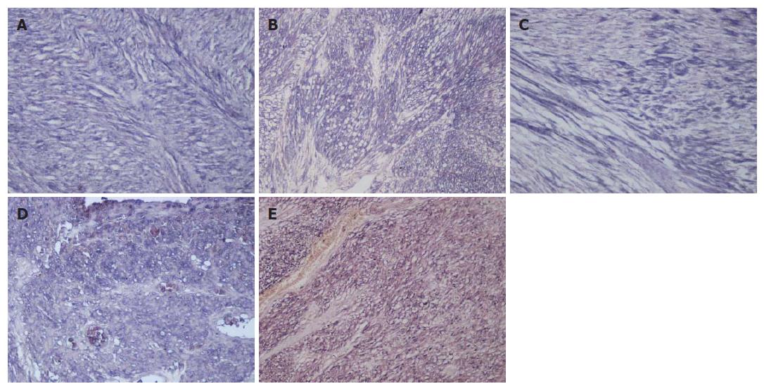
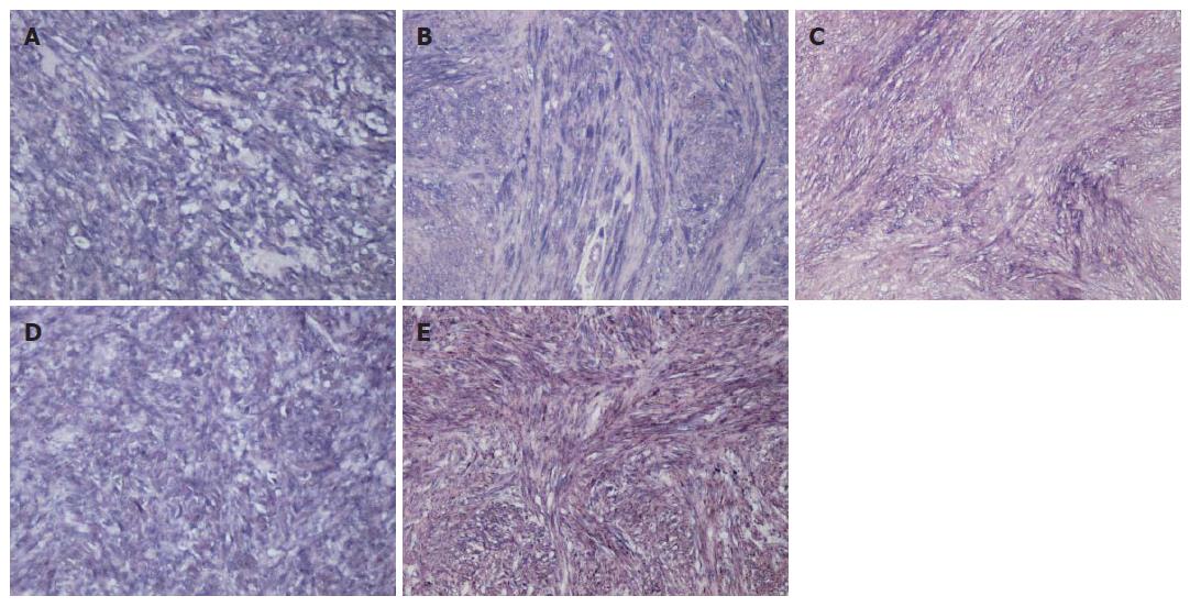
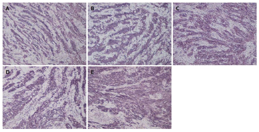
The results of immunohistochemical stainings for Ang-1, -2 and -4 and Tie-1 and -2 are summarized in Table 1. Ang-1, -2 and -4 and Tie-1 and -2 were heterogeneously expressed in GISTs, leiomyomas and schwannomas and localized to the cytoplasm and/or membrane of tumor cells. Immunohistochemical staining revealed Ang-1, -2 and -4 in the cytoplasm of GIST (Figure 1A-C), leiomyoma (Figure 2A-C) and schwannoma (Figure 3A-C) cells. Tie-1 and -2 were found in the membrane and cytoplasm of GIST (Figure 1D and E), leiomyoma (Figure 2D and E) and schwannoma (Figure 3D and E) cells. Immunohistochemical staining revealed that 66.7% of GISTs (20 of 30), 76.5% of leiomyomas (13 of 17) and 83.3% of schwannomas (5 of 6) were positive for Ang-1. 83.3% of GISTs (25 of 30), 82.4% of leiomyomas (14 of 17) and 100% of schwannomas (6 of 6) were positive for Ang-2. 36.7% of GISTs (11 of 30), 58.8% of leiomyomas (10 of 17) and 83.3% of schwannomas (5 of 6) were positive for Ang-4. There were no statistical differences in Ang-1, -2 or -4 expression between GISTs and leiomyomas or schwannomas. 60.0% of GISTs (18 of 30), 82.4% of leiomyomas and 100% of schwannomas (6 of 6) were positive for Tie-1. 10.0% of GISTs (3 of 30), 94.1% of leiomyomas (16 of 17) and 66.7% of schwannomas (2 of 6) were positive for Tie-2. Tie-2 expression was statistically different between GISTs and leiomyomas (P < 0.001). However, there was no correlation between Tie-1 expression and histological differences.
| Ang-1 | Ang-2 | Ang-4 | Tie-1 | Tie-2 | |||||||
| n | - | + | - | + | - | + | - | + | - | + | |
| GIST | 30 | 10 (33.3) | 20 (66.7) | 5 (16.7) | 25 (83.3) | 19 (63.3) | 11 (36.7) | 12 (40.0) | 18 (60.0) | 27 (90.0) | 3 (10.0)a |
| Leiomyoma | 17 | 4 (23.5) | 13 (76.5) | 3 (17.6) | 14 (82.4) | 7 (41.2) | 10 (58.8) | 3 (17.6) | 14 (82.4) | 1 (5.9) | 16 (94.1) |
| Schwannoma | 6 | 1 (16.7) | 5 (83.3) | 0 (0.0) | 6 (100) | 1 (16.7) | 5 (83.3) | 0 (0.0) | 6 (100) | 4 (66.7) | 2 (33.3) |
GISTs were classified by risk category, mitosis counts and tumor size in Table 2. All six cases within the high risk category expressed Ang-1 and -2 and Tie-1 and -2 proteins. All three cases with over 10 mitoses per 50 HPFs strongly expressed Ang-1, -2 and -4 and Tie-1 and -2. Finally, only two tumors that measured over 10 cm strongly expressed Ang-1, -2 and -4 and Tie-1 and -2. However, there was no correlation between Ang-1, -2 and -4 and Tie-1 and -2 expression and each classification.
| Ang-1 | Ang-2 | Ang-4 | Tie-1 | Tie-2 | |||||||
| n | - | + | - | + | - | + | - | + | - | + | |
| GIST | 30 | 10 (33.3) | 20 (66.7) | 5 (16.7) | 25 (83.3) | 19 (63.3) | 11 (36.7) | 12 (40.0) | 18 (60.0) | 27 (90.0) | 3 (10.0) |
| Risk categories | NS | NS | NS | NS | NS | ||||||
| High | 6 | 1 (16.7) | 5 (83.3) | 2 (33.3) | 4 (66.7) | 6 (100) | 0 (0.0) | 1 (16.7) | 5 (83.3) | 5 (83.3) | 1 (16.7) |
| Intermediate | 4 | 2 (50.0) | 2 (50.0) | 1 (25.0) | 3 (75.0) | 2 (50.0) | 2 (50.0) | 2 (50.0) | 2 (50.0) | 4 (100) | 0 (0.0) |
| Low | 15 | 6 (40.0) | 9 (60.0) | 2 (13.3) | 13 (86.7) | 8 (53.3) | 7 (46.7) | 5 (33.3) | 10 (66.7) | 13 (86.7) | 2 (13.3) |
| Very low | 5 | 1 (20.0) | 4 (80.0) | 0 (0.0) | 5 (100) | 3 (60.0) | 2 (40.0) | 4 (80.0) | 1 (20.0) | 5 (100) | 0 (0.0) |
| Mitosis counts (per 50 fields, HPF) | NS | NS | NS | NS | NS | ||||||
| < 2 | 19 | 7 (36.8) | 12 (63.2) | 3 (15.8) | 16 (84.2) | 11 (57.9) | 8 (42.1) | 9 (47.4) | 10 (52.6) | 17 (89.5) | 2 (10.5) |
| 2-5 | 7 | 2 (28.6) | 5 (71.4) | 1 (14.3) | 6 (85.7) | 4 (57.1) | 3 (42.9) | 2 (28.6) | 5 (71.4) | 6 (85.7) | 1 (14.3) |
| 6-10 | 2 | 1 (50.0) | 1 (50.0) | 1 (50.0) | 1 (50.0) | 2 (100) | 0 (0.0) | 0 (0.0) | 2 (100) | 2 (100) | 0 (0.0) |
| 10 < | 2 | 0 (0.0) | 2 (100) | 0 (0.0) | 2 (100) | 2 (100) | 0 (0.0) | 1 (50.0) | 1 (50.0) | 2 (100) | 0 (0.0) |
| Tumor size (cm in length) | NS | NS | NS | NS | NS | ||||||
| < 2 | 4 | 1 (25.0) | 3 (75.0) | 0 (0.0) | 4 (100) | 3 (75.0) | 1 (25.0) | 3 (75.0) | 1 (25.0) | 4 (100) | 0 (0.0) |
| 2-5 | 16 | 6 (37.5) | 10 (62.5) | 2 (12.5) | 14 (87.5) | 9 (56.3) | 7 (43.8) | 6 (37.5) | 10 (62.5) | 15 (93.8) | 1 (6.3) |
| 5-10 | 7 | 3 (42.9) | 4 (57.1) | 2 (28.6) | 5 (71.4) | 4 (57.1) | 3 (42.9) | 2 (28.6) | 5 (71.4) | 6 (85.7) | 1 (14.3) |
| > 10 | 3 | 0 (0.0) | 3 (100) | 1 (33.3) | 2 (66.7) | 3 (100) | 0 (0.0) | 1 (33.3) | 2 (66.7) | 2 (66.7) | 1 (33.3) |
The coexpression of Ang-1, -2 and -4 and Tie-1 and -2 has been reported in tumor cells, suggesting the presence of an autocrine and/or a paracrine angiopoietin/Tie growth pathway in solid tumors[35-38]. Angiopoietins (Angs) also have been shown to play a role in the proliferation and/or differentiation of stromal tumors and normal mesenchymal cells[41,42]. Angiopoietin expression in GISTs has not been reported yet. Further, there have been no studies of Tie receptor expression in GISTs, leiomyomas and schwannomas or of the potential roles of angiopoietins and its receptors in the growth of these tumors. This is the first study to determine the expression of Tie receptors in GIST and stromal tumors. Our results demonstrate substantial levels of angiopoietins and Tie receptors in the cytoplasm of GIST, leiomyoma and schwannoma cells. Therefore, we suggest that angiopoietins and its receptors may play an important role in growth and/or differentiation of intestinal stromal tumors via autocrine and/or paracrine pathways.
We did not find any statistical correlation between risk grade and the expression of Angs or Ties for GISTs. However, all four GISTs in the high risk category expressed Angs and Ties (Table 2). Further, all four GISTs that had higher mitosis counts (over ten per 50 HPFs) were positive for Angs and Ties. Our data suggest that high risk GISTs might express Angs and Ties at higher than normal levels. Future studies will examine angiopoietin pathway components in high risk GISTs.
Ang-2 induces a variety of enzymes and proteins important in the degradation process, including matrix-degrading metalloproteinases, metalloproteinase interstitial collagenase, and serine proteases, such as urokinase-type plasminogen activator (u-PA)[43,44]. In this study, we did not evaluate the invasive activities of GIST cells because all of the GISTs were solitary and showed clear margins. However, the activation of these various factors by angiopoietins may allow for the development of a prodegradative environment that facilitates migration and invasion of tumor cells.
There has been a growing interest in understanding the role of receptor tyrosine kinases (RTK), such as vascular endothelial growth factor receptor (VEGFR)[11], platelet-derived growth factor receptor (PDGFR)[9] and stem cell factor receptor (KIT)[3] in promoting tumor growth and metastasis. Joensuu et al[45] reported a patient in whom Imatinib (STI-571, Gleevec), a tyrosine kinase inhibitor, was effective against a GIST. Imatinib has proven to be remarkably efficacious in heavily pretreated GIST patients with advanced disease[46]. Further, anti-angiopoietin reagents are being used in clinical trials for the therapy of gastric, lung and breast cancer[47,48].
Sunitinib (sunitinib malate; SU11248; SUTENT®; Pfizer Inc, New York, NY, USA) is a novel multi-targeted tyrosine kinase inhibitor with high binding affinity for VEGFR and PDGFR that recently has shown anti-tumor and anti-angiogenic activities[49]. This drug recently received approval from the US Food and Drug Administration (FDA) for two applications: advanced gastrointestinal stromal tumors (GISTs)[50] and renal cell carcinoma[51], in patients who are resistant or intolerant to treatment with Imatinib. Since the Tie receptor is an RTK, Sunitinib may be suitable to down-regulate the activity of the angiopoietin pathway. In fact, this study presents data that supports the clinical validity of Sunitinib in GISTs.
We are grateful to Mr. Toshiyuki Kawada (Nagasaki University Graduate School of Biomedical Sciences) for his excellent immunohistochemical assistance.
S- Editor Liu Y L- Editor Lutze M E- Editor Liu Y
| 1. | Miettinen M, Lasota J. Gastrointestinal stromal tumors--definition, clinical, histological, immunohistochemical, and molecular genetic features and differential diagnosis. Virchows Arch. 2001;438:1-12. [RCA] [PubMed] [DOI] [Full Text] [Cited by in Crossref: 1185] [Cited by in RCA: 1178] [Article Influence: 49.1] [Reference Citation Analysis (0)] |
| 2. | Kindblom LG, Remotti HE, Aldenborg F, Meis-Kindblom JM. Gastrointestinal pacemaker cell tumor (GIPACT): gastrointestinal stromal tumors show phenotypic characteristics of the interstitial cells of Cajal. Am J Pathol. 1998;152:1259-1269. [PubMed] |
| 3. | Hirota S, Isozaki K, Moriyama Y, Hashimoto K, Nishida T, Ishiguro S, Kawano K, Hanada M, Kurata A, Takeda M. Gain-of-function mutations of c-kit in human gastrointestinal stromal tumors. Science. 1998;279:577-580. [RCA] [PubMed] [DOI] [Full Text] [Cited by in Crossref: 3215] [Cited by in RCA: 3114] [Article Influence: 115.3] [Reference Citation Analysis (0)] |
| 4. | Appelman HD. Mesenchymal tumors of the gut: historical perspectives, new approaches, new results, and does it make any difference? Monogr Pathol. 1990;220-246. [PubMed] |
| 5. | Sircar K, Hewlett BR, Huizinga JD, Chorneyko K, Berezin I, Riddell RH. Interstitial cells of Cajal as precursors of gastrointestinal stromal tumors. Am J Surg Pathol. 1999;23:377-389. [RCA] [PubMed] [DOI] [Full Text] [Cited by in Crossref: 353] [Cited by in RCA: 337] [Article Influence: 13.0] [Reference Citation Analysis (0)] |
| 6. | Sanders KM. A case for interstitial cells of Cajal as pacemakers and mediators of neurotransmission in the gastrointestinal tract. Gastroenterology. 1996;111:492-515. [RCA] [PubMed] [DOI] [Full Text] [Cited by in Crossref: 765] [Cited by in RCA: 747] [Article Influence: 25.8] [Reference Citation Analysis (0)] |
| 7. | Miettinen M, Virolainen M. Gastrointestinal stromal tumors--value of CD34 antigen in their identification and separation from true leiomyomas and schwannomas. Am J Surg Pathol. 1995;19:207-216. [RCA] [PubMed] [DOI] [Full Text] [Cited by in Crossref: 327] [Cited by in RCA: 292] [Article Influence: 9.7] [Reference Citation Analysis (0)] |
| 8. | Plaat BE, Hollema H, Molenaar WM, Torn Broers GH, Pijpe J, Mastik MF, Hoekstra HJ, van den Berg E, Scheper RJ, van der Graaf WT. Soft tissue leiomyosarcomas and malignant gastrointestinal stromal tumors: differences in clinical outcome and expression of multidrug resistance proteins. J Clin Oncol. 2000;18:3211-3220. [PubMed] |
| 9. | Heinrich MC, Corless CL, Duensing A, McGreevey L, Chen CJ, Joseph N, Singer S, Griffith DJ, Haley A, Town A. PDGFRA activating mutations in gastrointestinal stromal tumors. Science. 2003;299:708-710. [RCA] [PubMed] [DOI] [Full Text] [Cited by in Crossref: 1712] [Cited by in RCA: 1722] [Article Influence: 78.3] [Reference Citation Analysis (0)] |
| 10. | Hirota S, Ohashi A, Nishida T, Isozaki K, Kinoshita K, Shinomura Y, Kitamura Y. Gain-of-function mutations of platelet-derived growth factor receptor alpha gene in gastrointestinal stromal tumors. Gastroenterology. 2003;125:660-667. [RCA] [PubMed] [DOI] [Full Text] [Cited by in Crossref: 492] [Cited by in RCA: 478] [Article Influence: 21.7] [Reference Citation Analysis (0)] |
| 11. | Nakayama T, Cho YC, Mine Y, Yoshizaki A, Naito S, Wen CY, Sekine I. Expression of vascular endothelial growth factor and its receptors VEGFR-1 and 2 in gastrointestinal stromal tumors, leiomyomas and schwannomas. World J Gastroenterol. 2006;12:6182-6187. [PubMed] |
| 12. | Maisonpierre PC, Goldfarb M, Yancopoulos GD, Gao G. Distinct rat genes with related profiles of expression define a TIE receptor tyrosine kinase family. Oncogene. 1993;8:1631-1637. [PubMed] |
| 13. | Sato TN, Qin Y, Kozak CA, Audus KL. Tie-1 and tie-2 define another class of putative receptor tyrosine kinase genes expressed in early embryonic vascular system. Proc Natl Acad Sci USA. 1993;90:9355-9358. [RCA] [PubMed] [DOI] [Full Text] [Cited by in Crossref: 334] [Cited by in RCA: 325] [Article Influence: 10.2] [Reference Citation Analysis (0)] |
| 14. | Witzenbichler B, Maisonpierre PC, Jones P, Yancopoulos GD, Isner JM. Chemotactic properties of angiopoietin-1 and -2, ligands for the endothelial-specific receptor tyrosine kinase Tie2. J Biol Chem. 1998;273:18514-18521. [RCA] [PubMed] [DOI] [Full Text] [Cited by in Crossref: 323] [Cited by in RCA: 317] [Article Influence: 11.7] [Reference Citation Analysis (0)] |
| 15. | Hayes AJ, Huang WQ, Mallah J, Yang D, Lippman ME, Li LY. Angiopoietin-1 and its receptor Tie-2 participate in the regulation of capillary-like tubule formation and survival of endothelial cells. Microvasc Res. 1999;58:224-237. [RCA] [PubMed] [DOI] [Full Text] [Cited by in Crossref: 130] [Cited by in RCA: 128] [Article Influence: 4.9] [Reference Citation Analysis (0)] |
| 16. | Sato TN, Tozawa Y, Deutsch U, Wolburg-Buchholz K, Fujiwara Y, Gendron-Maguire M, Gridley T, Wolburg H, Risau W, Qin Y. Distinct roles of the receptor tyrosine kinases Tie-1 and Tie-2 in blood vessel formation. Nature. 1995;376:70-74. [RCA] [PubMed] [DOI] [Full Text] [Cited by in Crossref: 1319] [Cited by in RCA: 1251] [Article Influence: 41.7] [Reference Citation Analysis (0)] |
| 17. | Eklund L, Olsen BR. Tie receptors and their angiopoietin ligands are context-dependent regulators of vascular remodeling. Exp Cell Res. 2006;312:630-641. [RCA] [PubMed] [DOI] [Full Text] [Cited by in Crossref: 237] [Cited by in RCA: 244] [Article Influence: 12.2] [Reference Citation Analysis (0)] |
| 18. | Davis S, Aldrich TH, Jones PF, Acheson A, Compton DL, Jain V, Ryan TE, Bruno J, Radziejewski C, Maisonpierre PC. Isolation of angiopoietin-1, a ligand for the TIE2 receptor, by secretion-trap expression cloning. Cell. 1996;87:1161-1169. [RCA] [PubMed] [DOI] [Full Text] [Cited by in Crossref: 1496] [Cited by in RCA: 1404] [Article Influence: 48.4] [Reference Citation Analysis (0)] |
| 19. | Maisonpierre PC, Suri C, Jones PF, Bartunkova S, Wiegand SJ, Radziejewski C, Compton D, McClain J, Aldrich TH, Papadopoulos N. Angiopoietin-2, a natural antagonist for Tie2 that disrupts in vivo angiogenesis. Science. 1997;277:55-60. [RCA] [PubMed] [DOI] [Full Text] [Cited by in Crossref: 2548] [Cited by in RCA: 2531] [Article Influence: 90.4] [Reference Citation Analysis (0)] |
| 20. | Suri C, Jones PF, Patan S, Bartunkova S, Maisonpierre PC, Davis S, Sato TN, Yancopoulos GD. Requisite role of angiopoietin-1, a ligand for the TIE2 receptor, during embryonic angiogenesis. Cell. 1996;87:1171-1180. [RCA] [PubMed] [DOI] [Full Text] [Cited by in Crossref: 2170] [Cited by in RCA: 2047] [Article Influence: 70.6] [Reference Citation Analysis (0)] |
| 21. | Valenzuela DM, Griffiths JA, Rojas J, Aldrich TH, Jones PF, Zhou H, McClain J, Copeland NG, Gilbert DJ, Jenkins NA. Angiopoietins 3 and 4: diverging gene counterparts in mice and humans. Proc Natl Acad Sci USA. 1999;96:1904-1909. [RCA] [PubMed] [DOI] [Full Text] [Cited by in Crossref: 326] [Cited by in RCA: 319] [Article Influence: 12.3] [Reference Citation Analysis (0)] |
| 22. | Kanda S, Miyata Y, Mochizuki Y, Matsuyama T, Kanetake H. Angiopoietin 1 is mitogenic for cultured endothelial cells. Cancer Res. 2005;65:6820-6827. [RCA] [PubMed] [DOI] [Full Text] [Cited by in Crossref: 61] [Cited by in RCA: 60] [Article Influence: 3.0] [Reference Citation Analysis (0)] |
| 23. | Reinmuth N, Liu W, Jung YD, Ahmad SA, Shaheen RM, Fan F, Bucana CD, McMahon G, Gallick GE, Ellis LM. Induction of VEGF in perivascular cells defines a potential paracrine mechanism for endothelial cell survival. FASEB J. 2001;15:1239-1241. [PubMed] |
| 24. | Brindle NP, Saharinen P, Alitalo K. Signaling and functions of angiopoietin-1 in vascular protection. Circ Res. 2006;98:1014-1023. [RCA] [PubMed] [DOI] [Full Text] [Full Text (PDF)] [Cited by in Crossref: 394] [Cited by in RCA: 374] [Article Influence: 19.7] [Reference Citation Analysis (0)] |
| 25. | Holash J, Wiegand SJ, Yancopoulos GD. New model of tumor angiogenesis: dynamic balance between vessel regression and growth mediated by angiopoietins and VEGF. Oncogene. 1999;18:5356-5362. [RCA] [PubMed] [DOI] [Full Text] [Cited by in Crossref: 553] [Cited by in RCA: 535] [Article Influence: 20.6] [Reference Citation Analysis (0)] |
| 26. | Lee HJ, Cho CH, Hwang SJ, Choi HH, Kim KT, Ahn SY, Kim JH, Oh JL, Lee GM, Koh GY. Biological characterization of angiopoietin-3 and angiopoietin-4. FASEB J. 2004;18:1200-1208. [RCA] [PubMed] [DOI] [Full Text] [Cited by in Crossref: 122] [Cited by in RCA: 129] [Article Influence: 6.5] [Reference Citation Analysis (0)] |
| 27. | Saito M, Hamasaki M, Shibuya M. Induction of tube formation by angiopoietin-1 in endothelial cell/fibroblast co-culture is dependent on endogenous VEGF. Cancer Sci. 2003;94:782-790. [RCA] [PubMed] [DOI] [Full Text] [Cited by in Crossref: 58] [Cited by in RCA: 57] [Article Influence: 2.6] [Reference Citation Analysis (0)] |
| 28. | DeBusk LM, Hallahan DE, Lin PC. Akt is a major angiogenic mediator downstream of the Ang1/Tie2 signaling pathway. Exp Cell Res. 2004;298:167-177. [RCA] [PubMed] [DOI] [Full Text] [Cited by in Crossref: 91] [Cited by in RCA: 98] [Article Influence: 4.7] [Reference Citation Analysis (0)] |
| 29. | Fujikawa K, de Aos Scherpenseel I, Jain SK, Presman E, Christensen RA, Varticovski L. Role of PI 3-kinase in angiopoietin-1-mediated migration and attachment-dependent survival of endothelial cells. Exp Cell Res. 1999;253:663-672. [RCA] [PubMed] [DOI] [Full Text] [Cited by in Crossref: 105] [Cited by in RCA: 106] [Article Influence: 4.1] [Reference Citation Analysis (0)] |
| 30. | Chen-Konak L, Guetta-Shubin Y, Yahav H, Shay-Salit A, Zilberman M, Binah O, Resnick N. Transcriptional and post-translation regulation of the Tie1 receptor by fluid shear stress changes in vascular endothelial cells. FASEB J. 2003;17:2121-2123. [PubMed] |
| 31. | McCarthy MJ, Burrows R, Bell SC, Christie G, Bell PR, Brindle NP. Potential roles of metalloprotease mediated ectodomain cleavage in signaling by the endothelial receptor tyrosine kinase Tie-1. Lab Invest. 1999;79:889-895. [PubMed] |
| 32. | Tsiamis AC, Morris PN, Marron MB, Brindle NP. Vascular endothelial growth factor modulates the Tie-2: Tie-1 receptor complex. Microvasc Res. 2002;63:149-158. [RCA] [PubMed] [DOI] [Full Text] [Cited by in Crossref: 35] [Cited by in RCA: 35] [Article Influence: 1.5] [Reference Citation Analysis (0)] |
| 33. | Saharinen P, Kerkelä K, Ekman N, Marron M, Brindle N, Lee GM, Augustin H, Koh GY, Alitalo K. Multiple angiopoietin recombinant proteins activate the Tie1 receptor tyrosine kinase and promote its interaction with Tie2. J Cell Biol. 2005;169:239-243. [RCA] [PubMed] [DOI] [Full Text] [Full Text (PDF)] [Cited by in Crossref: 150] [Cited by in RCA: 150] [Article Influence: 7.5] [Reference Citation Analysis (0)] |
| 34. | Tang D, Nagano H, Yamamoto H, Wada H, Nakamura M, Kondo M, Ota H, Yoshioka S, Kato H, Damdinsuren B. Angiogenesis in cholangiocellular carcinoma: expression of vascular endothelial growth factor, angiopoietin-1/2, thrombospondin-1 and clinicopathological significance. Oncol Rep. 2006;15:525-532. [PubMed] |
| 35. | Nakayama T, Yoshizaki A, Kawahara N, Ohtsuru A, Wen CY, Fukuda E, Nakashima M, Sekine I. Expression of Tie-1 and 2 receptors, and angiopoietin-1, 2 and 4 in gastric carcinoma; immunohistochemical analyses and correlation with clinicopathological factors. Histopathology. 2004;44:232-239. [RCA] [PubMed] [DOI] [Full Text] [Cited by in Crossref: 24] [Cited by in RCA: 25] [Article Influence: 1.2] [Reference Citation Analysis (0)] |
| 36. | Nakayama T, Hatachi G, Wen CY, Yoshizaki A, Yamazumi K, Niino D, Sekine I. Expression and significance of Tie-1 and Tie-2 receptors, and angiopoietins-1, 2 and 4 in colorectal adenocarcinoma: Immunohistochemical analysis and correlation with clinicopathological factors. World J Gastroenterol. 2005;11:964-969. [PubMed] |
| 37. | Lind AJ, Wikström P, Granfors T, Egevad L, Stattin P, Bergh A. Angiopoietin 2 expression is related to histological grade, vascular density, metastases, and outcome in prostate cancer. Prostate. 2005;62:394-399. [RCA] [PubMed] [DOI] [Full Text] [Cited by in Crossref: 62] [Cited by in RCA: 58] [Article Influence: 2.9] [Reference Citation Analysis (0)] |
| 38. | Mitsutake N, Namba H, Takahara K, Ishigaki K, Ishigaki J, Ayabe H, Yamashita S. Tie-2 and angiopoietin-1 expression in human thyroid tumors. Thyroid. 2002;12:95-99. [RCA] [PubMed] [DOI] [Full Text] [Cited by in Crossref: 33] [Cited by in RCA: 35] [Article Influence: 1.5] [Reference Citation Analysis (0)] |
| 39. | Fletcher CD, Berman JJ, Corless C, Gorstein F, Lasota J, Longley BJ, Miettinen M, O'Leary TJ, Remotti H, Rubin BP. Diagnosis of gastrointestinal stromal tumors: a consensus approach. Int J Surg Pathol. 2002;10:81-89. [RCA] [PubMed] [DOI] [Full Text] [Cited by in Crossref: 263] [Cited by in RCA: 275] [Article Influence: 12.0] [Reference Citation Analysis (0)] |
| 40. | Yu Y, Varughese J, Brown LF, Mulliken JB, Bischoff J. Increased Tie2 expression, enhanced response to angiopoietin-1, and dysregulated angiopoietin-2 expression in hemangioma-derived endothelial cells. Am J Pathol. 2001;159:2271-2280. [RCA] [PubMed] [DOI] [Full Text] [Cited by in Crossref: 112] [Cited by in RCA: 115] [Article Influence: 4.8] [Reference Citation Analysis (0)] |
| 41. | Takahara K, Iioka T, Furukawa K, Uchida T, Nakashima M, Tsukazaki T, Shindo H. Autocrine/paracrine role of the angiopoietin-1 and -2/Tie2 system in cell proliferation and chemotaxis of cultured fibroblastic synoviocytes in rheumatoid arthritis. Hum Pathol. 2004;35:150-158. [RCA] [PubMed] [DOI] [Full Text] [Cited by in Crossref: 18] [Cited by in RCA: 23] [Article Influence: 1.1] [Reference Citation Analysis (0)] |
| 42. | Gläsker S, Li J, Xia JB, Okamoto H, Zeng W, Lonser RR, Zhuang Z, Oldfield EH, Vortmeyer AO. Hemangioblastomas share protein expression with embryonal hemangioblast progenitor cell. Cancer Res. 2006;66:4167-4172. [RCA] [PubMed] [DOI] [Full Text] [Cited by in Crossref: 71] [Cited by in RCA: 72] [Article Influence: 3.8] [Reference Citation Analysis (0)] |
| 43. | Hu B, Jarzynka MJ, Guo P, Imanishi Y, Schlaepfer DD, Cheng SY. Angiopoietin 2 induces glioma cell invasion by stimulating matrix metalloprotease 2 expression through the alphavbeta1 integrin and focal adhesion kinase signaling pathway. Cancer Res. 2006;66:775-783. [RCA] [PubMed] [DOI] [Full Text] [Full Text (PDF)] [Cited by in Crossref: 136] [Cited by in RCA: 130] [Article Influence: 6.8] [Reference Citation Analysis (0)] |
| 44. | Etoh T, Inoue H, Tanaka S, Barnard GF, Kitano S, Mori M. Angiopoietin-2 is related to tumor angiogenesis in gastric carcinoma: possible in vivo regulation via induction of proteases. Cancer Res. 2001;61:2145-2153. [PubMed] |
| 45. | Joensuu H, Roberts PJ, Sarlomo-Rikala M, Andersson LC, Tervahartiala P, Tuveson D, Silberman S, Capdeville R, Dimitrijevic S, Druker B. Effect of the tyrosine kinase inhibitor STI571 in a patient with a metastatic gastrointestinal stromal tumor. N Engl J Med. 2001;344:1052-1056. [RCA] [PubMed] [DOI] [Full Text] [Cited by in Crossref: 1430] [Cited by in RCA: 1326] [Article Influence: 55.3] [Reference Citation Analysis (0)] |
| 46. | Maki RG. Gastrointestinal Stromal Tumors Respond to Tyrosine Kinase-targeted Therapy. Curr Treat Options Gastroenterol. 2004;7:13-17. [RCA] [PubMed] [DOI] [Full Text] [Cited by in Crossref: 18] [Cited by in RCA: 15] [Article Influence: 0.7] [Reference Citation Analysis (0)] |
| 47. | Wang J, Wu KC, Zhang DX, Fan DM. Antisense angiopoietin-1 inhibits tumorigenesis and angiogenesis of gastric cancer. World J Gastroenterol. 2006;12:2450-2454. [PubMed] |
| 48. | Perry BN, Govindarajan B, Bhandarkar SS, Knaus UG, Valo M, Sturk C, Carrillo CO, Sohn A, Cerimele F, Dumont D. Pharmacologic blockade of angiopoietin-2 is efficacious against model hemangiomas in mice. J Invest Dermatol. 2006;126:2316-2322. [RCA] [PubMed] [DOI] [Full Text] [Cited by in Crossref: 91] [Cited by in RCA: 94] [Article Influence: 4.9] [Reference Citation Analysis (0)] |
| 49. | Deeks ED, Keating GM. Sunitinib. Drugs. 2006;66:2255-2266; discussion 2255-2266;. [RCA] [PubMed] [DOI] [Full Text] [Cited by in Crossref: 38] [Cited by in RCA: 47] [Article Influence: 2.6] [Reference Citation Analysis (0)] |
| 50. | Desai J, Yassa L, Marqusee E, George S, Frates MC, Chen MH, Morgan JA, Dychter SS, Larsen PR, Demetri GD. Hypothyroidism after sunitinib treatment for patients with gastrointestinal stromal tumors. Ann Intern Med. 2006;145:660-664. [RCA] [PubMed] [DOI] [Full Text] [Cited by in Crossref: 307] [Cited by in RCA: 275] [Article Influence: 14.5] [Reference Citation Analysis (0)] |
| 51. | Ronnen EA, Kondagunta GV, Ishill N, Spodek L, Russo P, Reuter V, Bacik J, Motzer RJ. Treatment outcome for metastatic papillary renal cell carcinoma patients. Cancer. 2006;107:2617-2621. [RCA] [PubMed] [DOI] [Full Text] [Cited by in Crossref: 70] [Cited by in RCA: 69] [Article Influence: 3.6] [Reference Citation Analysis (0)] |


