Published online Aug 14, 2007. doi: 10.3748/wjg.v13.i30.4147
Revised: April 13, 2007
Accepted: April 30, 2007
Published online: August 14, 2007
Dedifferentiated liposarcoma is a variant of liposarcoma with a more aggressive course. It occurs most commonly in the retroperitoneum and rarely in other anatomic locations. In the present report, we describe a case of dedifferentiated liposarcoma that occurred in an unusual location, sigmoid mesocolon, which has not yet been documented.
- Citation: Winn B, Gao J, Akbari H, Bhattacharya B. Dedifferentiated liposarcoma arising from the sigmoid mesocolon: A case report. World J Gastroenterol 2007; 13(30): 4147-4148
- URL: https://www.wjgnet.com/1007-9327/full/v13/i30/4147.htm
- DOI: https://dx.doi.org/10.3748/wjg.v13.i30.4147
Liposarcoma is a common sarcoma of the soft tissue in adults, occurring most commonly in the extremities and retroperitoneum[1]. Well-differentiated liposarcoma is the most common variant. Dedifferentiated liposarcoma is a variant of liposarcoma with a worse prognosis, and occurs most commonly in the retroperitoneum[1,2]. Although well-differentiated liposarcoma arising from the sigmoid mesocolon has been documented[3-5], to the best of our knowledge, dedifferentiated liposarcoma from this unusual location has not been reported. This report describes such a case in a 59-year-old male.
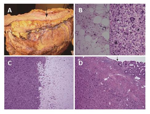
A 59-year-old previously healthy male presented as an outpatient with complaints of fatigue, recent 20 pounds weight loss, loss of appetite, and abdominal fullness. Physical examination revealed an abdominal mass in the left upper quadrant. CT scan of the abdomen showed a 19 cm × 17 cm × 12 cm mass with a necrotic center and an irregular soft tissue periphery. The mass was in close approximation to the splenic flexure of the colon, immediately anterior to the pancreas, and displaced the stomach medially. A left hemicolectomy, and splenectomy along with excision of the tail of the pancreas were performed. Gross examination revealed a 25 cm grey, fleshy mass with a necrotic center involving the wall of the left colon apparently arising from the sigmoid mesocolon (Figure 1A). An ulcer was present in the center of the large bowel segment, likely from pressure effect (Figure 1A). A 13 cm × 10 cm × 7 cm homogenous pale yellow, fatty mass was present at the periphery corresponding to the well differentiated component, composed of numerous lipoblasts and fibrous septa (Figure 1B). At the interface between the fatty and solid areas, the histology showed an abrupt transition from a well-differentiated component to a dedifferentiated component resembling a high-grade malignant fibrous histiocytoma (MFH) (Figure 1C). The dedifferentiated component was composed of pleomorphic, spindle cells with elongated, hyperchromatic nuclei, admixed with tumor giant cells, and abundant necrosis. The mitotic index was two per high power field and atypical mitoses were easily identified. The dedifferentiated component invaded into the wall of the colon (Figure 1D). Immunolabeling for S-100 was positive in the well-differentiated component (as would be expected of a liposarcoma). CD117 (C-KIT) and CD34 were negative in the dedifferentiated component, thus ruling out a gastrointestinal stromal tumor (GIST). Based on the histology and immunoprofile, a diagnosis of dedifferentiated liposarcoma was rendered.
Two and a half months postoperatively, a CT scan showed a recurrent mass for which the patient underwent resection of the posterior gastric wall and distal pancreatectomy. The pathologic examination revealed a mass (9.5 cm × 5.0 cm × 5.0 cm), which was morphologically identical to the patient’s previous tumor. Following adjuvant radiation therapy the patient was doing relatively well two years after the diagnosis without evidence of recurrent tumor.
Liposarcoma is divided into five subtypes according to the World Health Organization (well differentiated, dedifferentiated, myxoid, pleomorphic, and mixed type)[6]. Both well-differentiated and dedifferentiated liposarcomas have an equal sex predilection with the highest incidence in the 6th-7th decade of life. Dedifferentiated liposarcomas are defined histologically by a transition from well-differentiated liposarcoma to a non-lipogenic sarcoma with variable histological grade[6]. The dedifferentiated component can resemble any sarcoma, but often mimics MFH like our case[2]. Interestingly, dedifferentiated liposarcoma, despite its high-grade histology, has a less aggressive clinical course than other types of high-grade sarcoma, although the underlying mechanism is unclear[6]. Compared to well-differentiated liposarcoma, dedifferentiated liposarcoma has similar genetic changes, ring or giant marker chromosomes, but has a worse prognosis[6]. Approximately 40% of dedifferentiated liposarcomas will recur locally, 17% will metastasize, and 28% of the patients will ultimately die as a result of the tumor[6]. Therefore, it is important to thoroughly sample the resected mass in order to identify the non-lipogenic component, which may only comprise a small portion of the tumor. Also sampling of the usually adjacent innocuous looking fatty component is essential as this often contains the well-differentiated liposarcoma component.
Dedifferentiated liposarcoma has been reported most commonly in the retroperitoneum, rarely in other anatomic locations[2]. Five cases of dedifferentiated liposarcoma have been described from the small bowel mesentery[8]. Although well-differentiated liposarcoma has been documented in the sigmoid mesocolon[3-5], dedifferentiated liposarcoma from this unusual location has not been reported. Another unusual feature about our case was the dedifferentiated component transmurally invading the bowel wall.
The location of the lesion in our case raises the possibility of a GIST. GISTs usually resemble smooth muscle tumor with a variety of histological patterns, which does not consist of a well-differentiated liposarcoma component, necessary for the diagnosis of a dedifferentiated liposarcoma. GISTs typically stain with CD117 and CD34[9]. Immunohistochemically, dedifferentiated liposarcoma is usually negative for CD117 and CD34 in the dedifferentiated component and positive for S100 protein in the well-differentiated component. Dedifferentiated liposarcoma needs to be distinguished from other high-grade sarcomas such as MFH because these high-grade sarcomas have a much worse prognosis[7]. On needle biopsies the distinction of dedifferentiated liposarcoma from other high-grade sarcomas can be problematic as the samples are usually small, and the component of dedifferentiated liposarcoma can be identical to other high-grade sarcomas.
S- Editor Liu Y L- Editor Wang XL E- Editor Wang HF
| 1. | Kindblom LG, Meis-Kindblom JM, Enzinger FM. Variants of liposarcoma. Am J Surg Pathol. 1995;19:605-606; author reply 606-608. [RCA] [PubMed] [DOI] [Full Text] [Cited by in Crossref: 4] [Cited by in RCA: 4] [Article Influence: 0.1] [Reference Citation Analysis (0)] |
| 3. | Gutsu E, Ghidirim G, Gagauz I, Mishin I, Iakovleva I. Liposarcoma of the colon: a case report and review of literature. J Gastrointest Surg. 2006;10:652-656. [RCA] [PubMed] [DOI] [Full Text] [Cited by in Crossref: 30] [Cited by in RCA: 27] [Article Influence: 1.4] [Reference Citation Analysis (0)] |
| 4. | Chen KT. Liposarcoma of the colon: a case report. Int J Surg Pathol. 2004;12:281-285. [RCA] [PubMed] [DOI] [Full Text] [Cited by in Crossref: 25] [Cited by in RCA: 21] [Article Influence: 1.1] [Reference Citation Analysis (0)] |
| 5. | Amato G, Martella A, Ferraraccio F, Di Martino N, Maffettone V, Landolfi V, Fei L, Del Genio A. Well differentiated "lipoma-like" liposarcoma of the sigmoid mesocolon and multiple lipomatosis of the rectosigmoid colon. Report of a case. Hepatogastroenterology. 1998;45:2151-2156. [PubMed] |
| 6. | Fletcher CDM, Unni KK, Mertens F. World Health Organization Classification of Tumours. Pathology and Genetics of Tumors of Soft Tissue and Bone. Lyon: IARC Press 2002; 227-232. |
| 7. | Henricks WH, Chu YC, Goldblum JR, Weiss SW. Dedifferentiated liposarcoma: a clinicopathological analysis of 155 cases with a proposal for an expanded definition of dedifferentiation. Am J Surg Pathol. 1997;21:271-281. [RCA] [PubMed] [DOI] [Full Text] [Cited by in Crossref: 450] [Cited by in RCA: 433] [Article Influence: 15.5] [Reference Citation Analysis (0)] |
| 8. | Hasegawa T, Seki K, Hasegawa F, Matsuno Y, Shimodo T, Hirose T, Sano T, Hirohashi S. Dedifferentiated liposarcoma of retroperitoneum and mesentery: varied growth patterns and histological grades--a clinicopathologic study of 32 cases. Hum Pathol. 2000;31:717-727. [RCA] [PubMed] [DOI] [Full Text] [Cited by in Crossref: 106] [Cited by in RCA: 101] [Article Influence: 4.0] [Reference Citation Analysis (0)] |
| 9. | Miettinen M, Lasota J. Gastrointestinal stromal tumors: review on morphology, molecular pathology, prognosis, and differential diagnosis. Arch Pathol Lab Med. 2006;130:1466-1478. [PubMed] |


