Published online Jul 28, 2007. doi: 10.3748/wjg.v13.i28.3861
Revised: March 18, 2007
Accepted: March 28, 2007
Published online: July 28, 2007
AIM: To investigate the rate of complications of endoscopic retrograde cholangio-pancreatography (ERCP) performed immediately after endoscopic ultrasound fine needle aspiration (EUS-FNA) in a large series of patients.
METHODS: Patients with the following conditions were considered candidates for EUS-FNA and ERCP: diagnosis of locally advanced or metastatic pancreatic lesion not eligible for surgery, and patients with pancreatic lesion of unknown nature causing jaundice. Data were prospectively collected on the following parameters: indication for FNA, EUS findings, pathological diagnosis, procedure duration of EUS-FNA and combined EUS-FNA and ERCP, and immediate and late complications.
RESULTS: From January 2004 to October 2006, 72 patients were deemed eligible for combined EUS and ERCP. In 25/72 EUS-FNA was performed to obtain a pathology diagnosis of lesions causing biliary obstruction, and ERCP sequentially performed to drain the biliary system. No immediate complications occurred except for two mild bleeding episodes post sphincterotomy. No late complications were recorded except for one patient who experienced fever, promptly recovered with antibiotic therapy.
CONCLUSION: Simultaneous approach appears to be feasible and safe. When possible, this can be considered the reference standard to avoid double sedation and reduce duration of the procedure and hospital stay.
- Citation: Tarantino I, Barresi L, Di Pisa M, Traina M. Simultaneous endoscopic ultrasound fine needle aspiration and endoscopic retrograde cholangio-pancreatography: Evaluation of safety. World J Gastroenterol 2007; 13(28): 3861-3863
- URL: https://www.wjgnet.com/1007-9327/full/v13/i28/3861.htm
- DOI: https://dx.doi.org/10.3748/wjg.v13.i28.3861
Endoscopic ultrasound fine needle aspiration (EUS-FNA) is a proven method for diagnosis of pancreatic-biliary lesions causing jaundice with an overall complication rate of about 1% to 2%[1-4]. Endoscopic retrograde cholangio-pancreatography (ERCP) is the most appropriate technique for treatment of common bile duct stenosis due to benign and malignant diseases; many published studies report rates of complication of 5%-10%[5-9].
In some tertiary care centers, EUS-FNA and ERCP are performed sequentially, in order to avoid a double sedation and to reduce the procedure time and hospital stay. Two reports were published showing the occurrence of additional complications in patients undergoing ERCP after EUS-FNA on pancreatic mass, without delay between the two procedures[10,11]. No prospective studies or case series confirmed these results.
This report shows the rate of complications of ERCP done immediately after EUS-FNA in a large series of patients.
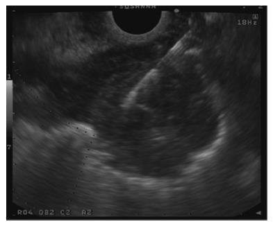
Patients with the following conditions were considered candidates for EUS plus ERCP: a diagnosis of locally advanced or metastatic pancreatic lesion considered not eligible for surgery but candidate for palliative biliary drainage and chemotherapy; a pancreatic lesion of unknown nature causing jaundice, and finally, patients showing signs of biliary obstruction of unknown origin despite other imaging tests. For patients with advanced pancreatic tumor and pancreatic lesions of unknown nature EUS-FNA was planned to obtain a pathology diagnosis (Figure 1).
Two separate informed consents for EUS-FNA and ERCP were obtained before the examination. Following our protocol for ERCP, coagulation tests and platelet counts were checked before the procedure and all patients received prophylactic antibiotics before the procedure. The procedures were done under sedation with propofol administered by an anesthesiologist.
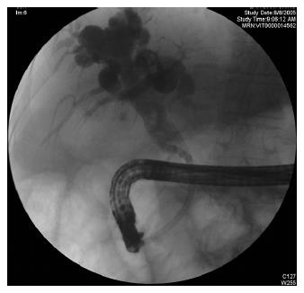
All procedures were performed in the ERCP suite with a dedicated endosonography processor system (EUS exera EU-C60, Olympus America Corp.; Melville, N.Y). The linear array echo-endoscope (GF UCT160-OL5, Olympus America Corp.; Melville, N.Y) and 22G needle for FNA (ECHO-1-22; Wilson Cook Medical, Inc, Winston-Salem, NC) were used. Sample adequacy was assessed in the room by a pathologist. After obtaining the sample, the ERCP, sphincterotomy and stent placement were performed using conventional techniques with either a TJF-140 or TJF-160 duodenoscope (Olympus America Corp, Melville, NY) (Figure 2).
Amylase and lipase were checked at 6 and 12 h after procedure. If no complications occurred the patient was discharged after 24 h.
After discharge, patients underwent close follow up for almost one week by our Nurse Coordinator and repeated laboratory tests (CBC, bilirubin, transaminase and alkaline phosphatase/γ-glutamyl transpeptidase levels) after one week.
Data were prospectively collected on the following parameters: indication for FNA, EUS findings, pathological diagnosis, time required for EUS-FNA and for combined procedures, and rate of immediate (during the procedure or within 24 h) and late complications (after 24 h, during the follow-up).
From January 2004 to October 2006, 72 patients were deemed eligible for combined EUS and ERCP. In only 25 of these patients, EUS-FNA was performed to obtain a pathological diagnosis of lesions causing biliary obstruction, and ERCP was sequentially performed to drain the biliary system. In the remaining 37 patients, EUS was done to assess the origin of the biliary obstruction, but no FNAs were done.
Of the 25 patients undergoing EUS-FNA, 19 were male and six female and the average age was 64.8 ± 14.5 (range 32-85) years. In 14/17 patients with suspected pancreatic tumor, EUS confirmed the diagnosis: in the remaining three, EUS findings indicated chronic pancreatitis. In the other cases, EUS findings confirmed the diagnosis done with previous imaging tests: one IPMT (intra-papillary mucinous tumor), one chronic pancreatitis, four biliary lesions, one large peri-biliary lymph node, and one tumor of the papilla invading the pancreatic parenchyma (Table 1).
| Indication for EUS-FNA | EUS findings | |
| Pancreatic cancer | 17 | 14 |
| IPMT | 1 | 1 |
| Chronic pancreatitis | 1 | 4 |
| Choledochus lesion | 4 | 4 |
| Lymph node | 1 | 1 |
| Papilla Tumor | 1 | 1 |
| Total | 25 | 25 |
The pathology assessment was obtained in 23/25 FNAs. In two cases, samples were unsatisfactory for a definitive diagnosis, both in patients with clear images of pancreatic tumor (Table 2).
| Tumor | Inflammation | Not satisfactory | Total | |
| Pancreatic cancer | 12 | 0 | 2 | 14 |
| IPMT | 1 | 0 | 0 | 1 |
| Chronic pancreatitis | 0 | 4 | 0 | 4 |
| Choledochus lesion | 2 | 2 | 0 | 4 |
| Lymph node | 0 | 1 | 0 | 1 |
| Papilla Tumor | 1 | 0 | 0 | 1 |
The average number of needle passes was two (range one-three) based on sample adequacy evaluated in the room by the pathologist.
The average time for EUS-FNA was 26.64 ± 9.31 (range 10-40) min and the total time for combined procedure was 58.6 ± 16.14 (range 30-91) min. Considering that in our Center the average time of ERCP with stent placement is about 45 min, the combined time of the two procedures was less than that for the two procedures separately. All ERCPs were completed with stent placement; one patient with pancreatic cancer needed pre-cut before stent placement due a difficult canulation.
No biliary leakages were seen at cholangiography. No immediate complications occurred except two mild bleeding episodes post sphincterotomy, self limited and not requiring any treatment.
Patients were strictly followed for almost one week after discharge by our Nurse Coordinator. No complications were recorded except for one patient who experienced fever, promptly recovered with antibiotic therapy and did not require hospitalization.
The ERCP is the most appropriate technique for treatment of common bile duct stenosis due to benign and malignant diseases. However, it is associated with a rate of complication of 5%-10%[5-9].
Extensive published data exists on the usefulness of EUS-FNA to evaluate suspected pancreatic lesions, before ERCP. In a series of 147 consecutive patients, the use of EUS with FNA as the initial approach to patients with obstructive jaundice, was studied by Erickson et al[2]. EUS-FNA proved useful not only as a diagnostic and staging modality, but also helped direct subsequent therapeutic endoscopic retrograde cholangiopancreatography (ERCP), saving approximately $1007 to $1313 per patient. EUS has the distinct advantage of allowing patients to undergo a therapeutic ERCP in the same setting. In 1998 Mergener et al[10] published a case of a 77-year old man with pneumoperitoneum complicating ERCP performed immediately after EUS-FNA on a peri-pancreatic lymph node. The authors concluded that the pneumoperitoneum probably resulted from insufflated air tracking through the FNA site, during the ERCP. Another recent report, by Di Matteo et al[11] shows two cases of biliary leakages complicating ERCP done the same day as EUS-FNA on lesions in the head of the pancreas. The authors discuss the possibility of causing small and sub clinical bile duct injury during FNA that could be aggravated by therapeutic maneuvers in the bile duct during ERCP, done on the same day. The present study series on 25 patients shows that no additional complications occurred when the two procedures were performed sequentially. Two mild post sphincterotomy bleeding episodes occurred, as immediate complications not requiring any treatment. The only late complication was one episode of fever after discharge home, but the patient recovered with antibiotics and did not require hospitalization.
In this series, the simultaneous approach EUS- FNA and ERCP, appears to be feasible and safe, providing an accurate diagnosis and, at the same time, an appropriate treatment of biliary stenosis when needed. At present, in centers without logistical difficulties, when the two procedures are indicated, their consecutive use can be considered the reference standard in order to avoid a double sedation and to reduce the procedure time and hospital stay.
S- Editor Zhu LH L- Editor Roberts SE E- Editor Liu Y
| 1. | Raut CP, Grau AM, Staerkel GA, Kaw M, Tamm EP, Wolff RA, Vauthey JN, Lee JE, Pisters PW, Evans DB. Diagnostic accuracy of endoscopic ultrasound-guided fine-needle aspiration in patients with presumed pancreatic cancer. J Gastrointest Surg. 2003;7:118-126; discussion 127-128. [RCA] [PubMed] [DOI] [Full Text] [Cited by in Crossref: 211] [Cited by in RCA: 192] [Article Influence: 8.7] [Reference Citation Analysis (0)] |
| 2. | Erickson RA, Garza AA. EUS with EUS-guided fine-needle aspiration as the first endoscopic test for the evaluation of obstructive jaundice. Gastrointest Endosc. 2001;53:475-484. [RCA] [PubMed] [DOI] [Full Text] [Cited by in Crossref: 42] [Cited by in RCA: 45] [Article Influence: 1.9] [Reference Citation Analysis (0)] |
| 3. | Wiersema MJ, Vilmann P, Giovannini M, Chang KJ, Wiersema LM. Endosonography-guided fine-needle aspiration biopsy: diagnostic accuracy and complication assessment. Gastroenterology. 1997;112:1087-1095. [RCA] [PubMed] [DOI] [Full Text] [Cited by in Crossref: 874] [Cited by in RCA: 736] [Article Influence: 26.3] [Reference Citation Analysis (0)] |
| 4. | O'Toole D, Palazzo L, Arotçarena R, Dancour A, Aubert A, Hammel P, Amaris J, Ruszniewski P. Assessment of complications of EUS-guided fine-needle aspiration. Gastrointest Endosc. 2001;53:470-474. [RCA] [PubMed] [DOI] [Full Text] [Cited by in Crossref: 261] [Cited by in RCA: 234] [Article Influence: 9.8] [Reference Citation Analysis (0)] |
| 5. | Freeman ML, Nelson DB, Sherman S, Haber GB, Herman ME, Dorsher PJ, Moore JP, Fennerty MB, Ryan ME, Shaw MJ. Complications of endoscopic biliary sphincterotomy. N Engl J Med. 1996;335:909-918. [RCA] [PubMed] [DOI] [Full Text] [Cited by in Crossref: 1716] [Cited by in RCA: 1687] [Article Influence: 58.2] [Reference Citation Analysis (2)] |
| 6. | Huibregtse K. Complications of endoscopic sphincterotomy and their prevention. N Engl J Med. 1996;335:961-963. [RCA] [PubMed] [DOI] [Full Text] [Cited by in Crossref: 74] [Cited by in RCA: 79] [Article Influence: 2.7] [Reference Citation Analysis (0)] |
| 7. | Loperfido S, Angelini G, Benedetti G, Chilovi F, Costan F, De Berardinis F, De Bernardin M, Ederle A, Fina P, Fratton A. Major early complications from diagnostic and therapeutic ERCP: a prospective multicenter study. Gastrointest Endosc. 1998;48:1-10. [RCA] [PubMed] [DOI] [Full Text] [Cited by in Crossref: 801] [Cited by in RCA: 779] [Article Influence: 28.9] [Reference Citation Analysis (1)] |
| 8. | Hewitt PM, Krige JE, Bornman PC, Terblanche J. Choledochal cyst in pregnancy: a therapeutic dilemma. J Am Coll Surg. 1995;181:237-240. [PubMed] |
| 9. | Freeman ML. Complications of endoscopic biliary sphincterotomy: a review. Endoscopy. 1997;29:288-297. [RCA] [PubMed] [DOI] [Full Text] [Cited by in Crossref: 101] [Cited by in RCA: 105] [Article Influence: 3.8] [Reference Citation Analysis (0)] |
| 10. | Mergener K, Jowell PS, Branch MS, Baillie J. Pneumoperitoneum complicating ERCP performed immediately after EUS-guided fine needle aspiration. Gastrointest Endosc. 1998;47:541-542. [RCA] [PubMed] [DOI] [Full Text] [Cited by in Crossref: 22] [Cited by in RCA: 22] [Article Influence: 0.8] [Reference Citation Analysis (0)] |
| 11. | Di Matteo F, Shimpi L, Gabbrielli A, Martino M, Caricato M, Esposito A, De Cicco ML, Coppola R, Costamagna G. Same-day endoscopic retrograde cholangiopancreatography after transduodenal endoscopic ultrasound-guided needle aspiration: do we need to be cautious? Endoscopy. 2006;38:1149-1151. [RCA] [PubMed] [DOI] [Full Text] [Cited by in Crossref: 14] [Cited by in RCA: 17] [Article Influence: 0.9] [Reference Citation Analysis (0)] |


