Published online Jul 21, 2007. doi: 10.3748/wjg.v13.i27.3763
Revised: March 23, 2007
Accepted: March 31, 2007
Published online: July 21, 2007
We encountered a 65-year-old man with a carcinoid tumor of the minor duodenal papilla. Since he had liver cirrhosis and completely refused surgery, we performed an endoscopic snare papillectomy. The papillectomy was performed successfully without procedure-related complication. The specimens revealed a carcinoid tumor showing that the margin of the tumor was positive. One week later, upper GI endoscopy was performed and the biopsy specimens obtained from base of ulcer showed no neoplastic cells. We performed a duodenoscopy and CT 3, 6 and 18 mo later, and there was no macroscopic or microscopic evidence of tumor recurrence after more than 4 years.
- Citation: Itoi T, Sofuni A, Itokawa F, Tsuchiya T, Kurihara T, Moriyasu F. Endoscopic resection of carcinoid of the minor duodenal papilla. World J Gastroenterol 2007; 13(27): 3763-3764
- URL: https://www.wjgnet.com/1007-9327/full/v13/i27/3763.htm
- DOI: https://dx.doi.org/10.3748/wjg.v13.i27.3763
Recently, endoscopic papillectomy for not only adenoma, but also carcinoid of the major duodenal papilla is being increasingly performed as a minimally invasive alternative to conventional surgery[1-5]. However, it is controversial among endoscopists whether this technique can be adaptable for lesions of the minor duodenal papilla. We describe the successful endoscopic papillectomy of carcinoid of minor duodenal papilla and discuss the applications of the procedure.
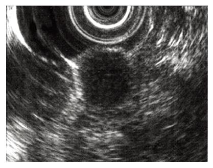
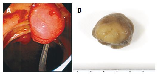
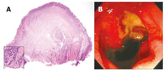
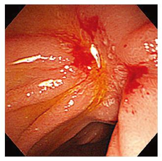
A 65-year-old asymptomatic man underwent esophago-gastroduodenal (EGD) endoscopy screening, which revealed a slightly swollen and yellowish minor duodenal papilla. A biopsy specimen from the lesion revealed a carcinoid tumor. EUS demonstrated a 12 mm × 11 mm hypoechoic mass located in the submucosa (Figure 1). CT revealed a slightly contrast-enhanced duodenal tumor without metastasis to the regional lymph nodes or liver. ERCP findings were normal, demonstrated dominant pancreatic drainage via the ventral duct and no effacement of the dorsal duct by tumor. The routine laboratory tests were normal, including tumor markers. Since he had liver cirrhosis and completely refused surgery, we then performed an endoscopic snare papillectomy after obtaining appropriate written informed consent. Snare excision was performed with a polypectomy snare forceps (SD-5U-1, 6U-1, Olympus Medical Systems, Tokyo, Japan) and a generator with an automatically controlled cut-out system (ICC200, Erbe Elektromedizin GmbH, Tubingen, Germany). The papillectomy was performed successfully without any procedure-related complications (Figure 2A and B). The specimens revealed a carcinoid tumor and showed that the margin of the tumor was positive (Figure 3A). One week later, EGD was performed and the biopsy specimens obtained from the base of ulcer showed no neoplastic cells (Figure 3B). We performed a duodenoscopy and CT 3, 6 and 18 mo later (Figure 4), and there was no macroscopic or microscopic evidence of tumor recurrence more than 4 years.
Carcinoid tumor of the gastrointestinal tract has a relatively high occurrence rate and often shows invasive growth[6]. Although Noda et al [7]reported that, microscopically, in the duodenal papilla, especially the minor duodenal papilla, carcinoids and endocrine cell micronests seemed to occur more frequently than generally thought, clinically, carcinoid tumors of the major or minor duodenal papilla are rare[6-8]. No definitive statement can be made regarding the optimal treatment of carcinoid of the major duodenal papilla, given the small number of cases in the literature. Hatzitheoklitos et al[9] reported that carcinoid of the papilla of less than 10 mm in size rarely metastasizes (2%), whereas 20% of tumors 10 to 20 mm and 75% of tumors greater than 20 mm in diameter metastatisize. Radical surgery, such as local excision or pancreaticoduodenectomy, has generally been preferred as the treatment of choice for carcinoid tumors of the papilla[6,8,9].
Endoscopic snare papillectomy has been accepted as a safe and feasible treatment for adenoma of the major duodenal papilla because of lower operative mortality and morbidity, provided that certain criteria are strictly observed[1-4]. Furthermore, recently, endoscopic papillectomy has been performed for not only adenoma of major duodenal papilla but also ampullary adenoma with intraductal extension and invasive carcinoma[3,10] or carcinoid tumor[3,5]. To the best of our knowledge, this is first report of a successful papillectomy for carcinoid tumor of the minor duodenal papilla. The growth pattern of carcinoids, however, is essentially frequently submucosal invasive. We think that there were two fortunate points in this case. One was that the growth pattern of this tumor showed mainly protrusion to the duodenal lumen, and the other was that follow-up biopsy was negative, perhaps because the cauterizing effect resulted in elimination any remaining carcinoid tumor cells despite the positive cut margin.
In conclusion, although further studies with long-term follow-up are needed to determine the ultimate outcome, this case, with a 4-year disease-free follow-up period, suggests that endoscopic resection of carcinoid of the minor duodenal papilla is one option when surgery cannot be applied in patients with minor duodenal papilla tumors.
S- Editor Zhu LH L- Editor Alpini GD E- Editor Liu Y
| 1. | Binmoeller KF, Boaventura S, Ramsperger K, Soehendra N. Endoscopic snare excision of benign adenomas of the papilla of Vater. Gastrointest Endosc. 1993;39:127-131. [RCA] [PubMed] [DOI] [Full Text] [Cited by in Crossref: 244] [Cited by in RCA: 201] [Article Influence: 6.3] [Reference Citation Analysis (0)] |
| 2. | Norton ID, Gostout CJ, Baron TH, Geller A, Petersen BT, Wiersema MJ. Safety and outcome of endoscopic snare excision of the major duodenal papilla. Gastrointest Endosc. 2002;56:239-243. [RCA] [PubMed] [DOI] [Full Text] [Cited by in Crossref: 176] [Cited by in RCA: 151] [Article Influence: 6.6] [Reference Citation Analysis (0)] |
| 3. | Cheng CL, Sherman S, Fogel EL, McHenry L, Watkins JL, Fukushima T, Howard TJ, Lazzell-Pannell L, Lehman GA. Endoscopic snare papillectomy for tumors of the duodenal papillae. Gastrointest Endosc. 2004;60:757-764. [RCA] [PubMed] [DOI] [Full Text] [Cited by in Crossref: 189] [Cited by in RCA: 151] [Article Influence: 7.2] [Reference Citation Analysis (0)] |
| 4. | Catalano MF, Linder JD, Chak A, Sivak MV, Raijman I, Geenen JE, Howell DA. Endoscopic management of adenoma of the major duodenal papilla. Gastrointest Endosc. 2004;59:225-232. [RCA] [PubMed] [DOI] [Full Text] [Cited by in Crossref: 258] [Cited by in RCA: 217] [Article Influence: 10.3] [Reference Citation Analysis (0)] |
| 5. | Niido T, Itoi T, Harada Y, Haruyama K, Ebihara Y, Tsuchida A, Kasuya K. Carcinoid of major duodenal papilla. Gastrointest Endosc. 2005;61:106-107. [RCA] [PubMed] [DOI] [Full Text] [Cited by in Crossref: 19] [Cited by in RCA: 23] [Article Influence: 1.2] [Reference Citation Analysis (0)] |
| 6. | Modlin IM, Sandor A. An analysis of 8305 cases of carcinoid tumors. Cancer. 1997;79:813-829. [RCA] [PubMed] [DOI] [Full Text] [Cited by in RCA: 14] [Reference Citation Analysis (0)] |
| 7. | Noda Y, Watanabe H, Iwafuchi M, Furuta K, Ishihara N, Satoh M, Ajioka Y. Carcinoids and endocrine cell micronests of the minor and major duodenal papillae. Their incidence and characteristics. Cancer. 1992;70:1825-1833. [RCA] [PubMed] [DOI] [Full Text] [Cited by in RCA: 1] [Reference Citation Analysis (0)] |
| 8. | Dixon JM, Chapman RW, Berry AR. Carcinoid tumour of the ampulla of Vater presenting as acute pancreatitis. Gut. 1987;28:1296-1297. [RCA] [PubMed] [DOI] [Full Text] [Cited by in Crossref: 11] [Cited by in RCA: 14] [Article Influence: 0.4] [Reference Citation Analysis (0)] |
| 9. | Hatzitheoklitos E, Büchler MW, Friess H, Poch B, Ebert M, Mohr W, Imaizumi T, Beger HG. Carcinoid of the ampulla of Vater. Clinical characteristics and morphologic features. Cancer. 1994;73:1580-1588. [RCA] [PubMed] [DOI] [Full Text] [Cited by in RCA: 4] [Reference Citation Analysis (0)] |
| 10. | Small AJ, Baron TH. Successful endoscopic resection of ampullary adenoma with intraductal extension and invasive carcinoma (with video). Gastrointest Endosc. 2006;64:148-151. [RCA] [PubMed] [DOI] [Full Text] [Cited by in Crossref: 12] [Cited by in RCA: 13] [Article Influence: 0.7] [Reference Citation Analysis (0)] |


