Copyright
©2007 Baishideng Publishing Group Co.
World J Gastroenterol. Apr 14, 2007; 13(14): 2066-2071
Published online Apr 14, 2007. doi: 10.3748/wjg.v13.i14.2066
Published online Apr 14, 2007. doi: 10.3748/wjg.v13.i14.2066
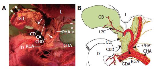
Figure 1 Innervation of the gallbladder (GB) from the ventral aspect (A) in a cadaver and a schematic representation of it (B).
The branches innervating the GB originate from the anterior hepatic plexus, and run along the cystic duct (CD) and the cystic artery (CA). The hepatic divisions (*) of the vagus join in the anterior hepatic plexus in the proper hepatic artery (PHA). Arrows indicate nerve branches. CBD: common bile duct; CHA: common hepatic artery; D: duodenum; GDA: gastroduodenal artery; L: liver; RGA: right gastric artery.
-
Citation: Yi SQ, Ohta T, Tsuchida A, Terayama H, Naito M, Li J, Wang HX, Yi N, Tanaka S, Itoh M. Surgical anatomy of innervation of the gallbladder in humans and
Suncus murinus with special reference to morphological understanding of gallstone formation after gastrectomy. World J Gastroenterol 2007; 13(14): 2066-2071 - URL: https://www.wjgnet.com/1007-9327/full/v13/i14/2066.htm
- DOI: https://dx.doi.org/10.3748/wjg.v13.i14.2066


