Published online Dec 21, 2006. doi: 10.3748/wjg.v12.i47.7690
Revised: October 20, 2006
Accepted: October 28, 2006
Published online: December 21, 2006
AIM: To study the prolonged colonic motility under normal conditions with a novel capsule-style micro-system and to assess its clinical significance.
METHODS: A single use telemetry capsule (10 mm in diameter, 20 mm in length) embedded with a pressure sensor was ingested by the subjects. The sensor is capable of transmitting colonic pressure wirelessly for more than 130 h. The time of capsule entering the segmental colon was detected by ultrasound. The ultrasonic electrodes were mounted on the surface of the ileocecum and navel and at the junction of the left and rectosigmoid colon of the subjects in sequence, which were identified by abdominal X-rays with radiopaque markers. To verify the accuracy and reliability of ultrasonic detection of telemetry capsules at key points of colon, the segmental colonic transit time was simultaneously recorded by using radiopaque markers.
RESULTS: The signal lamp showed that all recorders could receive the radio signal transmitted by the telemetry capsule. The X-rays showed that all telemetry capsules were detected successfully when they were passing through the key points of colon. There was a significant correlation between the transit results obtained by ultrasonic detection or by radiopaque markers. Colorectal recording was obtained from 20 healthy subjects during 613 h (411 h during waking, 202 h during sleep). Compared to waking, the number of pressure contractions and the area under pressure contractions were significantly (P < 0.05) decreased during sleep (21 ± 5 h-1vs 15 ± 4 h-1, 463 ± 54 mmHg·s/min vs 342 ± 45 mmHg·s/min). The colonic motility exhibited significant regional variations both in the circadian behavior and in response to waking and meal.
CONCLUSION: The capsule-style micro-system is reliable and noninvasive, and may represent a useful tool for the study of physiology and pathology of colonic motor disorders.
- Citation: Zhang WQ, Yan GZ, Yu LZ, Yang XQ. Non-invasive measurement of pan-colonic pressure over a whole digestive cycle: Clinical applications of a capsule-style manometric system. World J Gastroenterol 2006; 12(47): 7690-7694
- URL: https://www.wjgnet.com/1007-9327/full/v12/i47/7690.htm
- DOI: https://dx.doi.org/10.3748/wjg.v12.i47.7690
Colonic motility disorders are frequently encountered in clinical practice. Intraluminal manometry (via perfused and solid-state catheters) allows a direct study of colonic contractile activity over prolonged periods. However, catheter studies have generally been performed in the left and rectosigmoid colon, because of the technical difficulties in accessing the right colon[1].
Hagger et al[2] have reported a pan-colonic manometry by combined antegrade/retrograde intubation. However, due to sensor failure and/or catheter displacement, they could not assess the amplitude of pressure activities in the right colon.
Recent technological advances facilitate prolonged pan-colonic investigations. The success of Israeli M2A capsule endoscope has led to the research and development of other capsules aimed at monitoring either functional or organic diseases[3]. The American SmartPill capsule is capable of transmitting pressure, temperature and pH data continuously for 72 h, and suitable for diagnosing small intestine motility or delayed emptying of the stomach[4-6].
To satisfy the need of prolonged and noninvasive monitoring colonic pressures, the authors have developed a novel capsule-style manometric micro-system with its prototype reported[7]. The system was approved by the Food and Drug Administration of China in 2005. The system is composed of a one-off telemetry capsule, a data recorder, an ultrasonic location detector and a computer with analysis software. The capsule-style manometric system is capable of acquiring and transmitting intralu-minal pressure data continuously for more than 130 h.
The aim of this study was to evaluate the pan-colonic motor activity under the physiological conditions with a capsule-style manometric system. A study period of a whole digestive cycle (26-53 h) was used to characterize the infrequent patterns of motor activity of the colorectum.
Twenty healthy subjects (12 females and 8 males, median age 35 years, range 23-52 years) were studied. The subjects had a normal bowel habit which was defined as a stool frequency of ≤ 3 stools per day and ≥ 3 stools per week. The subjects were not on any medication and had no gastrointestinal symptoms and no history of previous gastrointestinal disorder or gastrointestinal surgery. Female subjects had a negative pregnancy test. An informed written consent was obtained from all subjects. The study protocol was approved by the Ethics Committee of Capital Medical University Chaoyang Hospital.
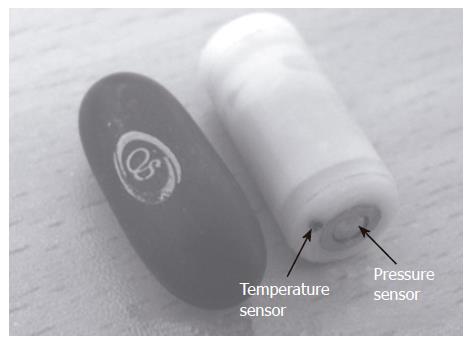
Telemetry capsule: The newly-developed telemetry capsule (10 mm in diameter, 20 mm in length, and weighing 2.9 g ) developed by the authors is shown in Figure 1. The effective range could reach 3-5 m between the capsule and the recorder.
The pressure sensor can be used from -60 to 200 mmHg with its error ≤± 1.5 mmHg, and the temperature sensor is sensitive in range of 33-42°C with its error ≤± 0.25°C. Changes in pressure sensor output due to fluctuations of temperature can be compensated automatically.
Data recorder: The data recorder consists of three functional blocks: a wireless receiver module, a micro-controller and an outer pressure transducer. The outer pressure transducer records the pressure changes induced by the surrounding altitude. The timer in the micro-controller records the time of received data. The obtained pressure data, temperature data, ultrasonic location information and relevant time are stored in a flash memory card.
Ultrasonic detection of capsule location: When the telemetry capsule passes through the ultrasonic electrodes mounted on the surface of key points of colon, the echo from batteries in the capsule would be much stronger than other echoes. When the amplitude of an echo is higher than the preset threshold, it can be regarded as returning from the capsule. Detailed technical information about the ultrasonic detection of the capsule positions has been reported elsewhere[8].
Colon was divided into three segments by three key points (ileocecum, navel and the junction of the left and rectosigmoid colon), which were identified by an experienced doctor with the help of abdominal X-rays with radiopaque markers.
Analysis software: In addition to the novel manometric system, a Matlab-based (The MathWorks, Natick, MA) software program was developed to correct baseline shifts, eliminate artefacts, and calculate the number of pressure contractions, amplitude and area under curve (AUC) in a fully automated way. The software features an intuitive graphical user interface that supports graphical and statistical data analysis. The baseline pressures were determined by identifying the minimum pressure in each 30 min, which was then subtracted from the raw signals. Pressure contractions with an amplitude ≥ 10 mmHg from the baseline and a duration ≥ 6 s were included in the analysis.
To verify the accuracy and reliability of ultrasonic detection of telemetry capsules at the key points of colon, the segmental colonic transit time was simultaneously recorded by using radiopaque markers.
After an overnight fast, the subjects ingested 20 radiopaque markers contained in a gelatin capsule (Binhai Hospital, Tianjin, China) at 8:00 AM next morning. A daily abdominal X-ray was taken at the same time on the following consecutive days until the number of remained markers was ≤ 4. The total and segmental colonic transit time (CTT) was calculated using the following formula: CTT = 1.2 ×Σni, where 1.2 = time interval (24 h)/number of radiopaque markers (20), i = day of X-rays taken, and ni = the total number of markers present on a given film sector on day I[9,10].
The subjects took the telemetry capsule an hour after ingestion of the markers. The subjects wore the recorder and checked the signal lamp on the receiver.
To help mounting the ultrasonic electrodes on the surface of the ileocecum of the patients correctly, an abdominal X-ray was taken at 6 h after the telemetry capsule was taken.
The ultrasonic electrodes were first mounted on the surface of the ileocecum by an experienced doctor with the help of radiopaque markers and bony landmarks in the X-ray film. The subjects put on the waistcoat containing an ultrasonic signal processor and an alarm buzzer. When alarming occurred, an abdominal X-ray was taken to confirm the location of telemetry capsules.
After successful detection of the ileocecum, the ultrasonic sensors were then mounted on the surface of the navel and at the junction of the left and rectosigmoid colon in sequence.
The subjects stayed totally ambulatory during inspecting, but drastic sport activities were prohibited. The subjects ate 1000 kcal standard meals. Ingestion of alcohol and caffeine containing drinks was prohibited.
After defecation, the signal lamp on the receiver would indicate whether the capsule was still in body or not. All capsules were recovered to confirm the discharge.
The data were expressed as mean ± SD unless otherwise stated. High-amplitude contraction (HAC) analysis was made by visual inspection of the pressure trace. The HAC was defined as a pressure contraction with an amplitude ≥ 50 mmHg from the baseline, a duration ≥ 20 s and a peak-to-peak time interval ≥ 40 s.
The diurnal and postprandial differences in AUC and the number of contractions were compared using repeated measures of ANOVA. P < 0.05 was considered statistically significant in all analyses.
The signal lamp on recorders showed that all recorders could receive the radio signal transmitted by the telemetry capsule in the subjects. No discomfort was reported by any volunteers during the experiment.
The telemetry capsules passed through the gastroin-testinal (GI) tract without any difficulties. All capsules were recovered with the guidance of the signal lamp.
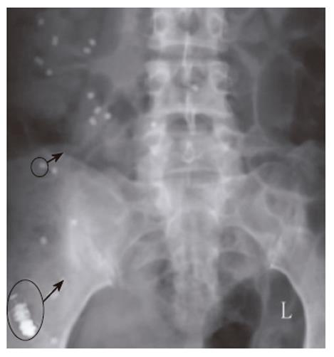
All telemetry capsules could be detected successfully when they were passing through the three key points of the colon. Additional X-rays confirmed the capsule location (Figure 2). The location of capsules could be recognized with the help of bony landmarks and radiopaque markers.
The total transit time and segmental colonic transit time obtained by ultrasonic detection and radiopaque markers are shown in Table 1. There was a significant correlation between the two methods for the detection of transit results. It verified the reliability and accuracy of ultrasonic detection for the segmental transit time.
| Transit time (h) | Total colon | Right colon | Left colon | Rectosigmoid |
| Markers | 31.5 ± 6.8 | 8.6 ± 3.3 | 11.4 ± 3.6 | 11.5 ± 3.3 |
| Ultrasonic | 29.7 ± 5.9 | 9.3 ± 3.5 | 10.8 ± 3.1 | 9.7 ± 2.9 |
| Correlation | 0.92 | 0.91 | 0.93 | 0.86 |
Colorectal recording was obtained from the 20 healthy subjects for a total of 613 h (411 h during waking, 202 h during sleep). In this study, the authors laid more emphasis on the colorectal motility response to meal ingestion and morning waking. During the colorectal pressure recording period, 26 subjects were waking in the morning and 78 had meal ingestions (1.3 and 3.9 per subject respectively).
Compared to waking, the number of pressure contractions and the AUC of pressure contractions during sleep were significantly decreased (21 ± 5 h-1vs 15 ± 4 h-1, 463 ± 54 mmHg·s·min-1vs 342 ± 45 mmHg·s·min-1, P < 0.05).
After waking and meal ingestion, both the number of pressure contractions and the AUC of pressure contractions were significantly increased. The contractile response to waking was significantly higher in the left/right colon than in the rectosigmoid colon (Figure 3), whereas the waking response was lower in the left colon than in the right colon, but was not significantly different. There were no significant differences in segmental colon motility response to meal ingestion. The details are shown in Table 2.
| Waking | Meal Ingestion | |||
| 1 h before | 1 h after | 2 h before | 2 h after | |
| No. of Contrctions (h-1) | ||||
| Right colon | 14 ± 4a | 45 ± 6a | 21 ± 6 | 44 ± 8b |
| Left colon | 13 ± 3a | 39 ± 4 | 22 ± 5 | 38 ± 6 |
| Rectosigmoid | 19 ± 5a | 29 ± 4a | 20 ± 6 | 32 ± 7b |
| AUC (mmHg·s·min-1) | ||||
| Right colon | 322 ± 44 | 984 ± 91a | 471 ± 59 | 862 ± 79 |
| Left colon | 307 ± 41b | 835 ± 82 | 465 ± 52 | 812 ± 74 |
| Rectosigmoid | 431 ± 52b | 725 ± 64a | 453 ± 52 | 773 ± 71 |
Regional variations in colonic contractile activity were apparent. During the daytime, the contractile frequency was significantly higher in the right colon than in the left and rectosigmoid colon. However, the contractile frequency was significantly higher in the rectosigmoid colon than in the left/right colon during the night. The HAC was recorded in all of the subjects, with 13.5 ± 6.1 times per subject (Figure 4). The regional variation was strongly associated with the incidence of HACs. The incidence of HACs was significantly higher in the right and rectosigmoid colon than in the left colon (39%, 44%, and 17%, respectively).
This study introduced a new technique for examining pan-colonic pressures under physiological conditions. Human studies demonstrated that the capsule-style manometric system was safe and easy to handle. The single-use telemetry capsule is encased in inert. The bio-compatible, medical-grade polycarbonate makes it safe for human consumption and easy to swallow. The sensors are surface mounted at the ends of the outer shell. All the other parts are sealed in the shell by medical silicone. The solid-state pressure transducer with a stainless cover is robust and biocompatible.
Location detection of telemetry capsules is a critical issue, since it can move freely through the digestive tract. Some telemetry capsules, such as M2A and Smartpill, can be localize at the capsule’s position in the upper digestive tract by detecting the angle and intensity of radio signal[11,12]. Radio signal detection can trace the capsule in real time, yet the precision is limited to 6-10 cm.
In this study, the authors verified that ultrasonic detection of telemetry capsules at the key points of colon was simple and accurate, suggesting that it is suitable for detection of telemetry capsules and segmental transit time in colon. However, it failed to trace the capsule in real time, indicating that a real-time tracing method based on magnetic field mapping should be developed.
The major advantages of telemetry capsule include noninvasiveness, long battery lifespan and low cost. During the process of inspecting, subjects even cannot feel the existence of the telemetry capsule which ensures the examination under physiological conditions. The lifespan of batteries in capsule and a memory capacity of 128 MB of recorder make it feasible to record telemetry capsules and segmental transit time in colon for more than 130 h. The cost of a single-use capsule is also relatively inexpensive.
The major disadvantage of the capsule-style system is hard to judge the propagation and direction of pressure contractions with a single recording point, which will be tested by a multi-capsule method in following studies.
Water-perfused and solid-state catheters are currently available for intraluminal manometry. Water-perfused catheters have the advantages of simplicity, relatively inexpensive components and applicability to the measurement of colon motor activity in several regions. Importantly, they are fully autoclavable, enabling simple sterilization. The major disadvantage of the system is that the catheter is linked to a pneumohydraulic infusion pump and a recording sensor, which almost precludes any ambulatory study.
Solid-state catheters allow measurements over a prolonged period from totally ambulant subjects. However, the transducers in solid state catheters are expensive, rather fragile and hard to be sterilized, and the maximum number of recording sensors available is notably reduced.
Compared to catheter manometry, capsule-style manometry has become a necessary supplement due to its noninvasiveness and long battery life.
Our results have confirmed in general the circadian behavior of the colonic motility, as well as its response to waking and meals[13-18].
There is no currently accepted gold standard for catheter manometry, because of the differences in catheter type and configurations, and methodology. As a result, the values of some parameters are significantly different. For example, it was reported that the amplitude threshold of HAC in catheter manometry ranges from 50 mmHg to 135 mmHg[19].
It was difficult to compare our results with previous results. However, the average amplitude of pressure contractions in colon obtained with the capsule in this study was lower than that obtained with water-perfused or solid-state catheters[20-25]. This may reflect the stimulation of catheters on colon.
In the present study, the colon also exhibited regional variations in the circadian behavior of the colonic motility, as well as its response to waking and meals, emphasizing the importance of studying the pancolonic motility activities.
In summary, the capsule-style manometric system is reliable and safe for the study of pancolonic motor activity under physiological conditions, representing a useful tool for the study of physiology and pathology of colonic motor disorders.
S- Editor Wang J L- Editor Wang XL E- Editor Ma WH
| 1. | Scott SM. Manometric techniques for the evaluation of colonic motor activity: current status. Neurogastroenterol Motil. 2003;15:483-513. [RCA] [PubMed] [DOI] [Full Text] [Cited by in Crossref: 87] [Cited by in RCA: 68] [Article Influence: 3.1] [Reference Citation Analysis (0)] |
| 2. | Hagger R, Kumar D, Benson M, Grundy A. Periodic colonic motor activity identified by 24-h pancolonic ambulatory manometry in humans. Neurogastroenterol Motil. 2002;14:271-278. [RCA] [PubMed] [DOI] [Full Text] [Cited by in Crossref: 56] [Cited by in RCA: 45] [Article Influence: 2.0] [Reference Citation Analysis (0)] |
| 3. | Arshak A, Arshak K, Waldron D, Morris D, Korostynska O, Jafer E, Lyons G. Review of the potential of a wireless MEMS and TFT microsystems for the measurement of pressure in the GI tract. Med Eng Phys. 2005;27:347-356. [RCA] [PubMed] [DOI] [Full Text] [Cited by in Crossref: 29] [Cited by in RCA: 14] [Article Influence: 0.7] [Reference Citation Analysis (0)] |
| 4. | Kuo B, Viazis N, Bahadur S; Non-invasive simultaneous measurement of intra-luminal pH and pressure: assessment of gastric emptying and upper GI manometry in healthy subjects. Abstracts for The 13th Biennial American Motility Society Meeting: 61; 2004, Rochester, MN. . |
| 5. | Kuo B, Urma D, Koch K, Sitrin M; Correlation between gastric emptying times of an indigestible capsule and scintigraphic meal. Abstracts for The 14th Annual Scientific Meeting of American Motility Society: 67; 2005, Santa Monica, California. . |
| 6. | Abstracts for The 14th Annual Scientific Meeting of American Motility Society: 68; 2005, Santa Monica, California. . |
| 7. | Wang WX, Yan GZ, Sun F, Jiang PP, Zhang WQ, Zhang GF. A non-invasive method for gastrointestinal parameter monitoring. World J Gastroenterol. 2005;11:521-524. [PubMed] |
| 8. | Jiang PP, Yan GZ. Localization of A robotic capsule for GI Motility Inspection with a portable ultrasonic system. Donghua Daxue Xuebao. 2004;3:190-194. |
| 9. | Bouchoucha M, Devroede G, Arhan P, Strom B, Weber J, Cugnenc PH, Denis P, Barbier JP. What is the meaning of colorectal transit time measurement. Dis Colon Rectum. 1992;35:773-782. [RCA] [PubMed] [DOI] [Full Text] [Cited by in Crossref: 147] [Cited by in RCA: 131] [Article Influence: 4.0] [Reference Citation Analysis (0)] |
| 10. | Arhan P, Devroede G, Jehannin B, Lanza M, Faverdin C, Dornic C, Persoz B, Tétreault L, Perey B, Pellerin D. Segmental colonic transit time. Dis Colon Rectum. 1981;24:625-629. [RCA] [PubMed] [DOI] [Full Text] [Cited by in Crossref: 372] [Cited by in RCA: 320] [Article Influence: 7.3] [Reference Citation Analysis (0)] |
| 11. | Iddan G, Meron G, Glukhovsky A, Swain P. Wireless capsule endoscopy. Nature. 2000;405:417. [RCA] [PubMed] [DOI] [Full Text] [Cited by in Crossref: 1994] [Cited by in RCA: 1386] [Article Influence: 55.4] [Reference Citation Analysis (1)] |
| 13. | Jameson JS, Misiewicz JJ. Colonic motility: practice or research. Gut. 1993;34:1009-1012. [RCA] [PubMed] [DOI] [Full Text] [Cited by in Crossref: 6] [Cited by in RCA: 7] [Article Influence: 0.2] [Reference Citation Analysis (0)] |
| 14. | Cook IJ, Furukawa Y, Panagopoulos V, Collins PJ, Dent J. Relationships between spatial patterns of colonic pressure and individual movements of content. Am J Physiol Gastrointest Liver Physiol. 2000;278:G329-G341. [PubMed] |
| 15. | Smout AJ. Manometry of the gastrointestinal tract: toy or tool. Scand J Gastroenterol Suppl. 2001;22-28. [RCA] [PubMed] [DOI] [Full Text] [Cited by in Crossref: 20] [Cited by in RCA: 18] [Article Influence: 0.8] [Reference Citation Analysis (0)] |
| 16. | Herbst F, Kamm MA, Morris GP, Britton K, Woloszko J, Nicholls RJ. Gastrointestinal transit and prolonged ambulatory colonic motility in health and faecal incontinence. Gut. 1997;41:381-389. [RCA] [PubMed] [DOI] [Full Text] [Cited by in Crossref: 111] [Cited by in RCA: 93] [Article Influence: 3.3] [Reference Citation Analysis (0)] |
| 17. | Furukawa Y, Cook IJ, Panagopoulos V, McEvoy RD, Sharp DJ, Simula M. Relationship between sleep patterns and human colonic motor patterns. Gastroenterology. 1994;107:1372-1381. [RCA] [PubMed] [DOI] [Full Text] [Cited by in Crossref: 88] [Cited by in RCA: 81] [Article Influence: 2.6] [Reference Citation Analysis (0)] |
| 18. | Kerlin P, Zinsmeister A, Phillips S. Motor responses to food of the ileum, proximal colon, and distal colon of healthy humans. Gastroenterology. 1983;84:762-770. [PubMed] |
| 19. | De Schryver AM, Samsom M, Smout AJ. In search of objective manometric criteria for colonic high-amplitude propagated pressure waves. Neurogastroenterol Motil. 2002;14:375-381. [RCA] [PubMed] [DOI] [Full Text] [Cited by in Crossref: 17] [Cited by in RCA: 14] [Article Influence: 0.6] [Reference Citation Analysis (0)] |
| 20. | Narducci F, Bassotti G, Gaburri M, Morelli A. Twenty four hour manometric recording of colonic motor activity in healthy man. Gut. 1987;28:17-25. [RCA] [PubMed] [DOI] [Full Text] [Cited by in Crossref: 308] [Cited by in RCA: 252] [Article Influence: 6.6] [Reference Citation Analysis (0)] |
| 21. | Bassotti G, Gaburri M. Manometric investigation of high-amplitude propagated contractile activity of the human colon. Am J Physiol. 1988;255:G660-G664. [PubMed] |
| 22. | Bampton PA, Dinning PG, Kennedy ML, Lubowski DZ, Cook IJ. Prolonged multi-point recording of colonic manometry in the unprepared human colon: providing insight into potentially relevant pressure wave parameters. Am J Gastroenterol. 2001;96:1838-1848. [RCA] [PubMed] [DOI] [Full Text] [Cited by in Crossref: 97] [Cited by in RCA: 90] [Article Influence: 3.8] [Reference Citation Analysis (0)] |
| 23. | Soffer EE, Scalabrini P, Wingate DL. Prolonged ambulant monitoring of human colonic motility. Am J Physiol. 1989;257:G601-G606. [PubMed] |
| 24. | Crowell MD, Bassotti G, Cheskin LJ, Schuster MM, Whitehead WE. Method for prolonged ambulatory monitoring of high-amplitude propagated contractions from colon. Am J Physiol. 1991;261:G263-G268. [PubMed] |
| 25. | Rao SS, Sadeghi P, Beaty J, Kavlock R, Ackerson K. Ambulatory 24-h colonic manometry in healthy humans. Am J Physiol Gastrointest Liver Physiol. 2001;280:G629-G639. [PubMed] |


