Copyright
©2006 Baishideng Publishing Group Co.
World J Gastroenterol. Dec 7, 2006; 12(45): 7292-7298
Published online Dec 7, 2006. doi: 10.3748/wjg.v12.i45.7292
Published online Dec 7, 2006. doi: 10.3748/wjg.v12.i45.7292
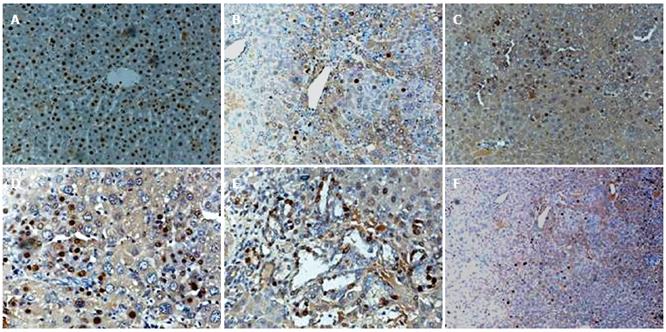
Figure 4 Proliferation and differentiation of FLEP cells in DEN-treated mice and normal mice.
A: Immunohistochemical positive control staining for sry protein; normal male mouse liver with nuclear positivity in the majority of hepatocytes (brown nuclei); B: One month after FLEP cell transplantation and DEN treatment, some cells stained for sry. C: Two months after FLEP cell transplantation and DEN treatment, sry+ cells were present in liver parenchyma. D and E: Three months after FLEP cell transplantation and DEN treatment, sry+ cells formed numerous mature hepatocytes showed canalicular structures (D), or bile duct epithelial cells in bile duct region, as evidenced by diffusely stained sry+ small epithelial cells in bile duct-like structures (E); F: Three months after FLEP cell transplantation without DEN treatment, scattered sry+ cells were observed. Original magnifications, x 200 (A-E) and x100 (D).
- Citation: Zheng JF, Liang LJ, Wu CX, Chen JS, Zhang ZS. Transplantation of fetal liver epithelial progenitor cells ameliorates experimental liver fibrosis in mice. World J Gastroenterol 2006; 12(45): 7292-7298
- URL: https://www.wjgnet.com/1007-9327/full/v12/i45/7292.htm
- DOI: https://dx.doi.org/10.3748/wjg.v12.i45.7292


