Editorial Open Access
Copyright ©2006 Baishideng Publishing Group Co., Limited. All rights reserved.
World J Gastroenterol. Sep 28, 2006; 12(36): 5767-5771
Published online Sep 28, 2006. doi: 10.3748/wjg.v12.i36.5767
Gastric carditis: Is it a histological response to high concentrations of luminal nitric oxide?
Katsunori Iijima, Tooru Shimosegawa, Division of Gastroenterology, Tohoku University Graduate School of Medicine, Sendai, Miyagi, Japan
Author contributions: All authors contributed equally to the work.
Correspondence to: Katsunori Iijima, MD, Division of Gastroenterology, Tohoku University Graduate School of Medicine, Seiryo-machi, Aobaku, Sendai 980-8574, Japan. jiijima@int3.med.tohoku.ac.jp
Telephone: +81-22-7177171 Fax: +81-22-7177177
Received: July 25, 2006
Revised: August 6, 2006
Accepted: August 11, 2006
Published online: September 28, 2006

Abstract

During the last decade, inflammation (carditis) and intestinal metaplasia localized to immediately below the human gastro-oesophageal junction have received much attention in relation to the rising incidence of cancer at this site. Since these histological findings are frequently observed even among those who are H pylori-negative, the causative factors for such histologic events at the human gastro-oesophageal junction remain obscure. A series of recent studies have demonstrated that a high level of salivary nitrite is sustained over several hours after the ingestion of a high nitrate meal, and that the nitrite in swallowed saliva is rapidly converted to nitric oxide by an acid catalyzed chemical reaction at the gastro-oesophageal junction. Eventually, a substantial amount of nitric oxide diffuses from the lumen into the adjacent tissue. Therefore, the human gastro-oesophageal junction is likely to be a region of high nitrosative stress. Considering the life-time exposure of the gastro-oesophageal junction to cytotoxic levels of nitric oxide, this may account for the high prevalence of inflammation, intestinal metaplasia, and subsequent development of neoplasia at this site. Although gastric acid, pepsin, and bile acid have been intensively investigated as a cause of adenocarcinoma at the gastro-oesophageal junction and the distal esophagus, nitric oxide and the related nitrosative stress should also be examined.

Key Words: Nitric oxide; Gastro-oesophageal junction; Dietary nitrate; Carditis; Intestinal metaplasia at gastro-oesophageal junction


CARDITIS AND INTESTINAL METAPLASIA AT THE GASTRO-ESOPHAGEAL JUNCTION

The incidence of adenocarcinoma of the cardia of the stomach and adjacent gastro-oesophageal (GO) junction has been rising over the past 25 years, especially in Western countries[1]. In recent years, a similar trend has been reported in Japan, although GO junction adenocarcinoma is still rare[2]. We recently reported that this type of cancer occurs in subjects with preserved gastric acid secretion and is not associated with H pylori infection[3]. Although gastric acid, pepsin, and bile have a deleterious effect on the epithelium at that site, the carcinogen for this type of cancer remains unknown. A sequence of histological events in which chronic inflammation progresses to metaplasia, dysplasia and, finally, carcinoma has been documented in distal gastric cancer, and is associated with H pylori infection in most cases. The sequence is also well defined for esophageal adenocarcinoma through the formation of intestinal metaplasia (IM) in the distal esophagus (Barrett’s esophagus). Although such a process has not been established in the cardia, a similar sequence of events would occur at that site as IM at the squamo-columnar junction which was shown to have a malignant potential similar to that of Barrett’s esophagus[4,5].

During the last decade, inflammation (carditis) and IM localized to the immediate vicinity of the squamo-columnar junction have received much attention in relation to the rising incidence of cancer at this site. These histological findings are reported to be highly prevalent (38%-79% for carditis[6-10], 10%-36% for IM[7,11-15]) even among healthy subjects. Of these two histological parameters, the junctional IM is usually associated with carditis[7,15], and hence the IM could be assumed to be a consequence of chronic inflammation at the cardia as in more distal areas of the stomach[18]. There are two distinct patterns of carditis[6,7,9,10]. One is associated with H pylori infection and is characterized by inflammation in other sites of the stomach. The other, which is unassociated with H pylori infection, comprises inflammation confined to the cardiac mucosa and is seen in patients with otherwise healthy stomachs. There are also differences in the grade and activity of the inflammatory infiltrate involving the columnar mucosa at the squamo-columnar junction, that is, patients with H pylori-associated carditis have more severe inflammation than patients with healthy stomachs and neutrophil infiltration is uncommon in the latter group[6,7,10,17]. Although carditis in H pylori-negative subjects is sometimes accompanied by the presence of gastro-oesophageal reflux disease[6-10,16,17], its cause remains obscure and it may well represent the histological response to many different types of insult. Considering the milder inflammation usually seen in the absence of neutrophil infiltration, chemical insult can be a probable candidate as a cause of the carditis.

LUMINAL NITRIC OXIDE GENERATION AT THE GASTRO-OESOPHAGEAL JUNCTION

Recently, the hypothesis that a high concentration of luminal nitric oxide (NO) at the human GO junction after nitrate ingestion may be relevant to various diseases at that site was made by a group of researchers in Glasgow[19,20]. Since then, that group and the present authors have been doing further research on this issue[21-24].

NO is an important radical that mediates a wide range of physiologic and pathologic events. It is generated at low concentrations by the enzyme constitutive nitric oxide synthase (cNOS) to modulate neuromuscular and vascular functions. Higher concentrations are generated by the inducible form of the enzyme as a part of the immune and inflammatory response. Sustained generation of NO by inducible NO synthase (iNOS) has been implicated in the aetiology of the mutagenesis and neoplasia related to chronic inflammation.

The highest concentrations of NO occurring in the body are not the result of enzymatic synthesis but rather by chemical reactions within the lumen of the stomach, especially in the most proximal area. A previous study reported that high, potentially mutagenic concentrations of NO are generated luminally at the GO junction through the entero-salivary re-circulation of dietary nitrate in humans[19]. This is explained as follows. Ingested nitrate as an ingredient of food is absorbed from the small intestine, and 25% of this is re-secreted into the mouth by the salivary glands. Bacteria on the dorsum of the tongue then reduce about 30% of this nitrate to nitrite. When salivary nitrite enters the stomach, the combination of the acidity and the ascorbic acid content of the gastric juice converts the nitrite to NO. Since this reaction between nitrite and ascorbic acid at an acidic pH is very rapid[21], the intraluminal concentration of NO generated by the reaction is maximal at the GO junction and cardia, where the nitrite in saliva first encounters gastric acid. Indeed, this was confirmed by the afore-mentioned study in healthy volunteers which reported that substantial amounts of NO are generated following nitrate ingestion at these anatomical locations, in some case in excess of 50 μmol/L[19].

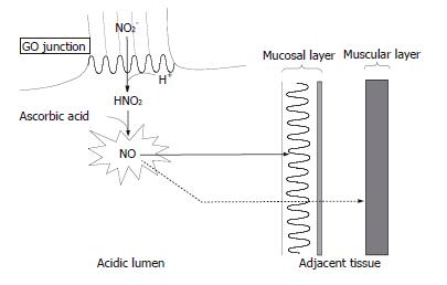
The entero-salivary re-circulation of dietary nitrate is sustained for several hours, during which period the adjacent epithelium of the GO junction is exposed to abundant amounts of NO generated in the lumen. Membranes in the tissues are not barriers to the diffusion of NO because of its gaseous and lipophilic properties. Since NO is known to have dual effects within the tissue, i. e. cytoprotective and cytotoxic depending on the gas level, determination of the NO level is required to evaluate its function in the tissue. We recently developed a rat animal model in which nitrite and acidified ascorbic acid were administered separately so that the generation of NO from nitrite would be maximal at the GO junction where the reactants first meet[24]. In that model, we demonstrated by means of electron paramagnetic resonance spectroscopy with an exogenously supplied NO trapping agent that the NO generated luminally diffused into adjacent tissues to a substantial degree and at a level comparable to that of iNOS derived-NO production and altered the integrity of the surrounding epithelium. As we applied a concentration of nitrite that has been observed in human saliva after the ingestion of a high nitrate meal, this finding may be applicable to the GO junction in humans[24] (Figure 1).

Figure 1
Figure 1 Nitric oxide chemistry at human gastro-oesophageal junction. Nitrite (NO2-) in swallowed saliva is converted to nitric oxide (NO) promptly at the gastro-oesophageal (GO) junction where encountering gastric acid containing ascorbic acid. NO thus formed diffuses into the adjacent tissue of the GO junction because of its gaseous and lipophilic properties. The majority of the NO arising from the lumen will be exhausted within the superficial mucosal layer by reacting with surrounding molecules while a small portion of the NO can reach the inner muscular layer. The luminal generation of NO is sustained for several hours after nitrate ingestion, during which period the GO junction is exposed to abundant amounts of NO.

Briefly, a high level of salivary nitrite is sustained over several hours after the ingestion of a high nitrate meal and the nitrite in swallowed saliva is rapidly converted to NO at the GO junction. Then, a substantial amount of NO eventually diffuses from the lumen into the adjacent tissue. Therefore, the human GO junction is likely to be a region of high nitrosative stress. Considering the life-time exposure of the GO junction to a high level of NO, this may account for the high prevalence of inflammation and IM at that site. Assuming the metaplasia-dysplasia-adenocarcinoma sequence at the GO junction, NO may eventually lead to the development of carcinoma at that site. In addition to being mediated through such a sequence, a high concentration of NO can directly exert a mutagenic and carcinogenic effect through the formation of higher oxides of nitrogen such as N2O3, which can damage DNA directly via deamination of bases and indirectly by forming N-nitrosocompounds. N2O3 is also known to inactivate DNA repair enzymes such as O6-alkylguanine DNA alkyltransferase[25-27].

POSSIBLE IMPLICATIONS OF NITRIC OXIDE AT THE GASTRO-OESOPHAGEAL JUNCTION

In this chemical reaction occurring at the GO junction, the presence of sufficient gastric acid is essential, and this condition is ensured only at the GO junction and the cardia after eating, where the acidic gastric juice largely escapes the buffering effect of food, which decreases the acidity of the gastric juice in the rest of the stomach[28]. The necessity of gastric acid for luminal NO generation is consistent with our recent clinical study which found that the preservation of gastric acid is necessary for the development of cancer at the GO junction[3]. In addition, a recent study has demonstrated that the site of NO generation shifted orally to the distal esophagus in cases with gastro-oesophageal reflux[23]. Hence, the NO thus generated in the distal esophagus may also be relevant to the formation of Barrett’s oesophagus and oesophageal adenocarcinoma.

Interestingly, although the majority of NO arising from the lumen would become exhausted within the superficial epithelial layer in our rat model, a small but significant amount of NO was detected even in the inner muscular layer of the GO junction, especially when a high concentration of nitrite was administered[24]. This suggests that a part of the nitric oxide arising from the lumen could escape from autoxidation occurring within the superficial layer and reach the inner tissue. It is well known that NO endogenously derived from cNOS localized to non-adrenergic, non-cholinergic nerves mediates the relaxation of the smooth muscle cells, including those of the lower esophageal sphincter[29]. An in vitro study using muscle strips from opossum lower esophageal sphincter demonstrated that a tiny amount of NO (in the range of nmol/L) was sufficient to induce relaxation of the muscle[30]. Therefore, the amount of NO formed in the lumen at the human GO junction may be sufficient to penetrate the epithelium and then affect the inner smooth muscle cell of the lower esophageal sphincter, leading to relaxation of the muscle and reflux of the acidic gastric content into the oesophagus. Actually, a recent report demonstrated in humans that NO generated luminally at the GO junction did affect the lower esophageal sphincter, leading to a significant increase in the transient relaxation of the lower esophageal sphincter[31]. Further studies are required to clarify to what extent this phenomenon is responsible for the pathogenesis of ordinary gastro-oesophageal reflux disease in humans. However, it may be related at least to the symptoms of heartburn that are known to be invoked after the ingestion of certain foods[32,33] (Figure 1).

Needless to say, NO is known to have some beneficial effects on the stomach as well. It has been shown that intragastric topical application or parental administration of nitric oxide-releasing substances protects the gastric mucosa from damage induced by injurious agents, suggesting that NO plays an important role in the protection of the gastric mucosa[34-36]. However, this protective effect is dose-sensitive; as some studies have reported that high doses of exogenous NO can exacerbate gastric mucosal injury[37,38]. Thus, a high concentration of local NO at the gastric cardia may be involved in the pathology of mucosal injury at that site as a potential cytotoxic mediator. On the other hand, a relatively low concentration of nitric oxide as seen in the distal stomach may function as a protective mediator to maintain the gastric mucosal integrity. Similarly, ascorbic acid, which is secreted in the healthy human stomach, could show even an undesirable effect on the GO junction by the rapid conversion of salivary nitrite to NO at that site[21,22], although the vitamin is well known to have beneficial effects as an antioxidant in the remaining stomach.

FUTURE STUDIES ON THE RELEVANCE OF NITRIC OXIDE TO THE VARIOUS DISEASES AT THE GASTRO-OESOPHAGEAL JUNCTION

Further studies to investigate the effect of the diffused NO on the cell biology in the tissue are required to clarify the pathologic relevance of NO to the various histological events occurring at the human GO junction. Accordingly, the characteristic features of NO, for example, the gas is generated abundantly by the unique chemical reaction in the acidic lumen, need to be considered with reference to other examples of pathological circumstances involving NO such as models of inflammation in humans. First, in inflammation, not only NO but other radicals such as superoxides are also generated from inflammatory cells and then the reaction products with NO (for example, peroxynitrite), not NO itself, may be exerting deleterious effects on the tissue[39]. In contrast, the effect of the diffused NO on the tissue at the GO junction should be mainly an NO-mediated process, at least at the early stage, although once the inflammatory process is initiated by the diffused NO other radicals are also formed. Second, since once the NO is diffused from the acidic lumen into the tissue at neutral pH it will be irreversibly metabolized to nitrite and nitrate, it is the very surface of the epithelium that is exposed to the highest concentration of NO, in contrast with examples of inflammation in which the immediate vicinity of inflammatory cells expressing iNOS would be exposed to the highest levels of endogenous NO[39]. Hence, the tight junction of the surface epithelium at the GO junction is likely to be the principal target of the NO arising from the lumen[40,41]. Third, as opposed to the inflammation that occurs only under specific circumstances, the exposure of the human GO junction to localized luminal NO occurs as long as the stomach is healthy and has sufficient acid secretion. In fact, it has been demonstrated that, after the ingestion of nitrate, the luminal NO concentration was maximal at the GO junction in 73% of subjects, although the gas level varied among individuals[19]. Thus, it appears reasonable that the locally generated NO is the cause of inflammation and IM at the cardia, both of which are frequently observed even in healthy subjects as previously mentioned. However, since neoplasia develops only in a small number of people, individual susceptibility to NO toxicity such as from mutations in cancer-related genes (for example p53) may be important in determining its ultimate contribution to carcinogenesis at the GO junction[42].

In conclusion, a cytotoxic concentration of NO is generated luminally at the human GO junction and the gas diffuses into the adjacent tissue. Although gastric acid, pepsin, and bile acid have been intensively investigated as a cause of adenocarcinoma at the GO junction and the distal esophagus, NO and the related nitrosative stress should also be examined.

Footnotes

S- Editor Liu Y L- Editor Alpini GD E- Editor Bi L

References
1.  Blot WJ, Devesa SS, Kneller RW, Fraumeni JF Jr. Rising incidence of adenocarcinoma of the esophagus and gastric cardia. JAMA. 1991;265:1287-1289.  [PubMed]  [DOI]  [Cited in This Article: ]  [Cited by in Crossref: 1287]  [Cited by in F6Publishing: 1132]  [Article Influence: 33.3]  [Reference Citation Analysis (0)]
2.  Okabayashi T, Gotoda T, Kondo H, Inui T, Ono H, Saito D, Yoshida S, Sasako M, Shimoda T. Early carcinoma of the gastric cardia in Japan: is it different from that in the West. Cancer. 2000;89:2555-2559.  [PubMed]  [DOI]  [Cited in This Article: ]  [Cited by in F6Publishing: 4]  [Reference Citation Analysis (0)]
3.  Inomata Y, Koike T, Ohara S, Abe Y, Sekine H, Iijima K, Ariizumi K, Yamagishi H, Kitagawa Y, Imatani A. Preservation of gastric acid secretion may be important for the development of gastroesophageal junction adenocarcinoma in Japanese people, irrespective of the H. pylori infection status. Am J Gastroenterol. 2006;101:926-933.  [PubMed]  [DOI]  [Cited in This Article: ]  [Cited by in Crossref: 34]  [Cited by in F6Publishing: 31]  [Article Influence: 1.6]  [Reference Citation Analysis (0)]
4.  Trudgill NJ, Suvarna SK, Royds JA, Riley SA. Cell cycle regulation in patients with intestinal metaplasia at the gastro-oesophageal junction. Mol Pathol. 2003;56:313-317.  [PubMed]  [DOI]  [Cited in This Article: ]  [Cited by in Crossref: 17]  [Cited by in F6Publishing: 18]  [Article Influence: 0.9]  [Reference Citation Analysis (0)]
5.  Ruol A, Parenti A, Zaninotto G, Merigliano S, Costantini M, Cagol M, Alfieri R, Bonavina L, Peracchia A, Ancona E. Intestinal metaplasia is the probable common precursor of adenocarcinoma in barrett esophagus and adenocarcinoma of the gastric cardia. Cancer. 2000;88:2520-2528.  [PubMed]  [DOI]  [Cited in This Article: ]  [Cited by in F6Publishing: 12]  [Reference Citation Analysis (0)]
6.  Bowrey DJ, Clark GW, Williams GT. Patterns of gastritis in patients with gastro-oesophageal reflux disease. Gut. 1999;45:798-803.  [PubMed]  [DOI]  [Cited in This Article: ]  [Cited by in Crossref: 33]  [Cited by in F6Publishing: 39]  [Article Influence: 1.5]  [Reference Citation Analysis (0)]
7.  Oksanen A, Sipponen P, Karttunen R, Rautelin H. Inflammation and intestinal metaplasia at the squamocolumnar junction in young patients with or without Helicobacter pylori infection. Gut. 2003;52:194-198.  [PubMed]  [DOI]  [Cited in This Article: ]  [Cited by in Crossref: 14]  [Cited by in F6Publishing: 18]  [Article Influence: 0.8]  [Reference Citation Analysis (0)]
8.  Lembo T, Ippoliti AF, Ramers C, Weinstein WM. Inflammation of the gastro-oesophageal junction (carditis) in patients with symptomatic gastro-oesophageal reflux disease: a prospective study. Gut. 1999;45:484-488.  [PubMed]  [DOI]  [Cited in This Article: ]  [Cited by in Crossref: 45]  [Cited by in F6Publishing: 44]  [Article Influence: 1.7]  [Reference Citation Analysis (0)]
9.  Voutilainen M, Färkkilä M, Mecklin JP, Juhola M, Sipponen P. Chronic inflammation at the gastroesophageal junction (carditis) appears to be a specific finding related to Helicobacter pylori infection and gastroesophageal reflux disease. The Central Finland Endoscopy Study Group. Am J Gastroenterol. 1999;94:3175-3180.  [PubMed]  [DOI]  [Cited in This Article: ]  [Cited by in Crossref: 41]  [Cited by in F6Publishing: 44]  [Article Influence: 1.7]  [Reference Citation Analysis (0)]
10.  Zentilin P, Mastracci L, Dulbecco P, Gambaro C, Bilardi C, Ceppa P, Spaggiari P, Iiritano E, Mansi C, Vigneri S. Carditis in patients with gastro-oesophageal reflux disease: results of a controlled study based on both endoscopy and 24-h oesophageal pH monitoring. Aliment Pharmacol Ther. 2004;19:1285-1292.  [PubMed]  [DOI]  [Cited in This Article: ]  [Cited by in Crossref: 9]  [Cited by in F6Publishing: 8]  [Article Influence: 0.4]  [Reference Citation Analysis (0)]
11.  McNamara D, Buckley M, Crotty P, Hall W, O'Sullivan M, O'Morain C. Carditis: all Helicobacter pylori or is there a role for gastro-oesophageal reflux. Scand J Gastroenterol. 2002;37:772-777.  [PubMed]  [DOI]  [Cited in This Article: ]
12.  Hackelsberger A, Günther T, Schultze V, Manes G, Dominguez-Muñoz JE, Roessner A, Malfertheiner P. Intestinal metaplasia at the gastro-oesophageal junction: Helicobacter pylori gastritis or gastro-oesophageal reflux disease. Gut. 1998;43:17-21.  [PubMed]  [DOI]  [Cited in This Article: ]  [Cited by in Crossref: 103]  [Cited by in F6Publishing: 114]  [Article Influence: 4.2]  [Reference Citation Analysis (0)]
13.  Trudgill NJ, Suvarna SK, Kapur KC, Riley SA. Intestinal metaplasia at the squamocolumnar junction in patients attending for diagnostic gastroscopy. Gut. 1997;41:585-589.  [PubMed]  [DOI]  [Cited in This Article: ]  [Cited by in Crossref: 91]  [Cited by in F6Publishing: 102]  [Article Influence: 3.6]  [Reference Citation Analysis (0)]
14.  Spechler SJ, Zeroogian JM, Antonioli DA, Wang HH, Goyal RK. Prevalence of metaplasia at the gastro-oesophageal junction. Lancet. 1994;344:1533-1536.  [PubMed]  [DOI]  [Cited in This Article: ]  [Cited by in Crossref: 409]  [Cited by in F6Publishing: 415]  [Article Influence: 13.4]  [Reference Citation Analysis (0)]
15.  Voutilainen M, Färkkilä M, Juhola M, Nuorva K, Mauranen K, Mäntynen T, Kunnamo I, Mecklin JP, Sipponen P. Specialized columnar epithelium of the esophagogastric junction: prevalence and associations. The Central Finland Endoscopy Study Group. Am J Gastroenterol. 1999;94:913-918.  [PubMed]  [DOI]  [Cited in This Article: ]  [Cited by in Crossref: 46]  [Cited by in F6Publishing: 46]  [Article Influence: 1.8]  [Reference Citation Analysis (0)]
16.  Oberg S, Peters JH, DeMeester TR, Chandrasoma P, Hagen JA, Ireland AP, Ritter MP, Mason RJ, Crookes P, Bremner CG. Inflammation and specialized intestinal metaplasia of cardiac mucosa is a manifestation of gastroesophageal reflux disease. Ann Surg. 1997;226:522-530; discussion 530-532.  [PubMed]  [DOI]  [Cited in This Article: ]  [Cited by in Crossref: 235]  [Cited by in F6Publishing: 267]  [Article Influence: 9.5]  [Reference Citation Analysis (0)]
17.  Chen YY, Antonioli DA, Spechler SJ, Zeroogian JM, Goyal RK, Wang HH. Gastroesophageal reflux disease versus Helicobacter pylori infection as the cause of gastric carditis. Mod Pathol. 1998;11:950-956.  [PubMed]  [DOI]  [Cited in This Article: ]
18.  Correa P. A human model of gastric carcinogenesis. Cancer Res. 1988;48:3554-3560.  [PubMed]  [DOI]  [Cited in This Article: ]
19.  Iijima K, Henry E, Moriya A, Wirz A, Kelman AW, McColl KE. Dietary nitrate generates potentially mutagenic concentrations of nitric oxide at the gastroesophageal junction. Gastroenterology. 2002;122:1248-1257.  [PubMed]  [DOI]  [Cited in This Article: ]  [Cited by in Crossref: 120]  [Cited by in F6Publishing: 122]  [Article Influence: 5.3]  [Reference Citation Analysis (0)]
20.  McColl KE. When saliva meets acid: chemical warfare at the oesophagogastric junction. Gut. 2005;54:1-3.  [PubMed]  [DOI]  [Cited in This Article: ]  [Cited by in Crossref: 74]  [Cited by in F6Publishing: 73]  [Article Influence: 3.7]  [Reference Citation Analysis (0)]
21.  Iijima K, Fyfe V, McColl KE. Studies of nitric oxide generation from salivary nitrite in human gastric juice. Scand J Gastroenterol. 2003;38:246-252.  [PubMed]  [DOI]  [Cited in This Article: ]
22.  Iijima K, Grant J, McElroy K, Fyfe V, Preston T, McColl KE. Novel mechanism of nitrosative stress from dietary nitrate with relevance to gastro-oesophageal junction cancers. Carcinogenesis. 2003;24:1951-1960.  [PubMed]  [DOI]  [Cited in This Article: ]  [Cited by in Crossref: 55]  [Cited by in F6Publishing: 54]  [Article Influence: 2.5]  [Reference Citation Analysis (0)]
23.  Suzuki H, Iijima K, Scobie G, Fyfe V, McColl KE. Nitrate and nitrosative chemistry within Barrett's oesophagus during acid reflux. Gut. 2005;54:1527-1535.  [PubMed]  [DOI]  [Cited in This Article: ]  [Cited by in Crossref: 70]  [Cited by in F6Publishing: 68]  [Article Influence: 3.4]  [Reference Citation Analysis (0)]
24.  Asanuma K, Iijima K, Sugata H, Ohara S, Shimosegawa T, Yoshimura T. Diffusion of cytotoxic concentrations of nitric oxide generated luminally at the gastro-oesophageal junction of rats. Gut. 2005;54:1072-1077.  [PubMed]  [DOI]  [Cited in This Article: ]  [Cited by in Crossref: 29]  [Cited by in F6Publishing: 29]  [Article Influence: 1.5]  [Reference Citation Analysis (0)]
25.  Wink DA, Fellisch M, Vodovotz Y, Fukuto J, Grisham MB. The chemical biology of nitric oxide. Reactive oxygen species in biological system. New York: Kluwer Academic/Plenum Publishers 1999; 245-292.  [PubMed]  [DOI]  [Cited in This Article: ]
26.  Ruiz F, Corrales FJ, Miqueo C, Mato JM. Nitric oxide inactivates rat hepatic methionine adenosyltransferase In vivo by S-nitrosylation. Hepatology. 1998;28:1051-1057.  [PubMed]  [DOI]  [Cited in This Article: ]  [Cited by in Crossref: 94]  [Cited by in F6Publishing: 96]  [Article Influence: 3.6]  [Reference Citation Analysis (0)]
27.  Jaiswal M, LaRusso NF, Burgart LJ, Gores GJ. Inflammatory cytokines induce DNA damage and inhibit DNA repair in cholangiocarcinoma cells by a nitric oxide-dependent mechanism. Cancer Res. 2000;60:184-190.  [PubMed]  [DOI]  [Cited in This Article: ]
28.  Fletcher J, Wirz A, Young J, Vallance R, McColl KE. Unbuffered highly acidic gastric juice exists at the gastroesophageal junction after a meal. Gastroenterology. 2001;121:775-783.  [PubMed]  [DOI]  [Cited in This Article: ]  [Cited by in Crossref: 225]  [Cited by in F6Publishing: 220]  [Article Influence: 9.2]  [Reference Citation Analysis (0)]
29.  Murray J, Du C, Ledlow A, Bates JN, Conklin JL. Nitric oxide: mediator of nonadrenergic noncholinergic responses of opossum esophageal muscle. Am J Physiol. 1991;261:G401-G406.  [PubMed]  [DOI]  [Cited in This Article: ]
30.  Knudsen MA, Svane D, Tøttrup A. Action profiles of nitric oxide, S-nitroso-L-cysteine, SNP, and NANC responses in opossum lower esophageal sphincter. Am J Physiol. 1992;262:G840-G846.  [PubMed]  [DOI]  [Cited in This Article: ]
31.  Manning JJ, Wirz A, McColl KEL. Chemicals formed from the gastric acidification of salivary nitrite influence oesophageal and gastric function. Gastroenterology. 2006;A-56.  [PubMed]  [DOI]  [Cited in This Article: ]
32.  Cranley JP, Achkar E, Fleshler B. Abnormal lower esophageal sphincter pressure responses in patients with orange juice-induced heartburn. Am J Gastroenterol. 1986;81:104-106.  [PubMed]  [DOI]  [Cited in This Article: ]
33.  Allen ML, Mellow MH, Robinson MG, Orr WC. The effect of raw onions on acid reflux and reflux symptoms. Am J Gastroenterol. 1990;85:377-380.  [PubMed]  [DOI]  [Cited in This Article: ]
34.  Kitagawa H, Takeda F, Kohei H. Effect of endothelium-derived relaxing factor on the gastric lesion induced by HCl in rats. J Pharmacol Exp Ther. 1990;253:1133-1137.  [PubMed]  [DOI]  [Cited in This Article: ]
35.  Miyoshi M, Kasahara E, Park AM, Hiramoto K, Minamiyama Y, Takemura S, Sato EF, Inoue M. Dietary nitrate inhibits stress-induced gastric mucosal injury in the rat. Free Radic Res. 2003;37:85-90.  [PubMed]  [DOI]  [Cited in This Article: ]  [Cited by in Crossref: 52]  [Cited by in F6Publishing: 54]  [Article Influence: 2.5]  [Reference Citation Analysis (0)]
36.  Björne H H, Petersson J, Phillipson M, Weitzberg E, Holm L, Lundberg JO. Nitrite in saliva increases gastric mucosal blood flow and mucus thickness. J Clin Invest. 2004;113:106-114.  [PubMed]  [DOI]  [Cited in This Article: ]
37.  Lopez-Belmonte J, Whittle BJ, Moncada S. The actions of nitric oxide donors in the prevention or induction of injury to the rat gastric mucosa. Br J Pharmacol. 1993;108:73-78.  [PubMed]  [DOI]  [Cited in This Article: ]  [Cited by in Crossref: 150]  [Cited by in F6Publishing: 153]  [Article Influence: 4.8]  [Reference Citation Analysis (0)]
38.  Wakulich CA, Tepperman BL. Role of glutathione in nitric oxide-mediated injury to rat gastric mucosal cells. Eur J Pharmacol. 1997;319:333-341.  [PubMed]  [DOI]  [Cited in This Article: ]  [Cited by in Crossref: 20]  [Cited by in F6Publishing: 21]  [Article Influence: 0.8]  [Reference Citation Analysis (0)]
39.  Grisham MB, Jourd'Heuil D, Wink DA. Nitric oxide. I. Physiological chemistry of nitric oxide and its metabolites: implications in inflammation. Am J Physiol. 1999;276:G315-G321.  [PubMed]  [DOI]  [Cited in This Article: ]
40.  Salzman AL, Menconi MJ, Unno N, Ezzell RM, Casey DM, Gonzalez PK, Fink MP. Nitric oxide dilates tight junctions and depletes ATP in cultured Caco-2BBe intestinal epithelial monolayers. Am J Physiol. 1995;268:G361-G373.  [PubMed]  [DOI]  [Cited in This Article: ]
41.  Mishima S, Xu D, Deitch EA. Increase in endotoxin-induced mucosal permeability is related to increased nitric oxide synthase activity using the Ussing chamber. Crit Care Med. 1999;27:880-886.  [PubMed]  [DOI]  [Cited in This Article: ]  [Cited by in Crossref: 34]  [Cited by in F6Publishing: 34]  [Article Influence: 1.3]  [Reference Citation Analysis (0)]
42.  Hofseth LJ, Hussain SP, Wogan GN, Harris CC. Nitric oxide in cancer and chemoprevention. Free Radic Biol Med. 2003;34:955-968.  [PubMed]  [DOI]  [Cited in This Article: ]  [Cited by in Crossref: 156]  [Cited by in F6Publishing: 145]  [Article Influence: 6.6]  [Reference Citation Analysis (0)]