Published online Jul 14, 2006. doi: 10.3748/wjg.v12.i26.4270
Revised: March 20, 2006
Accepted: March 27, 2006
Published online: July 14, 2006
In this report, we present computed tomographic findings of colonic trichuriasis. The patient was a 75-year-old man who complained of abdominal pain, and weight loss. Diagnosis was achieved by colonoscopic biopsy. Abdominal computed tomography showed irregular and nodular thickening of the wall of the cecum and ascending colon. Although these findings are nonspecific, they may be one of the findings of trichuriasis. These findings, confirmed by pathologic analysis of the biopsied tissue and Kato-Katz parasitological stool flotation technique, revealed adult Trichuris. To our knowledge, this is the first report of colonic trichuriasis indicated by computed tomography.
- Citation: Tokmak N, Koc Z, Ulusan S, Koltas IS, Bal N. Computed tomographic findings of trichuriasis. World J Gastroenterol 2006; 12(26): 4270-4272
- URL: https://www.wjgnet.com/1007-9327/full/v12/i26/4270.htm
- DOI: https://dx.doi.org/10.3748/wjg.v12.i26.4270
Trichuris trichiura (T. trichiura), a whipworm, is the third most common nematode worldwide after Ascaris and Enterobius[1]. First described by Roederer in 1761, the whipworm is found primarily in tropical climates characterized by poor sanitation[1]. Whipworm infection usually causes no clinical symptoms, although a severe infection can cause abdominal pain, diarrhea, constipation, weight loss, and anemia[2-5].
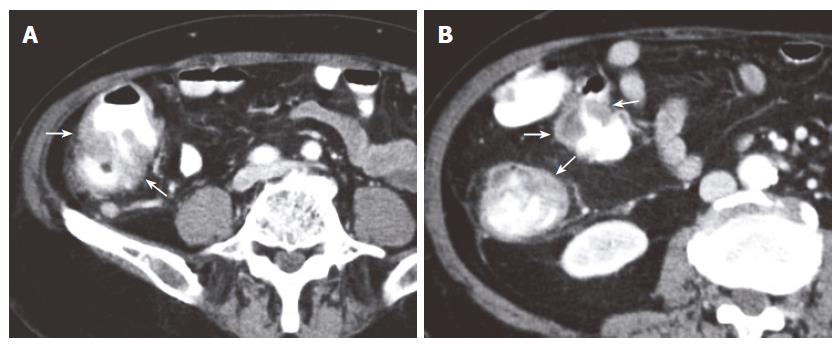
In the patient described in this report, we identified irregular and nodular marked thickening of the wall of the cecum and ascending colon as a CT finding of trichuriasis. To our knowledge, this is the first report of colonic trichuriasis revealed by that modality. A follow-up CT scan showed that the colonic manifestations of trichuriasis had resolved six months after the conclusion of the medical treatment.
A 75-year-old man with suspected colon carcinoma was referred to our radiology unit for evaluation by computed tomography (CT). He complained of abdominal pain, constipation, and a weight loss of 5 kg in the prior two months. Physical examination revealed mild tenderness in the right lower abdomen. The patient’s white blood cell count was 13.9 × 10⁹/L (reference intervals, 5-11 × 109/L) without eosinophilia. The results of other laboratory tests (including biochemical analysis) were within the normal range with the exception of moderate anemia (hemoglobin level, 90 g/L; reference intervals, 140-180 g/L).
CT scans were obtained using a four-channel MDCT scanner (Sensation 4; Siemens, Erlangen, Germany). A mixture of 1000 mL water and 40 mL contrast material was used for bowel opacification. Portal phase CT scans (2.5 mm collimation, 12.5 mm/0.5 s table speed) were acquired at 60 s after contrast material injection.
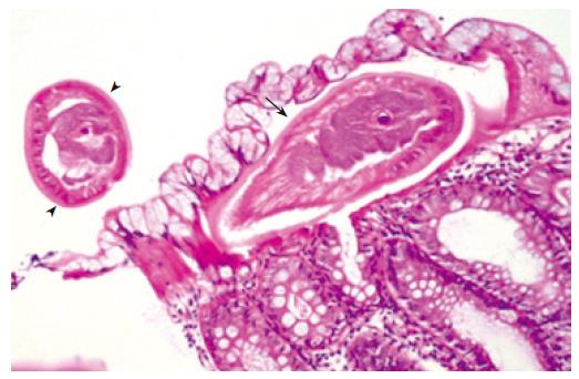
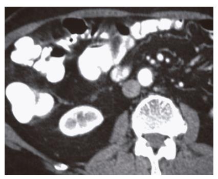
CT scan of the abdomen showed irregular and nodular marked thickening of the wall of the cecum and ascending colon (Figure 1A, B). Endoscopic examination revealed multiple ulcerated mucosal lesions in the ascending colon. The cecum could not be properly examined because of intestinal content. The results of pathologic analysis of the biopsied tissue, which was obtained colonoscopically from the ascending colon, revealed adult Trichuris and the signs of active colitis (Figure 2); these findings were confirmed by Kato-Katz parasitological stool flotation technique[6]. The patient was subsequently treated with mebendazole 100 mg twice daily for 3 d. On follow-up at the end of the treatment clinical findings had resolved, and a repeat CT scan obtained 6 mo after the treatment revealed a colonic wall thickness within the normal range (Figure 3) and no
T. trichiura eggs were seen in any stool specimens.
Trichuriasis is an intestinal infection found in human beings, which is caused by T. trichiura, more commonly known as whipworm because of its whip-like appearance. It is characterized by the invasion of the colonic mucosa by the adult Trichuris and produces minor inflammatory changes at the sites of localization. Trichuriasis remains a health problem in certain geographic areas. Infection is acquired by the ingestion of embryonated eggs from contaminated drinking water and food. The eggs hatch in the small intestine, and the larvae then enter the intestinal crypts[1]. After a brief period of maturation, the larvae may migrate to the proximal colon, where they reside and over 1 to 3 mo mature into adults[1,2,6,7]. The adult T. trichiura is a threadlike worm 30 to 50 mm in length. T. trichiura penetrates the intestinal mucosa and frequently triggers minor inflammatory changes in the cecum, appendix, and terminal ileum[7]. Diagnosis is usually based on the identification of typical T. trichiura eggs in stool specimens[6]. Adult whipworm is rarely seen during colonoscopy and colonoscopy can directly diagnose trichuriasis, confirming the threadlike form of worms with an attenuated end[7-11]. Literature research revealed that imaging findings sometimes similar with a tumor of the colon because of tumor-like appearance of the granulomatous tissue reaction due to T. trichiura[12].
A few reports have described the radiographic appearance of T. trichiura on double-contrast barium enema[13]. Characteristic findings revealed by that modality have included multiple tiny target-shaped or pinwheel-shaped collections of barium that are associated with peculiar s-shaped filling defects thought to be consistent with the shape of the adult male T. trichiura[13]. Colonic wall thickening is a common and nonspecific finding that occurs in many other conditions such as infections, inflammatory bowel disease and carcinoma. However, knowledge of this entity is helpful for differential diagnosis in the endemic areas of trichuriasis.
We concluded that colonoscopy with biopsy is the gold standard for such disease like trichuriasis. Irregular and nodular thickening of the cecum and the ascending colon on CT scan may be one of the findings of trichuriasis. Although that finding is nonspecific, parasitic infections including trichuriasis should be considered as differential diagnoses, especially in areas in which such infection is endemic.
S- Editor Wang J L- Editor Rampone B E- Editor Bi L
| 1. | Monroe LS. Gastrointestinal parasites. In : Haubrich WS, Schaffner F, Berk JE, eds. Bockus Gastroenterology. 5th ed. Philadelphia: Saunders 1995; 3113-3196. |
| 2. | Fishman JA, Perrone TL. Colonic obstruction and perforation due to Trichuris trichiura. Am J Med. 1984;77:154-156. [RCA] [PubMed] [DOI] [Full Text] [Cited by in Crossref: 19] [Cited by in RCA: 19] [Article Influence: 0.5] [Reference Citation Analysis (0)] |
| 3. | Bahon J, Poirriez J, Creusy C, Edriss AN, Laget JP, Dei Cas E. Colonic obstruction and perforation related to heavy Trichuris trichiura infestation. J Clin Pathol. 1997;50:615-616. [RCA] [PubMed] [DOI] [Full Text] [Cited by in Crossref: 17] [Cited by in RCA: 19] [Article Influence: 0.7] [Reference Citation Analysis (0)] |
| 4. | Huang NC, Fang HC, Chou KJ, Chung HM. Trichuris trichiura: an unusual cause of chronic diarrhoea in a renal transplant patient. Nephrol Dial Transplant. 2003;18:2434-2435. [RCA] [PubMed] [DOI] [Full Text] [Cited by in Crossref: 10] [Cited by in RCA: 10] [Article Influence: 0.5] [Reference Citation Analysis (0)] |
| 5. | Hong ST, Lim HS, Kim DH, Kim SJ. A case of gastroenteritis associated with gastric trichuriasis. J Korean Med Sci. 2003;18:429-432. [PubMed] |
| 6. | Santos FL, Cerqueira EJ, Soares NM. Comparison of the thick smear and Kato-Katz techniques for diagnosis of intestinal helminth infections. Rev Soc Bras Med Trop. 2005;38:196-198. [RCA] [PubMed] [DOI] [Full Text] [Cited by in Crossref: 33] [Cited by in RCA: 32] [Article Influence: 1.6] [Reference Citation Analysis (0)] |
| 7. | Chandra B, Long JD. Diagnosis of Trichuris trichiura (whipworm) by colonoscopic extraction. J Clin Gastroenterol. 1998;27:152-153. [RCA] [PubMed] [DOI] [Full Text] [Cited by in Crossref: 14] [Cited by in RCA: 13] [Article Influence: 0.5] [Reference Citation Analysis (0)] |
| 8. | Davis M, Matteson A, Williams WC. Radiographic and endoscopic findings in human whipworm infection (Trichuris trichiura). J Clin Gastroenterol. 1986;8:700-701. [RCA] [PubMed] [DOI] [Full Text] [Cited by in Crossref: 10] [Cited by in RCA: 9] [Article Influence: 0.2] [Reference Citation Analysis (0)] |
| 9. | Joo JH, Ryu KH, Lee YH, Park CW, Cho JY, Kim YS, Lee JS, Lee MS, Hwang SG, Shim CS. Colonoscopic diagnosis of whipworm infection. Hepatogastroenterology. 1998;45:2105-2109. [PubMed] |
| 10. | Yoshida M, Kutsumi H, Ogawa M, Soga T, Nishimura K, Tomita S, Kawabata K, Kinoshita Y, Chiba T, Fujimoto S. A case of Trichuris trichiura infection diagnosed by colonoscopy. Am J Gastroenterol. 1996;91:161-162. [PubMed] |
| 11. | Lorenzetti R, Campo SM, Stella F, Hassan C, Zullo A, Morini S. An unusual endoscopic finding: Trichuris trichiura. Case report and review of the literature. Dig Liver Dis. 2003;35:811-813. [RCA] [PubMed] [DOI] [Full Text] [Cited by in Crossref: 13] [Cited by in RCA: 13] [Article Influence: 0.6] [Reference Citation Analysis (0)] |
| 12. | Kojima Y, Sakuma H, Izumi R, Nakagawara G, Miyazaki I, Yoshimura H. A case of granuloma of the ascending colon due to penetration of Trichuris trichiura. Gastroenterol Jpn. 1981;16:193-196. [PubMed] |
| 13. | Manzano C, Thomas MA, Valenzuela C. Trichuriasis. Roentgenographic features and differential diagnosis with lymphoid hyperplasia. Pediatr Radiol. 1979;8:76-78. [RCA] [PubMed] [DOI] [Full Text] [Cited by in Crossref: 6] [Cited by in RCA: 4] [Article Influence: 0.1] [Reference Citation Analysis (0)] |


